catena
-Poly[[dimethylbis(thiocyanato-j
N
)tin(IV)]-
l
-(4,4
000-bipyridine-
j
2N
:
N
000)]
Ezzatollah Najafi,aMostafa M. Aminiaand Seik Weng Ngb*
aDepartment of Chemistry, General Campus, Shahid Beheshti University, Tehran
1983963113, Iran, andbDepartment of Chemistry, University of Malaya, 50603
Kuala Lumpur, Malaysia
Correspondence e-mail: seikweng@um.edu.my
Received 5 February 2011; accepted 14 February 2011
Key indicators: single-crystal X-ray study;T= 295 K; mean(C–C) = 0.005 A˚;
Rfactor = 0.023;wRfactor = 0.061; data-to-parameter ratio = 16.7.
The title dimethyltin diisothiocyanate adduct of 4,40 -bipyr-idine, [Sn(CH3)2(NCS)2(C10H8N2)]n, adopts a chain motif in which theN-heterocycle functions as a bridge to adjacent
all-transoctahedrally coordinated tin atoms. The SnIVatom lies on a special position of 2/msite symmetry, the methyl C atom on a special position of 2 site symmetry, and the thiocyanate and 4,40-bipyridine on a special position ofmsite symmetry.
Related literature
For the 4,40-bipyridine adducts of diorganotin dichlorides, see: Ma et al.(2004); Ng (1998). For the dimethyltin di(isothio-cyanate) adduct of 1,10-phenanthroline, see: Najafi et al.
(2011).
Experimental
Crystal data
[Sn(CH3)2(NCS)2(C10H8N2)]
Mr= 421.10
Monoclinic,C2=m a= 10.8697 (8) A˚
b= 7.7741 (6) A˚
c= 11.3979 (8) A˚
= 115.817 (1)
V= 867.0 (1) A˚3
Z= 2
MoKradiation
= 1.71 mm1
T= 295 K
0.300.200.10 mm
Data collection
Bruker SMART APEX diffractometer
Absorption correction: multi-scan (SADABS; Sheldrick, 1996)
Tmin= 0.628,Tmax= 0.847
4033 measured reflections 1066 independent reflections 1064 reflections withI> 2(I)
Rint= 0.021
Refinement
R[F2> 2(F2)] = 0.023
wR(F2) = 0.061
S= 1.08 1066 reflections
64 parameters
H-atom parameters constrained max= 0.49 e A˚
3 min=0.82 e A˚
3
Data collection:APEX2(Bruker, 2009); cell refinement:SAINT (Bruker, 2009); data reduction:SAINT; program(s) used to solve structure:SHELXS97(Sheldrick, 2008); program(s) used to refine structure: SHELXL97 (Sheldrick, 2008); molecular graphics: X-SEED (Barbour, 2001); software used to prepare material for publication:publCIF(Westrip, 2010).
We thank Shahid Beheshti University and the University of Malaya for supporting this study.
Supplementary data and figures for this paper are available from the IUCr electronic archives (Reference: SI2334).
References
Barbour, L. J. (2001).J. Supramol. Chem.1, 189–191.
Bruker (2009).APEX2andSAINT. Bruker AXS Inc., Madison, Wisconsin, USA.
Ma, C.-L., Zhang, J.-H. & Zhang, R.-F. (2004).Heteroatom. Chem.15, 338–346. Najafi, E., Amini, M. M. & Ng, S. W. (2011).Acta Cryst.E67. Submitted
(si2333).
Ng, S. W. (1998).Acta Cryst.C54, 1393–1395.
Sheldrick, G. M. (1996).SADABS. University of Go¨ttingen, Germany. Sheldrick, G. M. (2008).Acta Cryst.A64, 112–122.
Westrip, S. P. (2010).J. Appl. Cryst.43, 920–925.
Acta Crystallographica Section E Structure Reports Online
supporting information
Acta Cryst. (2011). E67, m350 [doi:10.1107/S1600536811005459]
catena
-Poly[[dimethylbis(thiocyanato-
κ
N
)tin(IV)]-
µ
-(4,4
′
-bipyridine-
κ
2N
:
N
′
)]
Ezzatollah Najafi, Mostafa M. Amini and Seik Weng Ng
S1. Comment
The 4,4′-bipyridine ligand forms a number of adducts with diorganotin dihalides; the adducts adopt linear chain structures
as the ligand functions in a bridging mode. The organotin dihalides include dimethyltin dichloride (Ng, 1998), dibutyltin
dichloride and dibenzyltin dichloride (Ma et al., 2004); no pseudohalides have been reported. The dimethyltin
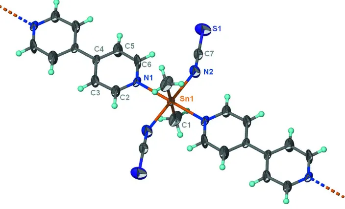
diisothio-cyanate adduct similarly adopts a chain motif (Scheme I, Fig. 1). Polymeric [Sn(NCS)2(CH3)2(C10H8N2)2]n has the N
-heterocycle functioning as a bridging to adjacent all-trans octahedrally coordinated tin atoms. The tin atom lies on a
special position of 2/m site symmetry, the methyl carbon on a special position of 2 site symmetry, and the isothiocyanate
and 4,4′-bipyridine on a special position of m site symmetry. The geometry of the tin atom in the dimethyltin
di(isothio-cyanate) adduct with 1,10-phenanthroline is a cis-octahedron (Najafi et al., 2011).
S2. Experimental
Dimethyltin diisothiocyanate (1 mmol, 0.26 g) and 4,4′-bipyridine (1 mmol, 0.16 g) were loaded into a convection tube.
The tube was filled with acetonitrile and methanol (v:v / 9:1) and kept at 333 K. Colorless crystals were collected from
the side arm after several days.
S3. Refinement
Hydrogen atoms were placed in calculated positions (C–H 0.93–0.96 Å) and were included in the refinement in the riding
Figure 1
Thermal ellipsoid plot (Barbour, 2001) of a portion of the Sn(NCS)2(CH3)2(C10H8N2) chain at the 50% probability level.
Hydrogen atoms are drawn as spheres of arbitrary radius.
catena-Poly[[dimethylbis(thiocyanato-κN)tin(IV)]- µ-(4,4′-bipyridine-κ2N:N′)]
Crystal data
[Sn(CH3)2(NCS)2(C10H8N2)] Mr = 421.10
Monoclinic, C2/m Hall symbol: -C 2y a = 10.8697 (8) Å b = 7.7741 (6) Å c = 11.3979 (8) Å β = 115.817 (1)° V = 867.0 (1) Å3
Z = 2
F(000) = 416 Dx = 1.613 Mg m−3
Mo Kα radiation, λ = 0.71073 Å Cell parameters from 3765 reflections θ = 3.8–28.3°
µ = 1.71 mm−1 T = 295 K Prism, colorless 0.30 × 0.20 × 0.10 mm
Data collection Bruker SMART APEX
diffractometer
Radiation source: fine-focus sealed tube Graphite monochromator
ω scans
Absorption correction: multi-scan
(SADABS; Sheldrick, 1996)
Tmin = 0.628, Tmax = 0.847
4033 measured reflections 1066 independent reflections 1064 reflections with I > 2σ(I) Rint = 0.021
θmax = 27.5°, θmin = 3.4°
h = −14→13 k = −10→10 l = −14→14
Refinement Refinement on F2 Least-squares matrix: full R[F2 > 2σ(F2)] = 0.023 wR(F2) = 0.061 S = 1.08 1066 reflections
64 parameters 0 restraints
Primary atom site location: structure-invariant direct methods
Hydrogen site location: inferred from neighbouring sites
H-atom parameters constrained
w = 1/[σ2(Fo2) + (0.048P)2 + 0.1158P] where P = (Fo2 + 2Fc2)/3
(Δ/σ)max = 0.001 Δρmax = 0.49 e Å−3
Δρmin = −0.82 e Å−3
Fractional atomic coordinates and isotropic or equivalent isotropic displacement parameters (Å2)
x y z Uiso*/Ueq Occ. (<1)
Sn1 0.5000 0.5000 0.5000 0.03309 (11)
S1 0.6834 (2) 0.5000 0.18029 (16) 0.0967 (5)
N1 0.3011 (3) 0.5000 0.2981 (2) 0.0404 (5)
N2 0.6379 (4) 0.5000 0.3960 (4) 0.0763 (12)
C1 0.5000 0.2290 (5) 0.5000 0.0713 (12)
H1A 0.5254 0.1878 0.5869 0.107* 0.50
H1B 0.5644 0.1878 0.4697 0.107* 0.50
H1C 0.4103 0.1878 0.4434 0.107* 0.50
C2 0.1780 (4) 0.5000 0.2946 (3) 0.0713 (14)
H2 0.1717 0.5000 0.3734 0.086*
C3 0.0584 (4) 0.5000 0.1812 (3) 0.0721 (15)
H3 −0.0254 0.5000 0.1849 0.087*
C4 0.0628 (3) 0.5000 0.0623 (3) 0.0388 (6)
C5 0.1893 (4) 0.5000 0.0663 (4) 0.102 (3)
H5 0.1985 0.5000 −0.0112 0.122*
C6 0.3047 (4) 0.5000 0.1840 (4) 0.099 (2)
H6 0.3896 0.5000 0.1826 0.119*
C7 0.6565 (3) 0.5000 0.3071 (4) 0.0501 (8)
Atomic displacement parameters (Å2)
U11 U22 U33 U12 U13 U23
Sn1 0.02266 (15) 0.04968 (17) 0.02072 (15) 0.000 0.00366 (10) 0.000
S1 0.1031 (11) 0.1490 (14) 0.0594 (8) 0.000 0.0554 (8) 0.000
N1 0.0246 (11) 0.0643 (15) 0.0225 (11) 0.000 0.0011 (9) 0.000
N2 0.0413 (17) 0.146 (4) 0.0449 (19) 0.000 0.0216 (16) 0.000
C1 0.064 (3) 0.0532 (19) 0.068 (3) 0.000 0.002 (2) 0.000
C2 0.0298 (16) 0.154 (5) 0.0201 (15) 0.000 0.0018 (13) 0.000
C3 0.0252 (16) 0.157 (5) 0.0280 (17) 0.000 0.0057 (14) 0.000
C4 0.0231 (13) 0.0624 (16) 0.0215 (13) 0.000 0.0009 (12) 0.000
C5 0.0260 (17) 0.253 (8) 0.0205 (17) 0.000 0.0053 (14) 0.000
C6 0.0210 (16) 0.244 (8) 0.0231 (17) 0.000 0.0016 (14) 0.000
C7 0.0300 (15) 0.077 (2) 0.0387 (17) 0.000 0.0110 (13) 0.000
Geometric parameters (Å, º)
Sn1—C1i 2.107 (4) C1—H1B 0.9600
Sn1—C1 2.107 (4) C1—H1C 0.9600
Sn1—N2i 2.280 (3) C2—C3 1.378 (5)
Sn1—N1i 2.374 (2) C3—C4 1.376 (4)
Sn1—N1 2.374 (2) C3—H3 0.9300
S1—C7 1.595 (4) C4—C5 1.356 (5)
N1—C6 1.318 (5) C4—C4ii 1.480 (5)
N1—C2 1.321 (5) C5—C6 1.381 (5)
N2—C7 1.116 (5) C5—H5 0.9300
C1—H1A 0.9600 C6—H6 0.9300
C1i—Sn1—C1 180.0 H1A—C1—H1B 109.5
C1i—Sn1—N2i 90.0 Sn1—C1—H1C 109.5
C1—Sn1—N2i 90.000 (1) H1A—C1—H1C 109.5
C1i—Sn1—N2 90.000 (1) H1B—C1—H1C 109.5
C1—Sn1—N2 90.0 N1—C2—C3 123.9 (3)
N2i—Sn1—N2 180.000 (1) N1—C2—H2 118.1
C1i—Sn1—N1i 90.000 (1) C3—C2—H2 118.1
C1—Sn1—N1i 90.000 (1) C4—C3—C2 120.0 (3)
N2i—Sn1—N1i 91.34 (12) C4—C3—H3 120.0
N2—Sn1—N1i 88.66 (12) C2—C3—H3 120.0
C1i—Sn1—N1 90.0 C5—C4—C3 115.9 (3)
C1—Sn1—N1 90.000 (1) C5—C4—C4ii 122.0 (3)
N2i—Sn1—N1 88.66 (12) C3—C4—C4ii 122.1 (4)
N2—Sn1—N1 91.34 (12) C4—C5—C6 120.7 (3)
N1i—Sn1—N1 180.0 C4—C5—H5 119.6
C6—N1—C2 115.8 (3) C6—C5—H5 119.6
C6—N1—Sn1 123.4 (2) N1—C6—C5 123.6 (3)
C2—N1—Sn1 120.8 (2) N1—C6—H6 118.2
C7—N2—Sn1 153.1 (3) C5—C6—H6 118.2
Sn1—C1—H1A 109.5 N2—C7—S1 179.8 (4)
Sn1—C1—H1B 109.5
C1i—Sn1—N1—C6 90.0 N1—Sn1—N2—C7 0.000 (2)
C1—Sn1—N1—C6 −90.0 C6—N1—C2—C3 0.0
N2i—Sn1—N1—C6 180.0 Sn1—N1—C2—C3 180.0
N2—Sn1—N1—C6 0.0 N1—C2—C3—C4 0.0
C1i—Sn1—N1—C2 −90.0 C2—C3—C4—C5 0.0
C1—Sn1—N1—C2 90.0 C2—C3—C4—C4ii 180.0
N2i—Sn1—N1—C2 0.0 C3—C4—C5—C6 0.0
N2—Sn1—N1—C2 180.0 C4ii—C4—C5—C6 180.0
C1i—Sn1—N2—C7 −90.000 (1) C2—N1—C6—C5 0.0
C1—Sn1—N2—C7 90.000 (1) Sn1—N1—C6—C5 180.0
N1i—Sn1—N2—C7 180.000 (2) C4—C5—C6—N1 0.0
![Crystal structure and fluorescence properties of catena poly[[(2,2′ bi 1H imidazole κ2N,N′)cadmium] di μ chlorido]](data:image/gif;base64,R0lGODlhAQABAIAAAP///wAAACH5BAEAAAAALAAAAAABAAEAAAICRAEAOw==)