1-Oxoisoindoline-2-carboxamide
Bushra Maliha,aIshtiaq Hussain,aM. Nawaz Tahir,b Muhammad Ilyas Tariqcand Hamid Latif Siddiquia*
a
University of the Punjab, Institute of Chemistry, Lahore 54590, Pakistan,bUniversity of Sargodha, Department of Physics, Sargodha, Pakistan, andcUniversity of Sargodha, Department of Chemistry, Sargodha, Pakistan
Correspondence e-mail: [email protected]
Received 15 February 2008; accepted 20 February 2008
Key indicators: single-crystal X-ray study;T= 296 K; mean(C–C) = 0.004 A˚; Rfactor = 0.040;wRfactor = 0.139; data-to-parameter ratio = 10.1.
The title molecule, C9H8N2O2, is essentially planar. The crystal
structure is stabilized by hydrogen bonding. An intra-molecular N—H O hydrogen bond results in a six-membered ring. Each molecule interacts with two others through N—H O and C—H O hydrogen bonding, resulting in the formation of nine-membered rings. These hydrogen bonds generate a two-dimensional polymeric network. There are also – interactions between the aromatic and heterocyclic rings [centroid–centroid distance 3.638 (2) A˚ ].
Related literature
For related literature, see: Bergeret al.(1999); Cignarellaet al. (1981); Goddard (1977); Goddard & Levitt (1979); Malihaet al. (2007); Mancilla et al. (2007); Momose (1980); Zuman (2004).
Experimental
Crystal data
C9H8N2O2 Mr= 176.17
Orthorhombic,P212121 a= 3.9839 (3) A˚ b= 7.8732 (8) A˚ c= 25.651 (2) A˚
V= 804.58 (13) A˚3 Z= 4
MoKradiation
= 0.11 mm1 T= 296 (2) K 0.250.120.10 mm
Data collection
Bruker Kappa APEXII CCD diffractometer
Absorption correction: multi-scan (SADABS; Bruker, 2005) Tmin= 0.975,Tmax= 0.990
5461 measured reflections 1254 independent reflections 860 reflections withI> 2(I) Rint= 0.037
Refinement
R[F2> 2(F2)] = 0.040 wR(F2) = 0.138 S= 1.07 1254 reflections 124 parameters
H atoms treated by a mixture of independent and constrained refinement
max= 0.23 e A˚
3
min=0.22 e A˚
3
Table 1
Hydrogen-bond geometry (A˚ ,).
D—H A D—H H A D A D—H A
N2—H2A O1 0.95 (3) 1.91 (3) 2.710 (3) 140 (2)
N2—H2B O2i 0.88 (3) 2.08 (3) 2.943 (3) 167 (3) C8—H8A O2ii
0.97 2.57 3.447 (4) 151
Symmetry codes: (i)xþ1;y1 2;zþ
1
2; (ii)xþ1;yþ 1 2;zþ
1 2.
Data collection:APEX2(Bruker, 2007); cell refinement:APEX2; data reduction: SAINT (Bruker, 2007); program(s) used to solve structure:SHELXS97(Sheldrick, 2008); program(s) used to refine structure: SHELXL97 (Sheldrick, 2008); molecular graphics:
ORTEP-3 for Windows(Farrugia, 1997) andPLATON(Spek, 2003); software used to prepare material for publication:WinGX(Farrugia, 1999) andPLATON.
The authors acknowledge the Higher Education Commi-sion, Islamabad, Pakistan, for the purchase of the diffract-ometer.
Supplementary data and figures for this paper are available from the IUCr electronic archives (Reference: AT2545).
References
Berger, D., Citarella, R., Dutia, M., Grenberger, L., Hallett, W., Paul, R. & Poweel, D. (1999).J. Med. Chem.42, 2145–2161.
Bruker (2005).SADABS. Bruker AXS Inc. Madison, Wisconsion, USA. Bruker (2007).APEX2andSAINT. Bruker AXS Inc. Madison, Wisconsion,
USA.
Cignarella, G., Sanna, P., Miele, E., Anania, V. & Desole, M. S. (1981).J. Med. Chem.24, 1003–1010.
Farrugia, L. J. (1997).J. Appl. Cryst.30, 565. Farrugia, L. J. (1999).J. Appl. Cryst.32, 837–838. Goddard, S. J. (1977). US Patent. No. 4 032 326.
Goddard, S. J. & Levitt, G. (1979). US Patent. No. 4 175 948.
Maliha, B., Hussain, I., Siddiqui, H. L., Tariq, M. I. & Parvez, M. (2007).Acta Cryst.E63, o4728.
Mancilla, T., Correa-Basurto, J. C., Carbajal, K. S. A., Escalante, E. T. J. S. & Ferrara, J. T. (2007).J. Mex. Chem. Soc.51, 96–102.
Momose, T. (1980).Talanta,27, 605–607. Sheldrick, G. M. (2008).Acta Cryst.A64, 112–122. Spek, A. L. (2003).J. Appl. Cryst.36, 7–13. Zuman, P. (2004).Chem. Rev.104, 3217–3238.
Acta Crystallographica Section E
Structure Reports
Online
supporting information
Acta Cryst. (2008). E64, o626 [doi:10.1107/S1600536808004923]
1-Oxoisoindoline-2-carboxamide
Bushra Maliha, Ishtiaq Hussain, M. Nawaz Tahir, Muhammad Ilyas Tariq and Hamid Latif
Siddiqui
S1. Comment
A number of isoindole type compounds are known due to their wide importance in pharmaceutical industry (Berger et al.,
1999; Cignarella et al., 1981). Several isoindoles have exhibited anti-inflammatory and analgesic activity (Mancilla et al.,
2007). Certain substituted isoindoles have wide applications as herbicides (Goddard, 1977; Goddard et al., 1979). In
continuation to our studies of ortho-phthaldehyde with various types of ureas (Maliha et al., 2007), the present compound
is isolated when simple urea is reacted as given in preparation. The estimation of urea present in the biological fluids is
determined with the help of color development (Momose, 1980; Zuman, 2004) when it is reacted with ortho
-phthaldehyde. This fact was utilized for the formation of the title compond (I).
For comparison the best molecule is of 1-oxo-N-phenylisoindoline-2- carboxamide (Maliha et al., 2007). The bond
distances in the aromatic ring (A) containing C3 are in the range of 1.379 (4) Å to 1.392 (4) Å. The formation of
heterocyclic ring (B: C1/N1/C8/C7/C2) containing carbonyl group (C1?O1) and attached to ring (A), affects the bond
angles in the aromatic ring. These bond angles vary in the range [118.1 (3)°-121.2 (3)°]. In this range there are three
values which are compareable for diagonal atoms. The range of the bond angles in the heterocyclic ring is [1.396 (3) Å -
1.500 (4) Å], in comparison to [1.3865 (17) Å - 1.5016 (18) Å] as reported in 1-oxo-N-phenylisoindoline-2-carboxamide.
The molecule is essentially planar with a maximum deviation of -0.028 (3) Å for N2. There exists an intramolecular
H-bond [N2—H2A···O1], thus forming a six membered ring as shown in Fig 1. The O1-atom is not involved in
intermolecular H-bonding. There exist intermolecular H-bond of N—H···O and C—H···O type as given in the Table 1.
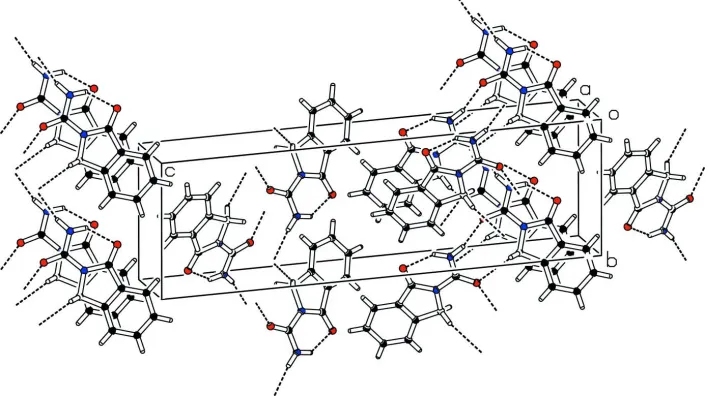
This kind of H-bond links each asymmetric unit at two places as shown in Fig 2. The distance between ring centroids of
aromatic and heterocyclic is 3.638 (2) Å along the a axis, which is indication of π-π interaction.
S2. Experimental
A mixture of o-phthaldehyde (0.67 g, 200 mmol) and urea (0.30 g, 200 mmol) in 100 ml of ethanol was refluxed for 6 h.
A blue color developed. The flask contents were allowed to stand for 24 h at room temperature. A white solid was
separated from the solution and was washed with ethanol,ether and hexane respectively, and dried in open air. The
crystals suitable for X-ray diffraction were grown in a mixture of acetone-ethanol (1:1) by slow evaporation at room
temperature. The compound is soluble in DMSO, DMF, acetone, ethyl acetate, and partially soluble in ethanol and
chloroform [m.p.: 493 K, yield: 55%].
S3. Refinement
H atoms were positioned geometrically, with C—H = 0.93, 0.97 Å for aromatic and methylene C-atoms and constrained
to ride on their parent atoms. The H-atoms attached to N2 were taken from fourier synthesis and their coordinates were
Figure 1
The ORTEP diagram of the title compound (I) with displacement ellipsoids at 50% probability level; intramolecular
interaction has been indicated by broken line. H-atoms are shown by small circles of arbitrary radii.
Figure 2
The packing figure (PLATON: Spek, 2003) which shows the H-bonding and the π-π interaction.
1-Oxoisoindoline-2-carboxamide
Crystal data
C9H8N2O2 Mr = 176.17
Orthorhombic, P212121 Hall symbol: P 2ac 2ab
a = 3.9839 (3) Å
b = 7.8732 (8) Å
c = 25.651 (2) Å
V = 804.58 (13) Å3 Z = 4
F(000) = 368
Dx = 1.454 Mg m−3
[image:3.610.129.484.360.562.2]Cell parameters from 1295 reflections
θ = 1.6–28.6°
µ = 0.11 mm−1
T = 296 K Needle, colourless 0.25 × 0.12 × 0.10 mm
Data collection
Bruker KappaAPEXII CCD diffractometer
Radiation source: fine-focus sealed tube Graphite monochromator
Detector resolution: 7.40 pixels mm-1 ω scans
Absorption correction: multi-scan (SADABS; Bruker, 2005)
Tmin = 0.975, Tmax = 0.990
5461 measured reflections 1254 independent reflections 860 reflections with I > 2σ(I)
Rint = 0.037
θmax = 28.6°, θmin = 1.6°
h = −3→5
k = −9→10
l = −34→22
Refinement
Refinement on F2 Least-squares matrix: full
R[F2 > 2σ(F2)] = 0.040 wR(F2) = 0.138 S = 1.07 1254 reflections 124 parameters 0 restraints
Primary atom site location: structure-invariant direct methods
Secondary atom site location: difference Fourier map
Hydrogen site location: inferred from neighbouring sites
H atoms treated by a mixture of independent and constrained refinement
w = 1/[σ2(F
o2) + (0.0804P)2] where P = (Fo2 + 2Fc2)/3 (Δ/σ)max < 0.001
Δρmax = 0.23 e Å−3 Δρmin = −0.22 e Å−3
Special details
Geometry. All e.s.d.'s (except the e.s.d. in the dihedral angle between two l.s. planes) are estimated using the full covariance matrix. The cell e.s.d.'s are taken into account individually in the estimation of e.s.d.'s in distances, angles and torsion angles; correlations between e.s.d.'s in cell parameters are only used when they are defined by crystal symmetry. An approximate (isotropic) treatment of cell e.s.d.'s is used for estimating e.s.d.'s involving l.s. planes.
Refinement. Refinement of F2 against ALL reflections. The weighted R-factor wR and goodness of fit S are based on F2, conventional R-factors R are based on F, with F set to zero for negative F2. The threshold expression of F2 > σ(F2) is used only for calculating R-factors(gt) etc. and is not relevant to the choice of reflections for refinement. R-factors based on F2 are statistically about twice as large as those based on F, and R- factors based on ALL data will be even larger.
Fractional atomic coordinates and isotropic or equivalent isotropic displacement parameters (Å2)
x y z Uiso*/Ueq
O1 0.1443 (8) 0.5881 (3) 0.09333 (8) 0.0599 (8)
O2 0.3962 (6) 0.7070 (2) 0.24721 (7) 0.0479 (7)
N1 0.1909 (7) 0.7618 (2) 0.16626 (8) 0.0335 (6)
N2 0.3940 (9) 0.4951 (3) 0.18736 (10) 0.0512 (8)
H2A 0.346 (10) 0.476 (4) 0.1514 (13) 0.061*
H2B 0.478 (10) 0.421 (4) 0.2092 (14) 0.061*
C1 0.1002 (9) 0.7240 (3) 0.11498 (10) 0.0380 (7)
C2 −0.0491 (8) 0.8806 (3) 0.09388 (10) 0.0351 (7)
C3 −0.1770 (10) 0.9115 (4) 0.04430 (11) 0.0450 (8)
H3 −0.1780 0.8269 0.0190 0.054*
C4 −0.3025 (9) 1.0715 (4) 0.03378 (12) 0.0491 (8)
C5 −0.2985 (9) 1.1968 (4) 0.07165 (12) 0.0489 (9)
H5 −0.3837 1.3038 0.0639 0.059*
C6 −0.1693 (9) 1.1655 (4) 0.12114 (11) 0.0427 (7)
H6 −0.1652 1.2504 0.1463 0.051*
C7 −0.0474 (8) 1.0053 (3) 0.13185 (10) 0.0343 (7)
C8 0.1037 (9) 0.9367 (3) 0.18109 (9) 0.0335 (7)
H8A 0.3014 1.0006 0.1912 0.040*
H8B −0.0569 0.9384 0.2095 0.040*
C9 0.3350 (8) 0.6532 (3) 0.20346 (10) 0.0350 (7)
Atomic displacement parameters (Å2)
U11 U22 U33 U12 U13 U23
O1 0.102 (2) 0.0398 (11) 0.0383 (10) 0.0154 (14) −0.0116 (14) −0.0118 (9)
O2 0.0734 (18) 0.0362 (11) 0.0340 (10) 0.0018 (12) −0.0113 (11) 0.0002 (8)
N1 0.0453 (16) 0.0263 (10) 0.0289 (10) 0.0045 (11) −0.0024 (10) −0.0006 (8)
N2 0.081 (2) 0.0327 (13) 0.0404 (13) 0.0173 (15) −0.0087 (15) 0.0015 (10)
C1 0.050 (2) 0.0358 (14) 0.0286 (12) −0.0004 (15) −0.0013 (13) −0.0047 (11)
C2 0.0382 (18) 0.0348 (14) 0.0323 (12) 0.0001 (13) 0.0018 (13) 0.0021 (11)
C3 0.050 (2) 0.0502 (17) 0.0345 (13) 0.0036 (18) −0.0019 (14) 0.0011 (13)
C4 0.047 (2) 0.064 (2) 0.0366 (13) 0.0042 (19) −0.0034 (14) 0.0140 (14)
C5 0.047 (2) 0.0477 (18) 0.0518 (17) 0.0100 (17) 0.0017 (16) 0.0159 (15)
C6 0.0473 (19) 0.0352 (14) 0.0455 (15) 0.0051 (16) 0.0040 (15) 0.0022 (12)
C7 0.0365 (18) 0.0344 (14) 0.0321 (12) 0.0019 (13) 0.0018 (12) 0.0016 (11)
C8 0.0437 (19) 0.0274 (12) 0.0293 (11) 0.0002 (14) 0.0001 (12) −0.0027 (10)
C9 0.0408 (18) 0.0314 (13) 0.0326 (12) −0.0014 (15) 0.0028 (13) 0.0020 (11)
Geometric parameters (Å, º)
O1—C1 1.218 (3) C3—C4 1.382 (4)
O2—C9 1.224 (3) C3—H3 0.9300
N1—C1 1.396 (3) C4—C5 1.385 (5)
N1—C9 1.404 (3) C4—H4 0.9300
N1—C8 1.470 (3) C5—C6 1.392 (4)
N2—C9 1.332 (3) C5—H5 0.9300
N2—H2A 0.95 (3) C6—C7 1.379 (4)
N2—H2B 0.87 (3) C6—H6 0.9300
C1—C2 1.472 (4) C7—C8 1.500 (4)
C2—C7 1.383 (4) C8—H8A 0.9700
C2—C3 1.392 (4) C8—H8B 0.9700
C1—N1—C9 128.0 (2) C4—C5—C6 121.2 (3)
C1—N1—C8 112.5 (2) C4—C5—H5 119.4
C9—N1—C8 119.4 (2) C6—C5—H5 119.4
C9—N2—H2A 114 (2) C7—C6—C5 118.3 (3)
C9—N2—H2B 120 (2) C7—C6—H6 120.8
H2A—N2—H2B 126 (3) C5—C6—H6 120.8
O1—C1—C2 128.8 (2) C6—C7—C8 129.7 (2)
N1—C1—C2 105.8 (2) C2—C7—C8 109.8 (2)
C7—C2—C3 121.4 (3) N1—C8—C7 102.36 (19)
C7—C2—C1 109.5 (2) N1—C8—H8A 111.3
C3—C2—C1 129.1 (2) C7—C8—H8A 111.3
C4—C3—C2 118.1 (3) N1—C8—H8B 111.3
C4—C3—H3 121.0 C7—C8—H8B 111.3
C2—C3—H3 121.0 H8A—C8—H8B 109.2
C3—C4—C5 120.6 (3) O2—C9—N2 124.9 (3)
C3—C4—H4 119.7 O2—C9—N1 119.6 (2)
C5—C4—H4 119.7 N2—C9—N1 115.5 (2)
C9—N1—C1—O1 −2.8 (5) C5—C6—C7—C8 −179.9 (3)
C8—N1—C1—O1 −179.9 (3) C3—C2—C7—C6 −0.9 (5)
C9—N1—C1—C2 178.1 (3) C1—C2—C7—C6 178.8 (3)
C8—N1—C1—C2 1.0 (3) C3—C2—C7—C8 179.9 (3)
O1—C1—C2—C7 −179.5 (3) C1—C2—C7—C8 −0.4 (4)
N1—C1—C2—C7 −0.4 (4) C1—N1—C8—C7 −1.2 (3)
O1—C1—C2—C3 0.2 (6) C9—N1—C8—C7 −178.5 (2)
N1—C1—C2—C3 179.3 (3) C6—C7—C8—N1 −178.1 (3)
C7—C2—C3—C4 0.2 (5) C2—C7—C8—N1 0.9 (3)
C1—C2—C3—C4 −179.5 (3) C1—N1—C9—O2 −179.3 (3)
C2—C3—C4—C5 0.3 (5) C8—N1—C9—O2 −2.4 (4)
C3—C4—C5—C6 −0.1 (5) C1—N1—C9—N2 0.6 (5)
C4—C5—C6—C7 −0.6 (5) C8—N1—C9—N2 177.5 (3)
C5—C6—C7—C2 1.1 (5)
Hydrogen-bond geometry (Å, º)
D—H···A D—H H···A D···A D—H···A
N2—H2A···O1 0.95 (3) 1.91 (3) 2.710 (3) 140 (2)
N2—H2B···O2i 0.88 (3) 2.08 (3) 2.943 (3) 167 (3)
C8—H8A···O2ii 0.97 2.57 3.447 (4) 151