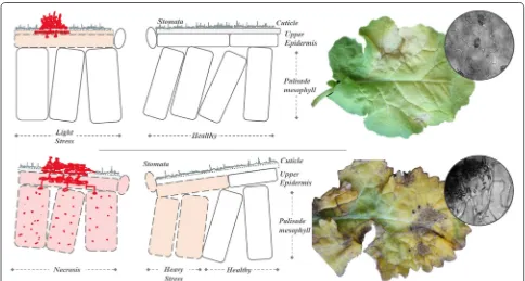
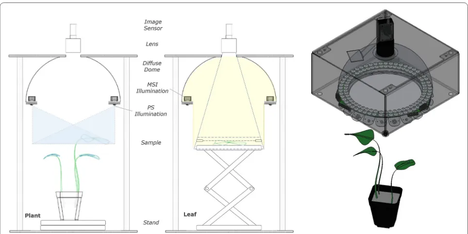
Multispectral imaging for presymptomatic analysis of light leaf spot in oilseed rape
Full text
Figure
![Fig. 5 Orientation effects. Variation in reflectance measured at high (blue) [ 50 ◦ ] and low (red) [ 10 ◦ ] inclination normalisation (left) PS reconstruction of control oilseed rape plant, at 03 DAI with excessive curvature of leaves highlighted with arrows (right)](https://thumb-us.123doks.com/thumbv2/123dok_us/571508.556671/10.595.57.539.87.240/orientation-variation-reflectance-inclination-normalisation-reconstruction-curvature-highlighted.webp)
Related documents
Duration of spring oilseed rape growth from sowing till 4–5 leaf stage, till the start and end of flowering and till seed development stage influenced nitrogen concentration
Effects of timing of Leptosphaeria maculans ascospore release and fungicide regime on phoma leaf spot and phoma stem canker development on winter oilseed rape ( Brassica
Figure 4 Phoma leaf spot severity ( Leptosphaeria maculans ) on leaves of oilseed rape cultivars Adriana [ Rlm4 + quantitative resistance (QR)], Bilbao ( Rlm4 ), Drakkar (no known
Ten leaves with phoma leaf spots were sampled randomly from 3-month-old winter oilseed rape plants in three replicate plots at three different sites in the UK (Cranwell, Harpenden
field conditions of the temperate climate zone, winter oilseed rape yield is mainly determined by agro-climatic conditions during the growing period, the level of
Figure 5 Assessment of light leaf spot symptoms (a) in a series of experiments in controlled-environment conditions with a subset (125 lines) from the N26 doubled haploid
The two maxima in the observed frequency distri- bution of plot disease incidence (Fig. 4J) reflect this observation. In the 1999–2000 season, only plants located south of the large
Simulated mean winter oilseed rape grain yield, N-balance, total plant N-uptake, grain N-uptake, N harvest index (NHI), N-concentration in the straw and the grain, and the harvest
