5,5-Dihydroxybarbituric acid
1,4-dioxane hemisolvate
Thomas Gelbrich,* Denise Rossi and Ulrich J. Griesser
Institute of Pharmacy, University of Innsbruck, Innrain 52, 6020 Innsbruck, Austria Correspondence e-mail: thomas.gelbrich@uibk.ac.at
Received 23 April 2010; accepted 26 April 2010
Key indicators: single-crystal X-ray study;T= 120 K; mean(C–C) = 0.003 A˚; Rfactor = 0.045;wRfactor = 0.131; data-to-parameter ratio = 10.3.
The asymmetric unit of the title compound,, C4H4N2O5 -0.5C4H8O2, contains one molecule of 5,5-dihydroxybarbituric acid with a nearly planar barbiturate ring and half a molecule of 1,4-dioxane. The geometry of the centrosymmetric dioxane molecule is close to an ideal chair conformation. The crystal structure exhibits a complex three-dimensional hydrogen-bonded network. Barbiturate molecules are connected to one
another via N—H O C, O—H O C and N—
H O(hydroxy) interactions, while the barbituric acid mol-ecule is linked to dioxane by an O—H O contact.
Related literature
For the crystal structure of unsolvated 5,5-dihydroxybarbituric acid, see: Singh (1965); Harrowfield et al. (1989). For the related monohydrate, see Lewis & Tocher (2004a). For the related trihydrate, see Mootz & Jeffrey (1965); Lewis & Tocher (2004b). For hydrogen-bond motifs, see: Bernstein et al.(1995).
Experimental
Crystal data
C4H4N2O50.5C4H8O2
Mr= 204.14 Triclinic,P1
a= 6.0232 (3) A˚
b= 8.3954 (4) A˚
c= 8.6858 (5) A˚ = 106.007 (4)
= 94.459 (3)
= 110.126 (3)
V= 389.09 (3) A˚3
Z= 2
MoKradiation = 0.16 mm1
T= 120 K
0.100.100.10 mm
Data collection
Bruker-Nonius Roper CCD camera on-goniostat diffractometer Absorption correction: multi-scan
(SADABS; Sheldrick, 2007)
Tmin= 0.984,Tmax= 0.984
5726 measured reflections 1529 independent reflections 1198 reflections withI> 2(I)
Rint= 0.049
Refinement
R[F2> 2(F2)] = 0.045
wR(F2) = 0.131
S= 1.01 1529 reflections 148 parameters 4 restraints
H atoms treated by a mixture of independent and constrained refinement
max= 0.25 e A˚
3
min=0.29 e A˚
3
Table 1
Hydrogen-bond geometry (A˚ ,).
D—H A D—H H A D A D—H A
N1—H1N O6i
0.89 (2) 2.39 (3) 3.068 (2) 134 (3) N1—H1N O7ii
0.89 (2) 2.44 (2) 3.180 (2) 141 (3) N3—H3N O2iii
0.88 (2) 1.93 (2) 2.810 (2) 172 (2) O7—H7O O1S 0.87 (2) 1.87 (2) 2.732 (2) 171 (3) O8—H8O O4iv
0.83 (2) 1.95 (2) 2.751 (2) 162 (3)
Symmetry codes: (i)xþ2;yþ2;zþ1; (ii)xþ1;y;z; (iii)xþ1;yþ1;z; (iv)x;yþ1;zþ1.
Data collection: COLLECT (Hooft, 1998); cell refinement: DENZO(Otwinowski & Minor, 1997) andCOLLECT; data reduc-tion:DENZO andCOLLECT; program(s) used to solve structure: SHELXS97(Sheldrick, 2008); program(s) used to refine structure: SHELXL97(Sheldrick, 2008); molecular graphics:XPinSHELXTL (Sheldrick, 2008) andMercury(Macraeet al., 2008); software used to prepare material for publication:publCIF(Westrip, 2010).
TG acknowledges financial support from the Lise Meitner Program of the Austrian Science Fund (FWF, project LM 1135-N17).
Supplementary data and figures for this paper are available from the IUCr electronic archives (Reference: JH2150).
References
Bernstein, J., Davis, R. E., Shimoni, L. & Chang, N.-L. (1995).Angew. Chem. Int. Ed. Engl.34, 1555–1573.
Harrowfield, J. M., Skelton, B. W., Soudi, A. A. & White, A. H. (1989).Aust. J. Chem.42, 1795–1798.
Hooft, R. W. W. (1998).COLLECT. Nonius BV, Delft, The Netherlands. Lewis, T. C. & Tocher, D. A. (2004a).Acta Cryst.E60, o1689–o1690. Lewis, T. C. & Tocher, D. A. (2004b).Acta Cryst.E60, o1748–o1750. Macrae, C. F., Bruno, I. J., Chisholm, J. A., Edgington, P. R., McCabe, P.,
Pidcock, E., Rodriguez-Monge, L., Taylor, R., van de Streek, J. & Wood, P. A. (2008).J. Appl. Cryst.41, 466–470.
Mootz, D. & Jeffrey, G. A. (1965).Acta Cryst.19, 717–725.
Otwinowski, Z. & Minor, W. (1997). Methods in Enzymology, Vol. 276,
Macromolecular Crystallography, Part A, edited by C. W. Carter Jr & R. M. Sweet, pp. 307–326. New York: Academic Press.
Sheldrick, G. M. (2007).SADABS. University of Go¨ttingen,Germany. Sheldrick, G. M. (2008).Acta Cryst.A64, 112–122.
Singh, C. (1965).Acta Cryst.19, 759–767. Westrip, S. P. (2010).J. Appl. Cryst.43. Submitted.
Acta Crystallographica Section E
Structure Reports Online
supporting information
Acta Cryst. (2010). E66, o1219 [https://doi.org/10.1107/S1600536810015321]
5,5-Dihydroxybarbituric acid 1,4-dioxane hemisolvate
Thomas Gelbrich, Denise Rossi and Ulrich J. Griesser
S1. Comment
The crystal structures of an unsolvated form (Singh, 1965; Harrowfield et al., 1989), a monohydrate (Lewis & Tocher,
2004a) and a trihydrate (Mootz & Jeffrey, 1965; Lewis & Tocher, 2004b) of 5,5-dihydroxybarbituric acid have been
reported previously. The asymmetric unit of the title structure consists of one molecule of the barbituric acid derivative
and one half of a dioxane moiety (Fig. 1). The six-membered C4N2 ring of the former is essentially planar, and its bond
distances and angles are in agreement with the parameters observed for the unsolvated and hydrate forms.
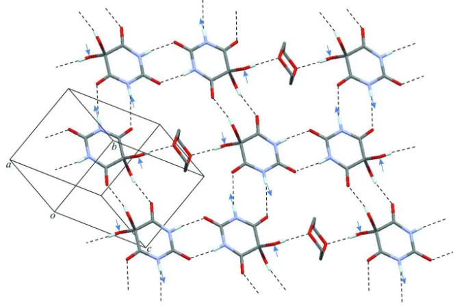
This crystal structure is characterized by extensive hydrogen bonding. Each dihydroxybarbituric acid molecule is linked
to two molecules of the same kind via two centrosymmetric N—H···O=C double bridges and a double bridge O—
H···O=C connects it to a third molecule. Joining these R2
2(8) and R22(10) motifs (Bernstein et al., 1995) gives a larger ring of six dihydroxybarbituric acid molecules. Two molecules of each such ring are additionally O—H···O bonded to a
dioxane molecule which lies in the centre of the ring. Fig. 2 shows the 2-dimensional H-bonded net parallel to (121)
which is obtained from these interactions. Additionally, one NH and one OH group of each dihydroxybarbituric acid
molecule are engaged as H-bond donor and acceptor, respectively, in N—H···O(hydroxy) interactions. These particular
contacts, indicated by arrows in Fig. 2, connect adjacent H-bonded 2D units of the kind discussed above to one another,
and an overall three-dimensional hydrogen bonded network is therefore formed. As expected, the two hydrogen bonds in
which the N1—H group is involved exhibit a much less favourable geometry than the single hydrogen bond in which the
N3—H group is employed.
S2. Experimental
A solution of 5,5-dibromobarbituric acid (Sigma-Aldrich) in dioxane was filled into an NMR tube for a crystallisation
experiment by slow evaporation of the solvent. After four months, an amber-coloured syrup had formed, indicating
decomposition of the original compound. This liquid contained a large colourless crystal that prooved to be composed of
the title compound.
S3. Refinement
All H atoms were identified in a difference map. H atoms bonded to secondary CH2 (C—H = 0.99 Å) carbon atoms were
positioned geometrically, and hydrogen atoms attached to N and O were refined with restrained distances [N—H =
Figure 1
The molecular structures of (I) with displacement ellipsoids drawn at the 50% probability level. Hydrogen atoms are
shown as spheres of arbitrary size. Symmetry code: (i) -x+1, -y+2, -z+2.
Figure 2
Portion of a hydrogen bonded sheet parallel to (1-21) showing N—H···O=C and O—H···O=C bonds between barbituric
acid molecules and O—H···O bonds between barbituric acid and dioxane. N—H···O(hydroxy) interactions linking to two
[image:3.610.139.470.371.595.2]5,5-dihydroxybarbituric acid 1,4-dioxane hemisolvate
Crystal data
C4H4N2O5·0.5C4H8O2
Mr = 204.14 Triclinic, P1 Hall symbol: -P 1
a = 6.0232 (3) Å
b = 8.3954 (4) Å
c = 8.6858 (5) Å
α = 106.007 (4)°
β = 94.459 (3)°
γ = 110.126 (3)°
V = 389.09 (3) Å3
Z = 2
F(000) = 212
Dx = 1.742 Mg m−3
Mo Kα radiation, λ = 0.71073 Å Cell parameters from 3190 reflections
θ = 2.9–26.0°
µ = 0.16 mm−1
T = 120 K Block, colourless 0.10 × 0.10 × 0.10 mm
Data collection
Bruker-Nonius Roper CCD camera on κ -goniostat
diffractometer
Radiation source: Bruker–Nonius FR591 rotating anode
Graphite monochromator
Detector resolution: 9.091 pixels mm-1
φ & ω scans
Absorption correction: multi-scan (SADABS; Sheldrick, 2007)
Tmin = 0.984, Tmax = 0.984 5726 measured reflections 1529 independent reflections 1198 reflections with I > 2σ(I)
Rint = 0.049
θmax = 26.0°, θmin = 3.6°
h = −7→7
k = −10→10
l = −10→10
Refinement
Refinement on F2 Least-squares matrix: full
R[F2 > 2σ(F2)] = 0.045
wR(F2) = 0.131
S = 1.01 1529 reflections 148 parameters 4 restraints
Primary atom site location: structure-invariant direct methods
Secondary atom site location: difference Fourier map
Hydrogen site location: inferred from neighbouring sites
H atoms treated by a mixture of independent and constrained refinement
w = 1/[σ2(F
o2) + (0.0787P)2 + 0.0767P] where P = (Fo2 + 2Fc2)/3
(Δ/σ)max < 0.001 Δρmax = 0.25 e Å−3 Δρmin = −0.29 e Å−3
Extinction correction: SHELXL97 (Sheldrick, 2008), Fc*=kFc[1+0.001xFc2λ3/sin(2θ)]-1/4 Extinction coefficient: 0.062 (16)
Special details
Geometry. All esds (except the esd in the dihedral angle between two l.s. planes) are estimated using the full covariance matrix. The cell esds are taken into account individually in the estimation of esds in distances, angles and torsion angles; correlations between esds in cell parameters are only used when they are defined by crystal symmetry. An approximate (isotropic) treatment of cell esds is used for estimating esds involving l.s. planes.
Refinement. Refinement of F2 against ALL reflections. The weighted R-factor wR and goodness of fit S are based on F2, conventional R-factors R are based on F, with F set to zero for negative F2. The threshold expression of F2 > σ(F2) is used only for calculating R-factors(gt) etc. and is not relevant to the choice of reflections for refinement. R-factors based on F2 are statistically about twice as large as those based on F, and R- factors based on ALL data will be even larger.
Fractional atomic coordinates and isotropic or equivalent isotropic displacement parameters (Å2)
x y z Uiso*/Ueq
H1N 0.934 (3) 0.835 (4) 0.398 (4) 0.061 (10)*
N3 0.4114 (3) 0.5803 (2) 0.2073 (2) 0.0175 (4)
H3N 0.342 (4) 0.506 (3) 0.107 (2) 0.029 (6)*
O2 0.7627 (3) 0.65461 (19) 0.11484 (17) 0.0242 (4)
O4 0.0683 (3) 0.4836 (2) 0.29980 (18) 0.0264 (4)
O6 0.8029 (3) 0.90452 (19) 0.65175 (17) 0.0265 (4)
O7 0.3029 (2) 0.83394 (18) 0.53542 (18) 0.0212 (4)
H7O 0.360 (6) 0.895 (4) 0.638 (2) 0.064 (10)*
O8 0.3738 (3) 0.60819 (19) 0.60882 (17) 0.0221 (4)
H8O 0.228 (3) 0.574 (4) 0.615 (4) 0.060 (10)*
C2 0.6555 (4) 0.6706 (3) 0.2277 (2) 0.0189 (5)
C4 0.2802 (4) 0.5806 (3) 0.3280 (2) 0.0185 (5)
C6 0.6801 (3) 0.8039 (3) 0.5200 (2) 0.0186 (5)
C5 0.4070 (3) 0.7064 (3) 0.5010 (2) 0.0178 (5)
O1S 0.4460 (2) 1.04067 (18) 0.85656 (16) 0.0201 (4)
C1S 0.6766 (4) 1.1366 (3) 0.9647 (3) 0.0218 (5)
H1S1 0.6742 1.2450 1.0457 0.030 (6)*
H1S2 0.8027 1.1755 0.9014 0.013 (5)*
C2S 0.2647 (3) 0.9800 (3) 0.9481 (2) 0.0213 (5)
H2S1 0.1066 0.9113 0.8733 0.031 (6)*
H2S2 0.2530 1.0848 1.0287 0.036 (7)*
Atomic displacement parameters (Å2)
U11 U22 U33 U12 U13 U23
N1 0.0145 (9) 0.0210 (9) 0.0182 (10) 0.0021 (8) 0.0036 (7) 0.0005 (7)
N3 0.0161 (9) 0.0192 (9) 0.0127 (9) 0.0042 (7) 0.0020 (7) 0.0019 (7)
O2 0.0192 (8) 0.0258 (8) 0.0188 (8) 0.0030 (6) 0.0070 (6) −0.0003 (6)
O4 0.0159 (8) 0.0307 (8) 0.0211 (8) 0.0004 (7) 0.0042 (6) 0.0016 (6)
O6 0.0191 (8) 0.0319 (9) 0.0186 (8) 0.0056 (7) 0.0014 (6) −0.0007 (6)
O7 0.0190 (8) 0.0229 (8) 0.0200 (8) 0.0092 (6) 0.0031 (6) 0.0031 (6)
O8 0.0214 (8) 0.0279 (8) 0.0202 (8) 0.0096 (7) 0.0070 (6) 0.0116 (6)
C2 0.0179 (10) 0.0165 (10) 0.0187 (11) 0.0036 (8) 0.0040 (9) 0.0039 (8) C4 0.0170 (10) 0.0187 (10) 0.0189 (11) 0.0056 (8) 0.0042 (8) 0.0061 (8) C6 0.0166 (10) 0.0198 (10) 0.0180 (11) 0.0060 (8) 0.0036 (8) 0.0053 (8) C5 0.0162 (10) 0.0199 (10) 0.0182 (11) 0.0069 (8) 0.0054 (8) 0.0068 (8) O1S 0.0158 (7) 0.0249 (8) 0.0162 (8) 0.0054 (6) 0.0027 (6) 0.0044 (6) C1S 0.0152 (10) 0.0225 (10) 0.0205 (11) 0.0019 (8) −0.0004 (8) 0.0039 (8) C2S 0.0138 (10) 0.0290 (11) 0.0182 (11) 0.0059 (9) 0.0040 (8) 0.0060 (9)
Geometric parameters (Å, º)
N1—C6 1.362 (3) O8—H8O 0.834 (18)
N1—C2 1.374 (3) C4—C5 1.532 (3)
N1—H1N 0.886 (18) C6—C5 1.536 (3)
N3—C4 1.361 (3) O1S—C2S 1.435 (2)
N3—C2 1.373 (3) O1S—C1S 1.439 (2)
O2—C2 1.217 (2) C1S—H1S1 0.9900
O4—C4 1.216 (2) C1S—H1S2 0.9900
O6—C6 1.214 (2) C2S—C1Si 1.504 (3)
O7—C5 1.394 (2) C2S—H2S1 0.9900
O7—H7O 0.867 (18) C2S—H2S2 0.9900
O8—C5 1.392 (2)
C6—N1—C2 126.66 (17) O8—C5—C4 109.42 (16)
C6—N1—H1N 115 (2) O7—C5—C4 105.53 (15)
C2—N1—H1N 118 (2) O8—C5—C6 106.79 (16)
C4—N3—C2 125.94 (17) O7—C5—C6 108.58 (15)
C4—N3—H3N 119.4 (16) C4—C5—C6 114.24 (16)
C2—N3—H3N 114.2 (16) C2S—O1S—C1S 109.37 (15)
C5—O7—H7O 105 (2) O1S—C1S—C2Si 110.63 (16)
C5—O8—H8O 107 (2) O1S—C1S—H1S1 109.5
O2—C2—N3 122.08 (19) C2Si—C1S—H1S1 109.5
O2—C2—N1 120.85 (18) O1S—C1S—H1S2 109.5
N3—C2—N1 117.07 (17) C2Si—C1S—H1S2 109.5
O4—C4—N3 121.06 (19) H1S1—C1S—H1S2 108.1
O4—C4—C5 120.76 (18) O1S—C2S—C1Si 110.95 (16)
N3—C4—C5 118.18 (17) O1S—C2S—H2S1 109.4
O6—C6—N1 121.57 (18) C1Si—C2S—H2S1 109.4
O6—C6—C5 120.87 (17) O1S—C2S—H2S2 109.4
N1—C6—C5 117.43 (17) C1Si—C2S—H2S2 109.4
O8—C5—O7 112.40 (16) H2S1—C2S—H2S2 108.0
C4—N3—C2—O2 175.20 (18) N3—C4—C5—O7 112.76 (19)
C4—N3—C2—N1 −5.1 (3) O4—C4—C5—C6 173.36 (17)
C6—N1—C2—O2 −176.10 (18) N3—C4—C5—C6 −6.4 (2)
C6—N1—C2—N3 4.2 (3) O6—C6—C5—O8 −57.3 (2)
C2—N3—C4—O4 −173.22 (18) N1—C6—C5—O8 126.73 (18)
C2—N3—C4—C5 6.6 (3) O6—C6—C5—O7 64.2 (2)
C2—N1—C6—O6 179.18 (18) N1—C6—C5—O7 −111.85 (19)
C2—N1—C6—C5 −4.8 (3) O6—C6—C5—C4 −178.39 (17)
O4—C4—C5—O8 53.7 (2) N1—C6—C5—C4 5.6 (2)
N3—C4—C5—O8 −126.09 (18) C2S—O1S—C1S—C2Si 57.5 (2)
O4—C4—C5—O7 −67.4 (2) C1S—O1S—C2S—C1Si −57.7 (2)
Symmetry code: (i) −x+1, −y+2, −z+2.
Hydrogen-bond geometry (Å, º)
D—H···A D—H H···A D···A D—H···A
N1—H1N···O6ii 0.89 (2) 2.39 (3) 3.068 (2) 134 (3)
N1—H1N···O7iii 0.89 (2) 2.44 (2) 3.180 (2) 141 (3)
O7—H7O···O1S 0.87 (2) 1.87 (2) 2.732 (2) 171 (3)
O8—H8O···O4v 0.83 (2) 1.95 (2) 2.751 (2) 162 (3)