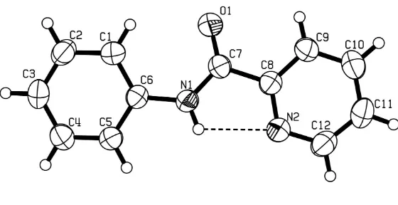
N
-Phenylpyridine-2-carbamide
Yu-Guo Zhuang,* Hua-Jiang Jiang, Zhi Hong and Fang-Li Qiu
School of Pharmaceutical and Chemical Engineering, Taizhou University, Linhai 317000, People’s Republic of China
Correspondence e-mail: [email protected]
Received 1 September 2008; accepted 4 September 2008
Key indicators: single-crystal X-ray study;T= 296 K; mean(C–C) = 0.003 A˚;
Rfactor = 0.030;wRfactor = 0.075; data-to-parameter ratio = 8.5.
In the title compound, C12H10N2O, the dihedral angle between the pyridine ring system and the phenyl ring is 1.8 (1). There
is an intramolecular N—H N hydrogen bond between the pyridine N atom and the amide NH function.
Related literature
For general background, see: Sousa & Filgueiras (1990); Gomeset al.(2007); Morsaliet al.(2003); Jacob & Mukherjee (2006); Marumotoet al.(1981); Piatnitski & Kiselyov (2004). For related structures, see: Qiet al.(2003); Zhanget al.(2006); Yinet al.(2007). For the synthesis, see: Chanet al.(2004).
Experimental
Crystal data
C12H10N2O
Mr= 198.22 Monoclinic,Pn a= 5.7469 (2) A˚
b= 6.2382 (2) A˚
c= 14.0158 (3) A˚ = 94.752 (2)
V= 500.74 (3) A˚3
Z= 2
MoKradiation = 0.09 mm 1
T= 296 (2) K 0.150.140.09 mm
Data collection
Bruker APEXII CCD diffractometer
Absorption correction: multi-scan (SADABS; Bruker, 2004)
Tmin= 0.968,Tmax= 0.992
5000 measured reflections 1162 independent reflections 1036 reflections withI> 2(I)
Rint= 0.017
Refinement
R[F2> 2(F2)] = 0.030
wR(F2) = 0.075
S= 1.00 1162 reflections
137 parameters
H-atom parameters constrained
max= 0.11 e A˚ 3
min= 0.12 e A˚ 3
Table 1
Hydrogen-bond geometry (A˚ ,).
D—H A D—H H A D A D—H A
N1—H101 N2 0.86 2.28 2.697 (2) 110
Data collection:APEX2(Bruker, 2004); cell refinement: SAINT-Plus(Bruker, 2004); data reduction:SAINT-Plus; program(s) used to solve structure:SHELXL97(Sheldrick, 2008); program(s) used to refine structure:SHELXL97(Sheldrick, 2008); molecular graphics:
ORTEP-3 for Windows(Farrugia, 1997); software used to prepare material for publication:WinGX(Farrugia,1999).
The authors are grateful for financial suport from the Key Discipline Open Foundation of Zhejiang University of Tech-nology (grant No. 20080604). The authors thank Mr Jian-Ming Gu (Testing and Analysis Center, Zhejiang University) for guidance in the structure analysis.
Supplementary data and figures for this paper are available from the IUCr electronic archives (Reference: IM2083).
References
Bruker (2004). APEX2, SAINT-Plus and SADABS. Bruker AXS Inc., Madison, Wisconsin, USA.
Chan, A. S. C., Qi, J. Y., Pai, C. C., Li, X. J., Deng, L. S., Li, W. Z. & Hu, J. Y. (2004). US Patent 6 680 385.
Farrugia, L. J. (1997).J. Appl. Cryst.30, 565. Farrugia, L. J. (1999).J. Appl. Cryst.32, 837–838.
Gomes, L., Low, J. N., Valente, M. A. D. C., Freire, C. & Castro, B. (2007).Acta Cryst.C63, m293–m296.
Jacob, W. & Mukherjee, R. (2006).Inorg. Chim. Acta,359, 4565–4573. Marumoto, R., Shunsuke, S. & Masao, T. (1981). Eur. Patent EP 38 161. Morsali, A., Ramazani, A. & Mahjoub, A. R. (2003).J. Coord. Chem.56, 1555–
1566.
Piatnitski, E. & Kiselyov, A. S. (2004). US Patent 0 017 248.
Qi, J. Y., Yang, Q. Y., Lam, K. H., Zhou, Z. Y. & Chan, A. S. C. (2003).Acta Cryst.E59, o374–o375.
Sheldrick, G. M. (2008).Acta Cryst.A64, 112–122.
Sousa, G. F. & Filgueiras, C. A. L. (1990).Transition Met. Chem.15, 286–289. Yin, X.-H., Zhao, K., Feng, Y. & Zhu, J. (2007).Acta Cryst.E63, o4617. Zhang, Q., Zhang, S.-P. & Shao, S.-C. (2006).Acta Cryst.E62, o4695–o4696.
Acta Crystallographica Section E
Structure Reports
Online
supporting information
Acta Cryst. (2008). E64, o1904 [doi:10.1107/S1600536808028274]
N
-Phenylpyridine-2-carbamide
Yu-Guo Zhuang, Hua-Jiang Jiang, Zhi Hong and Fang-Li Qiu
S1. Comment
Pyridine-containing amides continue to attract considerable interest as ligands for metals (Sousa & Filgueiras, 1990;
Gomes et al., 2007; Jacob & Mukherjee, 2006), building blocks in organic synthesis (Marumoto et al., 1981) and
physiologically active compounds (Piatnitski & Kiselyov, 2004). As part of our studies on the synthesis and
characterization of these compounds, we report here the crystal structure of the title compound.
The C7—O1 [1.223 (3) Å], N1—C7 [1.344 (3) Å] and N1—C6 [1.410 (2) Å] bond lengths indicate extensive electron
delocalization in the amide linkage. The pyridyl and phenyl rings of the title compound are almost coplanar, forming a
dihedral angle of 1.8 (1)°. In the crystal structure, there is an intramolecular hydrogen bond (N1—H101···N2) and no
intermolecular hydrogen bonds are observed (Table 1).
The reported monoclinic space-group is in a non-standard setting (Pn, #7). There is a strong feature (h + l = 2n) in hkl
data. Setting up the space group as Pc results in a β angle of 23° or 157°, respectively. Obviously such an unit-cell
division is inappropriate. Therefore, the non-standard setting Pn was chosen.
S2. Experimental
The title compound was synthesized from pyridine-2-carboxylic acid and aniline according to the procedure of Chan et
al. (2004). The crystal used for data collection was obtained by slow evaporation from a saturated ethanol/water solution
at room temperature.
S3. Refinement
All H atoms were placed in geometrically idealized positions and constrained to ride on their parent atoms, with N—H =
Figure 1
The molecular structure of the title compound, shown with 50% probability displacement ellipsoids.
N-Phenylpyridine-2-carbamide
Crystal data
C12H10N2O
Mr = 198.22
Monoclinic, Pn
Hall symbol: P -2yac
a = 5.7469 (2) Å
b = 6.2382 (2) Å
c = 14.0158 (3) Å
β = 94.752 (2)°
V = 500.74 (3) Å3
Z = 2
F(000) = 208
Dx = 1.315 Mg m−3
Mo Kα radiation, λ = 0.71073 Å
Cell parameters from 2226 reflections
θ = 2.9–25.1°
µ = 0.09 mm−1
T = 296 K
Block, colourless 0.15 × 0.14 × 0.09 mm
Data collection
Bruker APEXII CCD diffractometer
Radiation source: fine-focus sealed tube Graphite monochromator
Detector resolution: 10 pixels mm-1
ω scans
Absorption correction: multi-scan (SADABS; Bruker, 2004)
Tmin = 0.968, Tmax = 0.992
5000 measured reflections 1162 independent reflections 1036 reflections with I > 2σ(I)
Rint = 0.017
θmax = 27.7°, θmin = 2.9°
h = −7→7
k = −8→5
Refinement
Refinement on F2
Least-squares matrix: full
R[F2 > 2σ(F2)] = 0.030
wR(F2) = 0.075
S = 1.00
1162 reflections 137 parameters 0 restraints
Primary atom site location: structure-invariant direct methods
Secondary atom site location: difference Fourier map
Hydrogen site location: inferred from neighbouring sites
H-atom parameters constrained
w = 1/[σ2(F
o2) + (0.0365P)2 + 0.0548P]
where P = (Fo2 + 2Fc2)/3
(Δ/σ)max < 0.001
Δρmax = 0.11 e Å−3
Δρmin = −0.12 e Å−3
Special details
Geometry. All e.s.d.'s (except the e.s.d. in the dihedral angle between two l.s. planes) are estimated using the full covariance matrix. The cell e.s.d.'s are taken into account individually in the estimation of e.s.d.'s in distances, angles and torsion angles; correlations between e.s.d.'s in cell parameters are only used when they are defined by crystal symmetry. An approximate (isotropic) treatment of cell e.s.d.'s is used for estimating e.s.d.'s involving l.s. planes.
Refinement. Refinement of F2 against ALL reflections. The weighted R-factor wR and goodness of fit S are based on F2,
conventional R-factors R are based on F, with F set to zero for negative F2. The threshold expression of F2 > σ(F2) is used
only for calculating R-factors(gt) etc. and is not relevant to the choice of reflections for refinement. R-factors based on F2
are statistically about twice as large as those based on F, and R- factors based on ALL data will be even larger.
Fractional atomic coordinates and isotropic or equivalent isotropic displacement parameters (Å2)
x y z Uiso*/Ueq
N1 −0.0173 (3) 0.2086 (3) 0.59409 (11) 0.0452 (4)
H101 −0.1420 0.2510 0.6183 0.054*
C8 0.1414 (3) 0.5113 (3) 0.68241 (13) 0.0435 (4)
C6 −0.0401 (3) 0.0235 (3) 0.53631 (12) 0.0412 (4)
C1 0.1246 (4) −0.0375 (4) 0.47407 (15) 0.0509 (5)
H1 0.2570 0.0459 0.4685 0.061*
C7 0.1751 (4) 0.3270 (3) 0.61595 (15) 0.0475 (5)
C2 0.0901 (4) −0.2230 (4) 0.42049 (16) 0.0569 (5)
H2 0.1999 −0.2635 0.3787 0.068*
N2 −0.0537 (3) 0.5139 (3) 0.72828 (12) 0.0526 (4)
C5 −0.2373 (4) −0.1016 (3) 0.54213 (15) 0.0476 (5)
H5 −0.3507 −0.0595 0.5819 0.057*
O1 0.3656 (3) 0.2937 (3) 0.58575 (15) 0.0753 (6)
C10 0.2767 (5) 0.8384 (3) 0.75234 (17) 0.0575 (5)
H10 0.3863 0.9481 0.7597 0.069*
C11 0.0794 (4) 0.8441 (4) 0.80026 (16) 0.0612 (6)
H11 0.0523 0.9575 0.8410 0.073*
C3 −0.1044 (4) −0.3483 (4) 0.42826 (16) 0.0553 (5)
H3 −0.1255 −0.4731 0.3923 0.066*
C12 −0.0787 (4) 0.6788 (4) 0.78711 (17) 0.0629 (6)
H12 −0.2107 0.6823 0.8213 0.076*
C9 0.3106 (4) 0.6677 (3) 0.69302 (15) 0.0516 (5)
H9 0.4451 0.6581 0.6607 0.062*
C4 −0.2676 (4) −0.2879 (4) 0.48961 (17) 0.0553 (5)
Atomic displacement parameters (Å2)
U11 U22 U33 U12 U13 U23
N1 0.0458 (9) 0.0397 (9) 0.0515 (9) −0.0007 (7) 0.0121 (7) −0.0031 (7)
C8 0.0478 (10) 0.0408 (10) 0.0422 (10) 0.0013 (8) 0.0051 (8) 0.0021 (8)
C6 0.0458 (10) 0.0369 (10) 0.0408 (10) 0.0030 (8) 0.0042 (8) 0.0021 (8)
C1 0.0504 (11) 0.0521 (12) 0.0517 (11) −0.0041 (10) 0.0120 (9) −0.0028 (10)
C7 0.0497 (11) 0.0398 (11) 0.0539 (12) −0.0030 (9) 0.0092 (9) −0.0007 (9)
C2 0.0594 (13) 0.0609 (14) 0.0515 (12) 0.0060 (11) 0.0117 (9) −0.0103 (10)
N2 0.0477 (9) 0.0540 (10) 0.0566 (10) −0.0015 (8) 0.0067 (8) −0.0096 (8)
C5 0.0464 (10) 0.0458 (11) 0.0515 (10) −0.0004 (9) 0.0088 (8) −0.0024 (9)
O1 0.0531 (9) 0.0669 (11) 0.1095 (15) −0.0111 (9) 0.0283 (9) −0.0330 (10)
C10 0.0699 (14) 0.0471 (11) 0.0550 (11) −0.0102 (12) 0.0014 (10) −0.0007 (11)
C11 0.0714 (16) 0.0553 (14) 0.0559 (13) 0.0065 (12) −0.0002 (11) −0.0156 (11)
C3 0.0652 (13) 0.0459 (11) 0.0537 (11) 0.0014 (11) −0.0023 (10) −0.0119 (10)
C12 0.0561 (13) 0.0705 (15) 0.0635 (14) 0.0047 (11) 0.0122 (11) −0.0168 (12)
C9 0.0559 (11) 0.0486 (12) 0.0513 (11) −0.0086 (10) 0.0116 (9) −0.0018 (9)
C4 0.0550 (12) 0.0506 (13) 0.0598 (12) −0.0070 (10) 0.0027 (10) −0.0027 (11)
Geometric parameters (Å, º)
N1—C7 1.344 (3) N2—C12 1.333 (3)
N1—C6 1.410 (2) C5—C4 1.379 (3)
N1—H101 0.8600 C5—H5 0.9300
C8—N2 1.338 (2) C10—C11 1.365 (4)
C8—C9 1.377 (3) C10—C9 1.375 (3)
C8—C7 1.502 (3) C10—H10 0.9300
C6—C5 1.384 (3) C11—C12 1.377 (3)
C6—C1 1.392 (3) C11—H11 0.9300
C1—C2 1.385 (3) C3—C4 1.377 (3)
C1—H1 0.9300 C3—H3 0.9300
C7—O1 1.223 (3) C12—H12 0.9300
C2—C3 1.376 (4) C9—H9 0.9300
C2—H2 0.9300 C4—H4 0.9300
C7—N1—C6 128.07 (17) C4—C5—H5 119.6
C7—N1—H101 116.0 C6—C5—H5 119.6
C6—N1—H101 116.0 C11—C10—C9 118.9 (2)
N2—C8—C9 123.48 (19) C11—C10—H10 120.6
N2—C8—C7 117.62 (17) C9—C10—H10 120.6
C9—C8—C7 118.89 (18) C10—C11—C12 118.7 (2)
C5—C6—C1 119.06 (17) C10—C11—H11 120.7
C5—C6—N1 117.74 (17) C12—C11—H11 120.7
C1—C6—N1 123.19 (17) C2—C3—C4 119.6 (2)
C2—C1—C6 119.57 (19) C2—C3—H3 120.2
C2—C1—H1 120.2 C4—C3—H3 120.2
C6—C1—H1 120.2 N2—C12—C11 123.8 (2)
O1—C7—C8 120.61 (18) C11—C12—H12 118.1
N1—C7—C8 114.58 (18) C10—C9—C8 118.7 (2)
C3—C2—C1 120.8 (2) C10—C9—H9 120.6
C3—C2—H2 119.6 C8—C9—H9 120.6
C1—C2—H2 119.6 C3—C4—C5 120.1 (2)
C12—N2—C8 116.38 (18) C3—C4—H4 119.9
C4—C5—C6 120.77 (19) C5—C4—H4 119.9
Hydrogen-bond geometry (Å, º)
D—H···A D—H H···A D···A D—H···A