1-Deoxy-
D-arabinitol
Sarah F. Jenkinson,a* Filipa P. Cruz,aKathrine V. Booth,a George W. J. Fleet,aKen Izumori,b Chu-Yi Yucand David J. Watkind
aDepartment of Organic Chemistry, Chemical Research Laboratory, University of
Oxford, Mansfield Road, Oxford OX1 3TA, England,bRare Sugar Research Centre,
Kagawa University, 2393 Miki-cho, Kita-gun, Kagawa 761-0795, Japan,cLaboratory
of Molecular Recognition and Selective Synthesis, Institute of Chemistry, Chinese Academy of Sciences, Beijing 10080, People’s Republic of China, anddDepartment
of Chemical Crystallography, Chemical Research Laboratory, University of Oxford, Mansfield Road, Oxford OX1 3TA, England
Correspondence e-mail: [email protected]
Received 24 April 2008; accepted 29 April 2008
Key indicators: single-crystal X-ray study;T= 150 K; mean(C–C) = 0.005 A˚; Rfactor = 0.044;wRfactor = 0.124; data-to-parameter ratio = 10.4.
Addition of methyl lithium to d-erythrono-1,4-lactone
followed by acid deprotection was shown, by X-ray crystal-lography, to give 1-deoxy-d-arabinitol, C5H12O4, rather than
1-deoxy-d-ribitol as the major product. The crystal structure
exists as hydrogen-bonded chains of molecules running parallel to the c axis which are further linked together by hydrogen bonds. Each molecule is a donor and an acceptor for four hydrogen bonds.
Related literature
For related literature see: Izumori (2002, 2006); Granstromet al. (2004); Beadle et al. (1992); Skytte (2002); Levin (2002); Howling & Callagan (2000); Bertelsenet al.(1999); Takataet al.(2005); Menavuvuet al.(2006); Suiet al.(2005); Hossainet al.(2006); Zehneret al.(1994); Donneret al.(1999); Yoshihara
et al.(2008); Takai & Heathcock (1985); Zissis & Richtmyer (1954).
Experimental
Crystal data
C5H12O4 Mr= 136.15
Tetragonal,I41
a= 12.9873 (5) A˚
c= 8.3679 (3) A˚
V= 1411.41 (9) A˚3
Z= 8
MoKradiation
= 0.11 mm1 T= 150 K
0.250.250.25 mm
Data collection
Nonius KappaCCD area-detector diffractometer
Absorption correction: multi-scan (DENZO/SCALEPACK; Otwinowski & Minor, 1997)
Tmin= 0.93,Tmax= 0.97
3189 measured reflections 855 independent reflections 750 reflections withI> 2(I)
Rint= 0.020
Refinement
R[F2> 2(F2)] = 0.043
wR(F2) = 0.123 S= 1.00 855 reflections 82 parameters
1 restraint
H-atom parameters constrained max= 0.34 e A˚3
min=0.39 e A˚
3
Table 1
Hydrogen-bond geometry (A˚ ,).
D—H A D—H H A D A D—H A
O8—H8 O8i
0.96 1.76 2.698 (4) 164 O6—H6 O6ii
1.00 1.98 2.712 (4) 128 O4—H4 O1iii
0.98 1.77 2.718 (4) 162 O1—H1 O4iv
1.05 2.03 2.712 (3) 120
Symmetry codes: (i)yþ1
2;xþ1;z 1
4; (ii)y;xþ 3 2;zþ
1 4; (iii)yþ
3 2;x;z
1 4; (iv) yþ1;x1
2;zþ 1 4.
Data collection: COLLECT (Nonius, 2001); cell refinement: DENZO/SCALEPACK(Otwinowski & Minor, 1997); data reduc-tion: DENZO/SCALEPACK; program(s) used to solve structure: SIR92(Altomareet al., 1994); program(s) used to refine structure: CRYSTALS (Betteridge et al., 2003); molecular graphics: CAMERON(Watkinet al., 1996); software used to prepare material for publication:CRYSTALS.
Supplementary data and figures for this paper are available from the IUCr electronic archives (Reference: LH2622).
References
Altomare, A., Cascarano, G., Giacovazzo, G., Guagliardi, A., Burla, M. C., Polidori, G. & Camalli, M. (1994).J. Appl. Cryst.27, 435–435.
Beadle, J. R., Saunders, J. P. & Wajda, T. J. (1992). US Patent 5 078 796. Bertelsen, H., Jensen, B. B. & Buemann, B. (1999).World Rev. Nutr. Diet.85,
98–109.
Betteridge, P. W., Carruthers, J. R., Cooper, R. I., Prout, K. & Watkin, D. J. (2003).J. Appl. Cryst.36, 1487.
Donner, T. W., Wilber, J. F. & Ostrowski, D. (1999).Diabetes Obes. Metab.1, 285–291.
Granstrom, T. B., Takata, G., Tokuda, M. & Izumori, K. (2004).J. Biosci. Bioeng.97, 89–94.
Hossain, M. A., Wakabayashi, H., Izuishi, K., Okano, K., Yachida, S., Tokuda, M., Izumori, K. & Maeta, H. (2006).J. Biosci. Bioeng.101, 369–371. Howling, D. & Callagan, J. L. (2000). PCT Int. App. WO 2000 042 865. Izumori, K. (2002).Naturwissenschaften,89, 120–124.
Izumori, K. (2006).J. Biotechnol.124, 717–722. Levin, G. V. (2002).J. Med. Food,5, 23–36.
Menavuvu, B. T., Poonperm, W., Leang, K., Noguchi, N., Okada, H., Morimoto, K., Granstrom, T. B., Takada, G. & Izumori, K. (2006).J. Biosci. Bioeng.101, 340–345.
Nonius (2001).COLLECT. Nonius BV, Delft, The Netherlands.
Otwinowski, Z. & Minor, W. (1997). Methods in Enzymology, Vol. 276,
Macromolecular Crystallography, Part A, edited by C. W. Carter Jr & R. M. Sweet, pp. 307–326. New York: Academic Press.
Skytte, U. P. (2002).Cereal Foods World47, 224–224.
Sui, L., Dong, Y. Y., Watanabe, Y., Yamaguchi, F., Hatano, N., Tsukamoto, I., Izumori, K. & Tokuda, M. (2005).Intl. J. Ocology,27, 907-912.
Takai, K. & Heathcock, C. H. (1985).J. Org. Chem.50, 3247–3251.
organic compounds
o1010
Jenkinsonet al. doi:10.1107/S1600536808012555 Acta Cryst.(2008). E64, o1010–o1011Acta Crystallographica Section E Structure Reports
Online
Tsukamoto, I., Nagata, M., Izumori, K. & Tokuda, M. (2005). J. Biosci. Bioeng.100, 511–516.
Watkin, D. J., Prout, C. K. & Pearce, L. J. (1996).CAMERON. Chemical Crystallography Laboratory, Oxford, UK.
Jones, N., Jenkinson, S. F., Wormald, M. R., Dwek, R. A., Fleet, G. W. J. & Izumori, K. (2008).Tetrahedron Asymmetry,19, 1739–745.
Zehner, L. R., Levin, G. V., Saunders, J. P. & Beadle, J. R. (1994). US Patent 5 356 879.
supporting information
sup-1
Acta Cryst. (2008). E64, o1010–o1011
supporting information
Acta Cryst. (2008). E64, o1010–o1011 [doi:10.1107/S1600536808012555]
1-Deoxy-
D-arabinitol
Sarah F. Jenkinson, Filipa P. Cruz, Kathrine V. Booth, George W. J. Fleet, Ken Izumori, Chu-Yi Yu
and David J. Watkin
S1. Comment
The demand for the large scale production of rare sugars by biotechnological (Izumori, 2006; Izumori, 2002; Granstrom
et al., 2004) and chemical (Beadle et al., 1992) methods is driven by the demand for alternative foodstuffs (Skytte, 2002)
and D-tagatose itself is used as a low calorie sweetener (Levin, 2002; Howling & Callagan, 2000; Bertelsen et al. 1999)
Rare monosaccharides have been found to demonstrate interesting pharmaceutical properties, for example, D-psicose
(Takata et al., 2005; Menavuvu et al., 2006) and D-allose (Sui et al., 2005; Hossain et al., 2006) have significant
chemotherapeutic properties and D-tagatose has been found to be an anti-hyperglycemic agent (Zehner et al., 1994;
Donner et al., 1999) and therefore potentially useful in the treatment of diabetes.
The methodology developed by Izumori et al. (2002, 2006) for the interconversion of tetroses, pentoses and hexoses by
enzymatic oxidation, inversion at C3 with a single epimerase, and reduction to the aldose has been seen to be generally
applicable for the 1-deoxy ketohexoses (Yoshihara et al., 2008). In order to investigate the viability of this process to the
corresponding pentoses and thus to evaluate their therapeutic potential 1-deoxy-D-arabinitol was synthesized, in 3 steps,
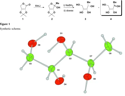
from 2,3-O-isopropylidene-D-erythronolactone 1 (Fig.1). It has previously been seen that the four diastereomeric tetraols
are very difficult to distinguish between by NMR spectroscopy (Takai & Heathcock, 1985). X-ray crystallography
confirmed that the major product was the arabinitol 4 rather than the ribitol 3 which differs only in the stereochemistry at
the C2 position (Fig. 2).
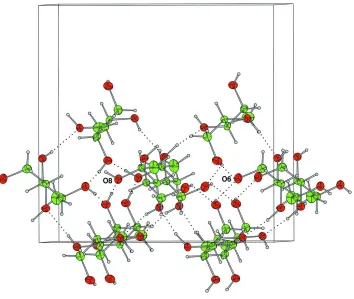
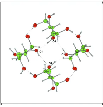
The molecules are linked by three hydrogen bonding systems and the structure consists of alternating spiral chains of
O6—H6···O6 or O8—H8···O8 hydrogen-bonded molecules running parallel to the c-axis (Fig. 3) interconnected by O1—
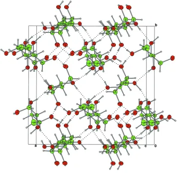
H1···O4—H4···O1 hydrogen bonds (Fig.4). Each molecule is a donor and acceptor for 4 hydrogen bonds (Fig. 5).
In summary, the stereochemistry at C2 of the title compound 1-deoxy-D-arabinitol 4 was firmly established by X-ray
crystallography, the absolute configuration is determined by the use of D-erythronolactone as the starting material. As
well as the potential biological properties of 1-deoxy ketoses, they are likely to provide a new set of building blocks for
the synthesis of a wide variety of complex biomolecules.
S2. Experimental
The title compound was recrystallized from hot methanol: m.p. 398–400 K; [α]D21 +0.8 (c, 8 in H2O) {Lit. (Zissis &
Richtmyer, 1954) m.p. 129–131°C; [α]D20 +0.7 (c, 10 in H2O; l, 4)}.
S3. Refinement
In the absence of significant anomalous scattering, Friedel pairs were merged and the absolute configuration assigned
The H atoms were initially refined with soft restraints on the bond lengths and angles to regularize their geometry (C—H
in the range 0.93–0.98, O—H = 0.82 Å) and Uiso(H) (in the range 1.2–1.5 times Ueq of the parent atom), after which the
positions were refined with riding constraints.
Figure 1
[image:4.610.61.492.137.470.2]Synthetic scheme.
Figure 2
The title compound with displacement ellipsoids drawn at the 50% probability level. H atoms are shown as spheres of
supporting information
sup-3
[image:5.610.130.484.69.368.2]Acta Cryst. (2008). E64, o1010–o1011 Figure 3
Figure 4
supporting information
sup-5
[image:7.610.128.483.71.418.2]Acta Cryst. (2008). E64, o1010–o1011 Figure 5
Packing diagram for the compound projected along the c-axis. Each molecule is a donor and an acceptor for 4
hydrogen-bonds.
1-Deoxy-D-arabinitol
Crystal data
C5H12O4 Mr = 136.15
Tetragonal, I41
Hall symbol: I 4bw
a = 12.9873 (5) Å
c = 8.3679 (3) Å
V = 1411.41 (9) Å3 Z = 8
F(000) = 592
Dx = 1.281 Mg m−3
Mo Kα radiation, λ = 0.71073 Å Cell parameters from 815 reflections
θ = 5–27°
µ = 0.11 mm−1 T = 150 K Block, colourless 0.25 × 0.25 × 0.25 mm
Data collection
Nonius KappaCCD area-detector diffractometer
Graphite monochromator
ω scans
Absorption correction: multi-scan
(DENZO/SCALEPACK; Otwinowski & Minor, 1997)
Rint = 0.020
θmax = 27.5°, θmin = 5.3°
k = −11→11
l = −10→10
Refinement
Refinement on F2
Least-squares matrix: full
R[F2 > 2σ(F2)] = 0.043 wR(F2) = 0.123 S = 1.00 855 reflections 82 parameters 1 restraint
Primary atom site location: structure-invariant direct methods
Hydrogen site location: inferred from neighbouring sites
H-atom parameters constrained
w = 1/[σ2(F2) + ( 0.07P)2 + 1.26P],
where P = (max(Fo2,0) + 2Fc2)/3
(Δ/σ)max = 0.002
Δρmax = 0.34 e Å−3
Δρmin = −0.39 e Å−3
Fractional atomic coordinates and isotropic or equivalent isotropic displacement parameters (Å2)
x y z Uiso*/Ueq
O1 0.64776 (13) 0.51955 (15) 0.6622 (3) 0.0211 C2 0.75127 (18) 0.5139 (2) 0.6068 (4) 0.0186 C3 0.7537 (2) 0.4842 (2) 0.4296 (4) 0.0187 O4 0.85700 (13) 0.48073 (16) 0.3723 (3) 0.0237 C5 0.6897 (2) 0.5564 (2) 0.3268 (4) 0.0235 O6 0.73116 (15) 0.65798 (14) 0.3242 (3) 0.0250 C7 0.8135 (2) 0.4417 (2) 0.7135 (4) 0.0208 O8 0.76689 (14) 0.34124 (13) 0.7126 (3) 0.0216 C9 0.8162 (3) 0.4788 (2) 0.8844 (4) 0.0371
H21 0.7853 0.5822 0.6286 0.0184*
H31 0.7208 0.4168 0.4126 0.0196*
H51 0.6985 0.5315 0.2238 0.0277*
H52 0.6191 0.5542 0.3475 0.0271*
H71 0.8827 0.4379 0.6604 0.0259*
H91 0.8413 0.4265 0.9544 0.0541*
H92 0.8595 0.5396 0.8958 0.0548*
H93 0.7474 0.4971 0.9202 0.0552*
H1 0.6194 0.4722 0.5703 0.0308*
H8 0.7975 0.2944 0.6379 0.0334*
H6 0.7418 0.6761 0.4388 0.0359*
H4 0.9070 0.5369 0.3651 0.0365*
Atomic displacement parameters (Å2)
U11 U22 U33 U12 U13 U23
supporting information
sup-7
Acta Cryst. (2008). E64, o1010–o1011
C7 0.0201 (13) 0.0204 (13) 0.0218 (16) −0.0035 (10) −0.0045 (13) −0.0004 (13) O8 0.0254 (10) 0.0189 (10) 0.0204 (12) 0.0010 (7) 0.0049 (10) 0.0000 (9) C9 0.053 (2) 0.0332 (16) 0.0253 (15) −0.0023 (15) −0.0149 (15) −0.0036 (13)
Geometric parameters (Å, º)
O1—C2 1.424 (3) C5—H51 0.927
O1—H1 1.051 C5—H52 0.934
C2—C3 1.532 (3) O6—H6 0.997
C2—C7 1.527 (4) C7—O8 1.438 (3)
C2—H21 1.008 C7—C9 1.510 (5)
C3—O4 1.425 (3) C7—H71 1.004
C3—C5 1.520 (4) O8—H8 0.959
C3—H31 0.985 C9—H91 0.954
O4—H4 0.978 C9—H92 0.974
C5—O6 1.425 (3) C9—H93 0.972
C2—O1—H1 93.6 C3—C5—H52 114.4
O1—C2—C3 110.4 (2) O6—C5—H52 113.8
O1—C2—C7 109.9 (3) H51—C5—H52 106.4
C3—C2—C7 113.6 (2) C5—O6—H6 104.9
O1—C2—H21 108.0 C2—C7—O8 109.4 (2)
C3—C2—H21 112.9 C2—C7—C9 111.7 (2)
C7—C2—H21 101.7 O8—C7—C9 107.7 (3)
C2—C3—O4 110.7 (2) C2—C7—H71 104.2
C2—C3—C5 112.4 (2) O8—C7—H71 109.3
O4—C3—C5 110.1 (2) C9—C7—H71 114.4
C2—C3—H31 110.8 C7—O8—H8 113.9
O4—C3—H31 109.4 C7—C9—H91 111.2
C5—C3—H31 103.2 C7—C9—H92 111.4
C3—O4—H4 128.4 H91—C9—H92 108.7
C3—C5—O6 111.9 (2) C7—C9—H93 110.4
C3—C5—H51 104.0 H91—C9—H93 107.4
O6—C5—H51 105.2 H92—C9—H93 107.6
Hydrogen-bond geometry (Å, º)
D—H···A D—H H···A D···A D—H···A
O8—H8···O8i 0.96 1.76 2.698 (4) 164
O6—H6···O6ii 1.00 1.98 2.712 (4) 128
O4—H4···O1iii 0.98 1.77 2.718 (4) 162
O1—H1···O4iv 1.05 2.03 2.712 (3) 120