Neuronal Circuits in C. elegans
Sleep
Thesis by Julie YoungHee Cho
In Partial Fulfillment of the Requirements for the degree of
Doctor of Philosophy
CALIFORNIA INSTITUTE OF TECHNOLOGY Pasadena, California
2013
2013
First and foremost, I would like to thank my advisor, Paul Sternberg. It is hard to find the words to properly describe my experience working for Paul. In addition to the art of rapid communication and wild gesticulation, he showed me the skill of tempering fearless experimentation and boundless enthusiasm with pragmatism and discipline. Paul pushed me harder than I expected and tested the limits of my endurance and ingenuity while being a good sport when I pushed back on the limits of his patience, support, and knowledge. I appreciate his enduring many awkward moments, answering countless questions (both scientific and random), working with me side by the side when I was on the brink of boredom or despair, and finding funding for use of special facilities and equipment.
I thank my committee: David Anderson for his patience, thoroughness, and constructive suggestions; David Prober for his generous time and advice, for his openness with both praise and criticism, and his constant thoughtfulness and concern throughout my time at Caltech; Thanos Siapas for providing perspective and a little bit of humor during my committee meetings; and Masakazu Konishi for joining my committee at the eleventh hour and making it complete. My graduate work would have been very different had it not been for members of my committee, and I have benefitted greatly from both their brilliance and kindness.
Most of the work described in this thesis involves techniques and tools new to the lab, and I could not have done any of it without the help of many people. My training prior to joining the Sternberg lab was pivotal in providing the technical training for large portions of my graduate work. Specifically, I thank members of the Erin Schuman group (Daniela Dietrech, Anne Taylor, Jenn Hodas) for training in imaging, microfluidics, and basic neuroscience methods. I thank the Richard Andersen group (He Cui) for knowledge of electrophysiology and programming. I could not have made any of my microfluidic devices in a timely manner without the help of the Michael Rourkes group (Trevor Fowler, Gustavo Rios, Alex Romero), the Watson facility, and Alireza Ghafferi. I additionally thank Cindy Chiu, Arya Khosravi, Oliver Loson, and Christopher Cronin for their help with all other technical issues.
Meenakshi Doma, Srimoyee Ghosh, Yen-Ping Hsueh, and Pei Shih for making it fun to come to work. I could not have made it through grad school without copious amounts of food and people to enjoy it with: Jennifer Hodas, Tammy Chow, Rebecca Denson, Melanie Lee, KJ-Tiffany Chang, Diane Lim, and Anh Pham.
Some of the work shown in my thesis could not have been done without the help of my summer students: Jordan Shaw, John Chen, and Elizabeth Ryan. I could not have finished imaging of my co-labeled strains in a timely manner without the help of John Demodena. Thanks to Shahla Gharib and Barbara Perry for keeping the lab running smoothly. Vivian Chiu was here to help test calcium channel mutants that are not mentioned in this thesis.
“You have brains in your head. You have feet in your shoes. You can steer yourself any direction you choose. You’re on your own. And you know what you know. And YOU are the one who’ll decide where to go…”
―Doctor Seuss, Oh the Places You’ll Go
It is always a joy to start something new. There is nothing like the initial excitement of getting a machine to work for the first time or the gratification of being surprised by the result of an experiment. It is the promise of something novel: another puzzle to solve.
In contrast, the real work is staying the course and finishing the story. Although I have provided most of the labor for the work shown here, its existence has to be attributed to others: partly to Paul who used both carrot and stick to drive me through my graduate career, partly to Michael who showed me how to start being an adult, and to my father who taught me responsibility. I hope that I’m not too far from the mark.
for my parents
Acknowledgements ... iii
Preface ...v
Abstract...vi
Table of Contents ... viii
List of Illustrations...ix
Chapter I: Overview of C. elegans...1
1.1 A brief description of C. elegans...2
1.2 C. elegans nervous system ...3
1.3 Signal transduction in neurons ...4
1.4 Functional circuits in C. elegans...4
1.5 C. elegans is a good model for state-specific modulation ...5
Figures...6
References...8
Chapter II: Delving into the Sleep State ...10
2.1 An introduction to sleep ...11
2.2 Circadian regulation of sleep...12
2.3 Homeostatic regulation of sleep...13
2.4 Molecular mechanisms of sleep ...14
2.5 Lethargus or C. elegans sleep...15
Figures...16
References...17
Chapter III: Toolkit...20
3.1 Electrophysiology in C. elegans...21
3.2 Calcium imaging in C. elegans...22
3.3 The optogenetic toolbox ...23
3.4 Microfluidics...24
References...26
Chapter IV: Multilevel Modulation in a Sensory Motor Circuit during Sleep ...28
4.1 Abstract...28
4.2 Introduction...28
4.3 ASH sensory neuron exhibits decreased sensory response ...31
4.4 Basal Activity of AVA is suppressed in lethargus...32
4.5 Synchrony between AVD and AVA is lost in lethargus ...32
4.6 Loss of synchrony is reversible ...33
4.7 Modulation also lies downstream of ASH depolarization...34
4.8 Activation of multiple command interneurons promotes awake behavior ...35
4.9 Increasing the extent of ASH depolarization can elicit immediate response ..36
4.10 Response delay to ASH depolarization is reversible ...37
4.11 Modulations in lethargus are general and dependent on arousal state ...37
4.12 Discussion...38
Figures...41
Methods ...57
References...60
5.3 Mechanisms for sensory depression...65
Figures...67
References...73
Chapter VI: Conserved Molecular Mechanisms of Sleep Homeostasis ...74
6.1 Abstract...75
6.2 Introduction...75
6.3 Adenosine receptor antagonist partially suppresses lethargus...76
6.4 Downstream effectors of A1 signaling promote hyperactivity in lethargus ...77
6.5 Suppression of sensory arousal is mediated through the nervous system...78
6.6 Interaction with other sleep regulators ...79
6.7 Discussion...80
Figures...81
References...87
Chapter VII: Findings and Discussion...89
Description of Findings ...90
Future Directions ...91
Number Page
1. Anatomy of the adult hermaphrodite...6
2. Structure of chemosensory organs...6
3. C. elegans head neurons ...7
4. Potential signal transduction pathway for nociception in ASH cilia ...7
5. Life cycle of C. elegans...17
6. Functional model of the avoidance circuit ...34
7. Schematic diagram of neurons manipulated to generate reversal...36
8. Schematic diagram of neurons manipulated ...37
9. Schematic of the feed forward loop...39
In order to understand the nature of neuronal modulation, it is essential not only to know where to look in the neural network, but to know what to expect in the unaltered state. The advantage of early mapping and a small nervous system1 is easily apparent, as characterization of functional circuits is both necessary and makes the following work possible. The nematode Caenorhabditis elegans with its 302 neurons and stereotyped connectivity is uniquely positioned for the study of state-dependent modulation, and the ease of handling and powerful genetics is an added bonus.
1.1 A BRIEF DESCRIPTION OF C. ELEGANS
C. elegans is a free-living roundworm in the Phylum nematoda. It lives in the soil-air interface and feeds on bacteria that grow on rotting fruit. C. elegans has several traits that make it a convenient and powerful tool for genetic analysis and behavioral testing. It grows from egg to adult in roughly three days and grows to 2-3mm in length (Figure 1). The adult hermaphrodite produces an average of two hundred progeny2, and in the absence of male worms, these progeny are genetically identical plus or minus any spontaneous mutations. Considering the variability of behavior, minimal genetic variation is an advantage.
The body of C. elegans is an unsegmented, tapered cylinder whose structure is maintained by a tube-like arrangement of muscles attached to the hypodermis and a tough cuticle cover. It uses a series of opposing muscles to deform the cuticle and initiate locomotion. C. elegans crawls or swims through its environment with a characteristic sinusoidal pattern. Its trajectory is a random walk, and C. elegans decelerates in the presence of food or potential mates. In contrast, the frequency of reversal and directional change increases in the absence of food, ensuring that the worm will be likely to spend more time in nutrient-rich areas2.
The C. elegans hermaphrodite nervous system comprises of 302 neurons. The male has an additional 79 neurons that are chiefly involved in the control of mating. The somatic nervous system is organized into ganglia in the head and tail (Figure 2a). The primary ganglia exists in the head of the worm (Figure 3), and the nerve ring is a synapse-rich band of processes that wraps around the phaynx anterior to the posterior bulb.
C. elegans uses chemosensation as its primary way to find food, avoid noxious conditions, find mates, and make appropriate decisions about development.2 The main sensory ganglia, the amphid, phasmid, and labial neurons penetrate the cuticle to sense the external environment.3 There are 32 sensory neurons in this group, and their cilia are either directly or indirectly exposed by openings made by the socket and sheath cells3 (Figure 3b). These neurons exist in pairs, and proper development of the cilia are key to proper sensory neuron function.4 Early studies with laser ablation allowed identification of specific sensory neurons and stimuli to which they respond.
A majority of sensory neurons reside in the head and tail ganglia. The exception is mechanosensory neurons whose cell bodies and processes are in relative proximity to areas that they innervate. The motor neurons that control locomotion are studded along the ventral midline, and can be characterized as cholinergic motor neurons that promote muscle contraction and GABAergic motor neurons that promote muscle relaxation. The two nerve cords, dorsal and ventral, carry the processes between the head and tail ganglia. It is approximated that this neural network has 6400 chemical synapses, 1500 neuromuscular junctions, and 900 gap junctions.5
The connections of these neurons are stereotyped and show little or no variation in the wildtype N2 strain. The computational components of the nervous system can be organized roughly into four layers: the sensory layer that is innervated by external stimuli, the first layer of interneurons that receive sensory information, the second layer of command interneurons that process sensory and interneuron input and convert them to a motor repetoire that is carried out by the fourth layer of motor neurons.6 Although these circuits are fixed anatomically, modulation of the function of these circuits occurs through the use of neurotransmitters and neuromodulators, such as dopamine, serotonin, and acetylcholine.7,8
environment, and absence or presence of other worms. The worm can respond to a variety of negative cues and will show quick avoidance behavior in response to harsh mechanical stimulation and aversive chemicals.2 C. elegans shows a type of associative learning, and when placed in a thermal, chemical, or electrical gradient, it migrates consistently and robustly towards zones associated with conditions previously associated with food or unstarved conditions.10,11 Furthermore, basic habituation and paired conditioning are observable and are modified by the presence of food.12,13
1.3 SIGNAL TRANSDUCTION IN NEURONS
The C. elegans candidate chemoreceptors are related to the family I (rhodopsin-related) G-protein coupled receptors. Well-conserved GPCRS also encode receptors that recognize serotonin, acetylcholine, and neuropeptides, but these genes are general and are not expressed preferentially in the chemosensory neurons.2 It is conjectured that individual sensory neurons would express a unique set of GPCRs that would bind to specific ligands and confer their functional identities. There are over a thousand predicted GPCRs, and few genes have been identified in the function of specific sensory neurons.
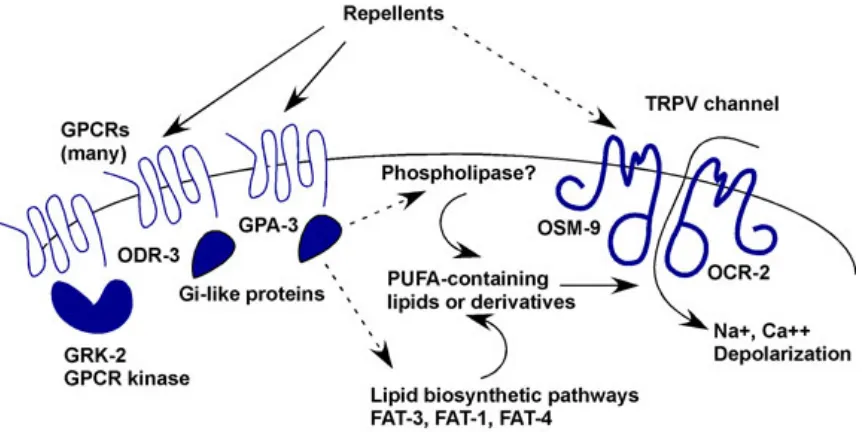
The ASH nocioceptive sensory neuron is one of best studied, and it is a polymodal sensory neuron that responds to mechanical stimuli as well as to metals and changes in osmolarity. Many amphid sensory neurons, in addition to the ASH, signal through transient receptor potential (TRP) channels encoded by osm-914 and ocr-215 genes (Figure 4). The TRP channel superfamily encodes
vertebrate channels that sense osmosensation, pain, and pressure2. It is believed that these channels are downstream of the odr-3 GPCR signaling and require synthesis of long-chain poly-unsaturated fatty acids (PUFAs).2 Loss of odr-3 and gpa-3 diminish calcium transients that are mediated by the TRP channels osm-9 and ocr-2 in the ASH.16
1.4 FUNCTIONAL CIRCUITS in C. ELEGANS
these studies indicate the necessity of both circuit and molecular components for specific behaviors, functional understanding was greatly improved by high-resolution data on the real-time processing done by these neurons.
Many functional circuits have been heavily studied over the last two decades. Of these, the best characterized are the avoidance circuit, the control of locomotion, and sensory coding.20 The ease of studying these circuits is obvious. The avoidance circuit is the most direct and simple of all circuits in this simple system. It consists of a handful of sensory neurons directly connected to the command interneurons16 and is easily observable as an immediate response. The control of locomotion is measurable and the role of various genes and neurons in the control of amplitude, speed and frequency of distinct features has been noted and characterized.21 However, mechanical stimuli are often manual, and precise consistent stimulation of the same area with the same pressure is not trivial. Therefore, many chemosensory neurons and their downstream components have been heavily studied with respect to their activity in response to each other and in the presence of modulating factors.
1.5 C. ELEGANS IS A GOOD CANDIDATE FOR STATE-SPECIFIC MODULATION
Figure 1. Anatomy of the Adult Hermaphrodite. A. DIC image of the adult hermaphrodite. Scale bar
is 1mm B. Schematic drawing of the anatomical structures. (Unmodified figure from Wormatlas.26)
Figure 2. Structure of chemosensory organs. a. Distribution of chemosensory neurons in the animal. Amphids contain 12 associated chemosensory or thermosensory neurons. Phasmids contain two chemosensory neurons, PHA and PHB. Each inner labial organ contains one IL2 chemosensory and one IL1 mechanosensory neuron. There are two URX neurons, one AQR neuron, and one PQR neuron.
[image:17.612.155.493.387.635.2][image:18.612.120.541.82.282.2]
Figure 3. C. elegans head neurons.Schematic drawing of all head neurons in the left and right sides of the
worm with respect to the pharyngeal muscle drawn in green. Note the ganglia drawn in beige in the top panel. (Unmodified figure from Wormatlas.26)
Figure 4. Potential signal transduction pathway for nociception in ASH cilia. A likely model is that
[image:18.612.114.545.381.597.2]
1 White, J. G., Southgate, E., Thomson, J. N. & Brenner, S. The structure of the nervous system of the nematode Caenorhabditis elegans. Philos. Trans. R. Soc. Lond. B. Biol. Sci. 314, 1-340 (1986).
2 Riddle, D. L., Blumenthal, T., Meyer, B. J. & Priess, J. R. Introduction to C. elegans. doi:NBK20183 [bookaccession] (1997).
3 Ward, S., Thomson, N., White, J. G. & Brenner, S. Electron microscopical reconstruction of the anterior sensory anatomy of the nematode Caenorhabditis elegans.?2UU. J. Comp. Neurol. 160, 313-337, doi:10.1002/cne.901600305 (1975).
4 Culotti, J. G. & Russell, R. L. Osmotic avoidance defective mutants of the nematode Caenorhabditis elegans. Genetics 90, 243-256 (1978).
5 Hall, D. H., Lints, R. & Altun, Z. Nematode neurons: anatomy and anatomical methods in Caenorhabditis elegans. Int. Rev. Neurobiol. 69, 1-35, doi:S0074-7742(05)69001-0 [pii] 10.1016/S0074-7742(05)69001-0 (2006).
6 Gray, J. M., Hill, J. J. & Bargmann, C. I. A circuit for navigation in Caenorhabditis elegans. Proc. Natl. Acad. Sci. U. S. A. 102, 3184-3191, doi:0409009101 [pii] 10.1073/pnas.0409009101 (2005).
7 Waggoner, L. E., Hardaker, L. A., Golik, S. & Schafer, W. R. Effect of a neuropeptide gene on behavioral states in Caenorhabditis elegans egg-laying. Genetics 154, 1181-1192 (2000).
8 Ezcurra, M., Tanizawa, Y., Swoboda, P. & Schafer, W. R. Food sensitizes C. elegans avoidance behaviours through acute dopamine signalling. EMBO J 30, 1110-1122, doi:10.1038/emboj.2011.22 (2011).
9 de Bono, M. & Maricq, A. V. Neuronal substrates of complex behaviors in C. elegans. Annu. Rev. Neurosci. 28, 451-501, doi:10.1146/annurev.neuro.27.070203.144259 (2005). 10 Hedgecock, E. M. & Russell, R. L. Normal and mutant thermotaxis in the nematode
Caenorhabditis elegans. Proc Natl Acad Sci U S A 72, 4061-4065 (1975).
11 Ward, S. Chemotaxis by the nematode Caenorhabditis elegans: identification of attractants and analysis of the response by use of mutants. Proc. Natl. Acad. Sci. U. S. A. 70, 817-821 (1973).
12 Giles, A. C. & Rankin, C. H. Behavioral and genetic characterization of habituation using Caenorhabditis elegans. Neurobiol. Learn. Mem. 92, 139-146, doi:S1074-7427(08)00148-2 [pii] 10.1016/j.nlm.2008.08.004 (2009).
13 Zhang, Y., Lu, H. & Bargmann, C. I. Pathogenic bacteria induce aversive olfactory learning in Caenorhabditis elegans. Nature 438, 179-184, doi:nature04216 [pii] 10.1038/nature04216 (2005).
14 Colbert, H. A., Smith, T. L. & Bargmann, C. I. OSM-9, a novel protein with structural similarity to channels, is required for olfaction, mechanosensation, and olfactory adaptation in Caenorhabditis elegans. J. Neurosci. 17, 8259-8269 (1997).
15 Tobin, D. et al. Combinatorial expression of TRPV channel proteins defines their sensory functions and subcellular localization in C. elegans neurons. Neuron 35, 307-318, doi:S0896627302007572 [pii] (2002).
16 Hilliard, M. A. et al. In vivo imaging of C. elegans ASH neurons: cellular response and adaptation to chemical repellents. EMBO J 24, 63-72, doi:10.1038/sj.emboj.7600493 (2005).
19 Chalfie, M. et al. The neural circuit for touch sensitivity in Caenorhabditis elegans. J. Neurosci. 5, 956-964 (1985).
20 Kaplan, J. M. & Horvitz, H. R. A dual mechanosensory and chemosensory neuron in Caenorhabditis elegans. Proc. Natl. Acad. Sci. U. S. A. 90, 2227-2231 (1993).
21 Cronin, C. J., Feng, Z. & Schafer, W. R. Automated imaging of C. elegans behavior. Methods Mol Biol 351, 241-251, doi:10.1385/1-59745-151-7:241 (2006).
22 Raizen, D. M. et al. Lethargus is a Caenorhabditis elegans sleep-like state. Nature 451, 569-572, doi:nature06535 [pii]10.1038/nature06535 (2008).
23 Ben Arous, J., Tanizawa, Y., Rabinowitch, I., Chatenay, D. & Schafer, W. R. Automated imaging of neuronal activity in freely behaving Caenorhabditis elegans. J. Neurosci. Methods 187, 229-234, doi:10.1016/j.jneumeth.2010.01.011 (2010).
24 Guo, Z. V., Hart, A. C. & Ramanathan, S. Optical interrogation of neural circuits in Caenorhabditis elegans. Nat. Methods 6, 891-896, doi:nmeth.1397 [pii] 10.1038/nmeth.1397 (2009).
25 Haspel, G., O'Donovan, M. J. & Hart, A. C. Motoneurons dedicated to either forward or backward locomotion in the nematode Caenorhabditis elegans. J. Neurosci. 30, 11151-11156, doi:30/33/11151 [pii] 10.1523/JNEUROSCI.2244-10.2010.
26 Hall, D. H. & Altun, Z. F. C. elegans atlas. (Cold Spring Harbor Laboratory Press, 2008).
10
CHAPTER 2:
11 INTRODUCTION
Sleep is essential for human and animal health1 and is behaviorally characterized by its circadian timing, behavioral quiescence, homeostasis, increased arousal threshold, and rapid reversibility.2 Sleep is a complex process involving many facets: hardwired circadian regulation, context and activity-dependent regulation (homeostasis), global neuronal activity, specific centers for sleep, specific centers for wakefulness, and numerous regulators or sleep factors. Specific areas of the brain such as the hippocampus, cortex, and hypothalamus have been studied in detail across various species, both vertebrates and invertebrates. These studies have been illuminating in understanding the purpose of sleep and some of the processes that occur during sleep. However, integrating all this information as an overview can be a nightmare.
Additional factors such as stress, health, and the effect of genetic variation in the response of individuals to sleep regulation, both circadian and homeostatic, complicate matters further. Studying these processes in the simplest systems can help tie in the disparate pieces of information and provide a more dynamic understanding of these processes and their interactions with each other. Conveniently, sleep is conserved and present in essentially all animal species3,4, and C. elegans exhibits a sleep-like state called lethargus.
Behavioral evidence suggests that our perception is dramatically dampened during sleep, and that there are physiological changes within individual neurons and their connections to each other.1 However, there are many ways to dampen arousal5, and despite studies spanning mammalian and non-mammalian species, the circuit modifications that promote sleep behavior are largely unknown. The conservation of neurotransmitters, several sleep regulating molecules, and sleep behaviors make anything found in C. elegans of potential interest in mammals, and the ease of genetics and a small nervous system allow for larger scale gene and circuit manipulations than previously possible.5
2.1 AN OVERVIEW OF SLEEP
Wakefulness depends on a network of cell groups in the brainstem, hypothalamus, and basal forebrain that activate the thalamus and the cerebral cortex.1 A switch in the hypothalamus promotes γ-aminobutyric acid (GABA)-containing neurons of the ventrolateral preoptic nucleus
12 arousal system fire fastest during wakefulness.6,7 Therefore, sleep can simply be thought of as an interplay between excitatory and inhibitory centers in the central nervous system.
The VLPO projects neuronal processes to the monoaminergic cell groups that show stereotyped and coordinated changes in firing patterns associated with sleep.8 The VLPO neurons are primarily active during sleep and contain the inhibitory neurotransmitters, galanin and GABA.9,10,11 Cell-specific lesions showed that neuronal subpopulations in the VLPO regulate REM and NREM sleep12 by inhibiting the monoaminergic cell groups. These cells in turn project to the VLPO and can inhibit the VLPO using both noradrenaline and serotonin.13
A circuit containing mutually inhibitory elements sets up a self-reinforcing loop, resulting in a “flip-flop switch”, which produces discrete states with sharp transitions. Hypocretin neurons in the lateral hypothalamus stabilize this switch, and loss of these neurons cause instability of and inappropriate switching between behavioral states.14 These neurons are mainly active during wakefulness, and especially during motor activity.14,15
The two-process model states that sleep results from the combination of both circadian and homeostatic processes.16 The circadian regulation of sleep ensures that an organism rests at the appropriate and evolutionarily beneficial time, whereas homeostatic regulation is driven by the need for rest induced by the duration and intensity of activity. The earliest circadian sleep studies depended on activity patterns.17 Now, sleep in humans and other mammals are characterized by electroencephalogram (EEG) as well as behavioral criteria to distinguish between wakefulness and sleep.18 However, the oscillatory patterns of the EEG data are a result of underlying neural architecture that cannot be reproduced in species with different neuroanatomy. Therefore, behavioral criteria (circadian timing, behavioral quiescence, homeostasis, and increased arousal threshold) are used to identify sleep or sleep-like states in non-mammalian systems, and sleep is conserved and present in essentially all animal species tested.3,4 Furthermore, these periods of quiescence have been shown to be associated with both circadian and homeostatic regulators of sleep.
2.2 CIRCADIAN REGULATION OF SLEEP
13 light detected by retina during the day, and by melatonin secretion from the pineal gland at night.20 The SCN has some projections to the VLPO and hypocretin neurons21, but the majority of its output is directed to the subparaventricular zone (SPZ) and the dorsomedial nucleus of the hypothalamus (DMH). Cell-specific lesions of the ventral and dorsal SPZ disrupt circadian rhythms of wakefulness and body temperature.22 The SPZ projects to the DMH, which in turn inhibits the sleep promoting cells in the VLPO and excites wake promoting cells in the lateral hypothalamic area (LHA).23 DMH integrates circadian input with physiological state, and DMH activity can be shifted by altered wake-sleep, activity, feeding, body temperature, and corticosteroid rhythms.24
The Period gene (per) drives the circadian clock, and PER, as well as its downstream components, has been shown to be consistent with sleep behavior. Disruption of this clock and lack of light-dark cues creates anachronistic sleep. However, a functioning circadian clock is not essential, and sleep still occurs even when circadian rhythms are disrupted or abolished.25
2.3 HOMEOSTATIC REGULATION OF SLEEP
Homeostatic regulation is driven by the need for rest induced by the duration and intensity of activity. Sleep homeostasis refers to the maintenance of sleep amount or depth following sleep deprivation, and is a reflection of the essential nature of sleep. The cause of this homeostasis is under debate, and there are three contending hypotheses: energy depletion hypothesis, neural plasticity hypothesis, and immune defense hypothesis.
The concept of energy metabolism follows the idea that the neuronal activity during waking consumes energy, and the energy is restored during sleep. According to experimental evidence, the degree of arousal is decreased after prolonged wakefulness, and the propensity to sleep and the intensity of delta EEG waves during sleep are proportional to the duration of prior wakefulness.26 One possible explanation for accumulation of sleep propensity is the accumulation of substances, sleep factors, during wakefulness. Then, logically, sleep factors should steadily increase during the waking period and decrease during sleep, their concentrations should be greater during waking than sleeping, and they should inhibit neuronal activity. Adenosine, interleukin-1, TNF-alpha, prostoglandin D2 (PDG2), and growth hormone-releasing factor (GnRH) fit these criteria.27
14 were shown to promote sleep.27 Extracellular adenosine in the basal forebrain (BF) increases during prolonged wakefulness and decreases during recovery sleep.28 Neuronal activity and consumption of ATP during waking is the cause of elevated adenosine concentration29, which in turn feeds back as an inhibitory neuromodulator to decrease activity30. It has been suggested that this is a self-controlling neuroprotective mechanism to preclude cell damage.31 Adenosine is formed from AMP, catalyzed by the enzyme 5’-nucleotidase, and inactivated either back to AMP by the enzyme adenosine kinase (AK) or further to inosine by the enzyme adenosine deaminase (ADA). The enzymes that metabolize adenosine undergo circadian variation, with maximal enzymatic activity during the night and minimal activity during the day.32
Four types of adenosine receptors are known: A1, A2, A3, and A4. Adenosine is generally an inhibitory neuromodulator that inhibits both excitatory and inhibitory neurons. A1 and A3 receptors decrease adenylyl cyclase and cAMP whereas A2 increases cAMP.27 The presynaptic action of adenosine is mediated by the Gi3 subtype of G-proteins that are coupled to inhibition of N-type Ca2+ channels and stimulation of K+ channels. G
i3 either inhibits adenylate cyclase or
activates phospholipase C (PLC) as effector pathways33. A1 receptors are widely distributed, whereas A2 receptors are restricted to the striatum, nucleus accumbens, and olfactory bulb. The main targets for vigilance state-modulating effects of adenosine are A1 receptors34 in the cholinergic cells in the BF, hypothalamus and cortex. Selective activation of cholinergic BF neurons using neurotensin promotes EEG gamma activity and state of wakefulness.35
2.4 MOLECULAR MECHANISMS OF SLEEP
Study of circadian rhythms in model organisms have helped find and understand several molecular mechanisms of sleep. Analysis of sleep in the fruit fly Drosophila showed that increased cAMP promotes wakefulness. Adenylate cyclase mutant rutabaga has reduced cAMP and exhibits increased sleep, whereas the phosphodiesterase mutant dunce increases cAMP and shows decreased sleep.36 cAMP targets CREB, and CREB activity has been correlated with activity in both flies and mice.36,37 However, there is not much mechanistic insight into how cAMP promotes wakefulness.
15 localized to the pars intercerebralis, an area that is developmentally and functionally analogous to the hypothalamus.39
Several other conserved molecular components of neuronal activity and signaling have been shown to affect sleep. In Drosophila, increased PKG activity is associated with more sleep.40 Additionally, preliminary reports suggest that PKG inhibition in the basal forebrain reduces subsequent sleep41. Dopamine, hypocretin, GABA, and 5’-hydroxytryptamine (5-HT) all affect the sleep wake cycle5. In addition, the activity of potassium channels has been shown to modulate neuronal excitability and sleep in both flies42 and mammals43.
2.5 LETHARGUS OR C. ELEGANS SLEEP
C. elegans exhibits four larval stages before it matures into the adult (Figure 1). The transition between each larval stage is marked by the molt, a process in which the worm synthesizes a new cuticle, then sheds its old one. C. elegans exhibits sleep-like behaviors immediately before the molt during a period called lethargus.40,44 Lethargus is a quiescent state during which locomotion and feeding are suppressed44 and sensory arousal is decreased40. Furthermore, this state shows a type of homeostasis that is similar to that seen in sleep homeostasis. Inducing spontaneous activity by increased sensory stimuli during lethargus results in anachronistic rebound quiescence.40
16 FIGURES
Figure 1. Life cycle of C. elegans at 22°C. 0 min is fertilization. Numbers in blue along the arrows
17
REFERENCES
1 Saper, C. B., Scammell, T. E. & Lu, J. Hypothalamic regulation of sleep and circadian
rhythms. Nature437, 1257-1263, doi:nature04284 [pii]10.1038/nature04284 (2005). 2 Saper, C. B., Cano, G. & Scammell, T. E. Homeostatic, circadian, and emotional
regulation of sleep. J. Comp. Neurol.493, 92-98, doi:10.1002/cne.20770 (2005).
3 Allada, R. & Siegel, J. M. Unearthing the phylogenetic roots of sleep. Curr. Biol. 18,
R670-R679, doi:10.1016/j.cub.2008.06.033 (2008).
4 Siegel, J. M. The REM sleep-memory consolidation hypothesis. Science294, 1058-1063,
doi:10.1126/science.1063049 (2001).
5 Zimmerman, J. E., Naidoo, N., Raizen, D. M. & Pack, A. I. Conservation of sleep:
insights from non-mammalian model systems. Trends Neurosci.31, 371-376, doi:S0166-2236(08)00133-1 [pii]10.1016/j.tins.2008.05.001 (2008).
6 Aston-Jones, G. & Bloom, F. E. Activity of norepinephrine-containing locus coeruleus
neurons in behaving rats anticipates fluctuations in the sleep-waking cycle. J. Neurosci.1, 876-886 (1981).
7 Fornal, C., Auerbach, S. & Jacobs, B. L. Activity of serotonin-containing neurons in
nucleus raphe magnus in freely moving cats. Exp. Neurol.88, 590-608 (1985).
8 Sherin, J. E., Shiromani, P. J., McCarley, R. W. & Saper, C. B. Activation of
ventrolateral preoptic neurons during sleep. Science271, 216-219 (1996).
9 Gaus, S. E., Strecker, R. E., Tate, B. A., Parker, R. A. & Saper, C. B. Ventrolateral
preoptic nucleus contains sleep-active, galaninergic neurons in multiple mammalian species. Neuroscience115, 285-294, doi:S0306452202003081 [pii] (2002).
10 Sherin, J. E., Elmquist, J. K., Torrealba, F. & Saper, C. B. Innervation of histaminergic
tuberomammillary neurons by GABAergic and galaninergic neurons in the ventrolateral preoptic nucleus of the rat. J. Neurosci.18, 4705-4721 (1998).
11 Szymusiak, R., Alam, N., Steininger, T. L. & McGinty, D. Sleep-waking discharge
patterns of ventrolateral preoptic/anterior hypothalamic neurons in rats. Brain Res. 803, 178-188, doi:S0006-8993(98)00631-3 [pii] (1998).
12 Lu, J. et al. Selective activation of the extended ventrolateral preoptic nucleus during
rapid eye movement sleep. J. Neurosci. 22, 4568-4576, doi:2002645522/11/4568 [pii] (2002).
13 Gallopin, T. et al. Identification of sleep-promoting neurons in vitro. Nature 404,
992-995, doi:10.1038/35010109 (2000).
14 Mileykovskiy, B. Y., Kiyashchenko, L. I. & Siegel, J. M. Behavioral correlates of
activity in identified hypocretin/orexin neurons. Neuron 46, 787-798, doi:S0896-6273(05)00395-8 [pii]10.1016/j.neuron.2005.04.035 (2005).
15 Lee, M. G., Hassani, O. K. & Jones, B. E. Discharge of identified orexin/hypocretin
neurons across the sleep-waking cycle. J. Neurosci. 25, 6716-6720, doi:25/28/6716 [pii]10.1523/JNEUROSCI.1887-05.2005 (2005).
16 Borbely, A. A. & Achermann, P. Sleep homeostasis and models of sleep regulation. J.
Biol. Rhythms14, 557-568 (1999).
17 Konopka, R. J. & Benzer, S. Clock mutants of Drosophila melanogaster. Proc. Natl.
Acad. Sci. U. S. A.68, 2112-2116 (1971).
18 Hendricks, J. C., Sehgal, A. & Pack, A. I. The need for a simple animal model to
understand sleep. Prog. Neurobiol.61, 339-351, doi:S0301-0082(99)00048-9 [pii] (2000). 19 Jin, X. et al. A molecular mechanism regulating rhythmic output from the
18 20 Cassone, V. M., Chesworth, M. J. & Armstrong, S. M. Entrainment of rat circadian
rhythms by daily injection of melatonin depends upon the hypothalamic suprachiasmatic nuclei. Physiol. Behav.36, 1111-1121 (1986).
21 Watts, A. G., Swanson, L. W. & Sanchez-Watts, G. Efferent projections of the
suprachiasmatic nucleus: I. Studies using anterograde transport of Phaseolus vulgaris leucoagglutinin in the rat. J. Comp. Neurol. 258, 204-229, doi:10.1002/cne.902580204 (1987).
22 Lu, J. et al. Contrasting effects of ibotenate lesions of the paraventricular nucleus and
subparaventricular zone on sleep-wake cycle and temperature regulation. J. Neurosci.21, 4864-4874, doi:21/13/4864 [pii] (2001).
23 Chou, T. C. et al. Critical role of dorsomedial hypothalamic nucleus in a wide range of
behavioral circadian rhythms. J Neurosci23, 10691-10702, doi:23/33/10691 [pii] (2003). 24 Saper, C. B., Lu, J., Chou, T. C. & Gooley, J. The hypothalamic integrator for circadian
rhythms. Trends. Neurosci. 28, 152-157, doi:S0166-2236(04)00395-9 [pii]10.1016/j.tins.2004.12.009 (2005).
25 Baker, F. C., Angara, C., Szymusiak, R. & McGinty, D. Persistence of sleep-temperature
coupling after suprachiasmatic nuclei lesions in rats. Am. J. Physiol. Regul. Integr. Comp.
Physiol.289, R827-838, doi:00093.2005 [pii]10.1152/ajpregu.00093.2005 (2005).
26 Borbely, A. A. A two process model of sleep regulation. Hum. Neurobiol. 1, 195-204
(1982).
27 Porkka-Heiskanen, T. & Kalinchuk, A. V. Adenosine, energy metabolism and sleep
homeostasis. Sleep Med. Rev. 15, 123-135, doi:S1087-0792(10)00066-3 [pii]10.1016/j.smrv.2010.06.005.
28 Porkka-Heiskanen, T. et al. Adenosine: a mediator of the sleep-inducing effects of
prolonged wakefulness. Science276, 1265-1268 (1997).
29 Mitchell, J. B., Lupica, C. R. & Dunwiddie, T. V. Activity-dependent release of
endogenous adenosine modulates synaptic responses in the rat hippocampus. J. Neurosci. 13, 3439-3447 (1993).
30 Illes, P., Klotz, K. N. & Lohse, M. J. Signaling by extracellular nucleotides and
nucleosides. Naunyn Schmiedebergs Arch. Pharmacol.362, 295-298 (2000).
31 Cunha, R. A. Neuroprotection by adenosine in the brain: From A(1) receptor activation to
A (2A) receptor blockade. Purinergic Signal1, 111-134, doi:10.1007/s11302-005-0649-1 (2005).
32 Chagoya de Sanchez, V. et al. Day-night variations of adenosine and its metabolizing
enzymes in the brain cortex of the rat--possible physiological significance for the energetic homeostasis and the sleep-wake cycle. Brain Res.612, 115-121 (1993).
33 Biber, K., Klotz, K. N., Berger, M., Gebicke-Harter, P. J. & van Calker, D. Adenosine A1
receptor-mediated activation of phospholipase C in cultured astrocytes depends on the level of receptor expression. J. Neurosci.17, 4956-4964 (1997).
34 Basheer, R., Strecker, R. E., Thakkar, M. M. & McCarley, R. W. Adenosine and
sleep-wake regulation. Prog. Neurobiol. 73, 379-396,
doi:10.1016/j.pneurobio.2004.06.004S0301-0082(04)00125-X [pii] (2004).
35 Cape, E. G., Manns, I. D., Alonso, A., Beaudet, A. & Jones, B. E. Neurotensin-induced
bursting of cholinergic basal forebrain neurons promotes gamma and theta cortical activity together with waking and paradoxical sleep. J. Neurosci. 20, 8452-8461, doi:20/22/8452 [pii] (2000).
36 Hendricks, J. C. et al. A non-circadian role for cAMP signaling and CREB activity in
19 37 Graves, L. A. et al. Genetic evidence for a role of CREB in sustained cortical arousal. J.
Neurophysiol.90, 1152-1159, doi:10.1152/jn.00882.200200882.2002 [pii] (2003).
38 Kushikata, T., Fang, J., Chen, Z., Wang, Y. & Krueger, J. M. Epidermal growth factor
enhances spontaneous sleep in rabbits. Am. J. Physiol.275, R509-514 (1998).
39 Foltenyi, K., Greenspan, R. J. & Newport, J. W. Activation of EGFR and ERK by
rhomboid signaling regulates the consolidation and maintenance of sleep in Drosophila.
Nat. Neurosci.10, 1160-1167, doi:nn1957 [pii]10.1038/nn1957 (2007).
40 Raizen, D. M. et al. Lethargus is a Caenorhabditis elegans sleep-like state. Nature451,
569-572, doi:nature06535 [pii]10.1038/nature06535 (2008).
41 Kalinchuk, A. V., Porkka-Heiskanen, T. & McCarley, R. W. Basal forebrain and saporin
cholinergic lesions: the devil dwells in delivery details. Sleep29, 1385-1387; discussion 1387-1389 (2006).
42 Cirelli, C. et al. Reduced sleep in Drosophila Shaker mutants. Nature 434, 1087-1092,
doi:nature03486 [pii]10.1038/nature03486 (2005).
43 Douglas, C. L. et al. Sleep in Kcna2 knockout mice. BMC Biol.5, 42,
doi:1741-7007-5-42 [pii]10.1186/1741-7007-5-doi:1741-7007-5-42 (2007).
44 Van Buskirk, C. & Sternberg, P. W. Epidermal growth factor signaling induces
behavioral quiescence in Caenorhabditis elegans. Nat. Neurosci. 10, 1300-1307, doi:nn1981 [pii]10.1038/nn1981 (2007).
45 Monsalve, G. C., Van Buskirk, C. & Frand, A. R. LIN-42/PERIOD controls cyclical and
developmental progression of C. elegans molts. Curr. Biol. 21, 2033-2045, doi:10.1016/j.cub.2011.10.054 (2011).
20
CHAPTER 3:
21 INTRODUCTION
The extensive number of tools available in the C. elegans toolkit helps make it an attractive
model system for the study of neuroscience. The most obvious is the number of genetic tools available for use. The ease of maintainence, small size, fast generation time, large numbers of identical progeny, and relatively small genome has allowed the characterization of a large mutant library, genetic mapping, and high degree of genetic control over targeting and characterizing
specific neurons.1 The transparent body of C. elegans allows live imaging of neuronal activity
and development in live, intact animals2, and electrophysiological methods have been developed
to study neuronal activity in the form of graded potentials. In addition, optical stimulation techniques allow temporal control as wells as extent of activation in the intact freely behaving animal.2,3
3.1 ELECTROPHYSIOLOGY IN C. ELEGANS
The first nematode measured with electrophysiological techniques was Ascaris lumbrocoides,
a worm with large neurons and graded active synaptic responses.4 Despite their difference in size,
there is striking anatomical similarity between Ascaris and C. elegans1, and the studies of the Ascaris locomotor circuit provided insight into the excitatory and inhibitory function of specific
C. elegans motor neurons.
Electrophysiology in C. elegans requires puncture of the external cover or cuticle, hence
measurement of intact circuits is challenging. The body of C. elegans is an unsegmented, tapered
cylinder whose structure is maintained by a tube-like arrangement of muscles attached to the hypodermis and a tough cuticle cover. The shape of the body is maintained by its high internal pressure, much like a balloon, and like a balloon, disruption of cuticle integrity results in
increased likelihood of rupture. In the case of C. elegans, a large enough rupture also means an
extrusion of organs and eventual death.
Early electrophysiological measurements in C. elegans focused on the neuromuscular
functions of the pharynx, and these electropharyngeograms were recordings of extracellular
potentials.5 Eventually, techniques were developed and modified to record synaptic activity at
the neuromuscular junction6 and then to record individual sensory and interneurons7. These are
high-resolution measurements that allow calculations such as transfer functions of individual
22 of neuropeptides and signaling factors of the recorded neurons, it is not ideal for the study of state modulation. Measurements in the intact worm are necessary and ideal.
3.2 CALCIUM IMAGING IN C. ELEGANS
The discovery and use of green fluorescent protein, or GFP, has been most useful in serving as a visual marker for temporal and spatial expression of genes, identification of structure and function, as well as in opening up the field for development of a variety of new tools. Derivatives of the original GFP have been used for a variety of functions, including the fusion to proteins in order to identify compartment-specific localization and dynamics, identify co-localization of different proteins using variants that modify the excitation and emission spectra of the fluorescent protein, and split derivatives serving as detectors for the presence of various molecules or for two molecules’ proximity to each other. Of course, the expression of any of these protein and products can serve as buffers and compete with the natural processes that are studied. This should be kept in mind in the context of behavior.
The calcium indicators are fluorescent detectors that increase their fluorescence in the presence of available calcium. Small molecule calcium dyes are an indirect indicator of neuronal
activity, but these require injection and lack cell specificity.8 Genetically encoded calcium
indicators allow visualization in an intact animal and availability of a large number of known
promoters provide cell specificity. These indicators are based on the protein calmodulin, which
changes its conformation in response to binding of calcium. The calcium binding region of calmodulin is used to link two proteins, of which one or both are fluorescent molecules, and the changing distance between the fluorescent particles either shifts the spectra or changes the intensity of the wavelengths of light emitted.
GCamP is a calcium indicator that is a fusion of GFP, calmodulin, and M13. In the absence of calcium, the conformation of the circularized GFP allows quenching of the fluorophore, which
is altered upon calcium binding.9 Therefore, there is a dramatic and fast increase in fluorescence
in response to calcium. Cameleon uses foster resonance energy transfer (FRET). FRET works through the use of two fluorophores: the donor which is excited with an external light source and an acceptor whose excitation spectra corresponds to the emission spectra of the donor. FRET requires the proximity of the donor and acceptor. Therefore, the ratio of the donor and acceptor emission that can be detected changes with alterations of the calmodulin conformation, and the
23 Ratiometric imaging has its drawbacks and advantages. The non-FRET indicators have better dynamic range, and cameleon often shows a smaller change in response to the same stimulus in a given neuron when compared to GCamP. However, there are also many advantages to ratiometric imaging: movement and lighting artifacts can be detected and cancel out in the ratiometric calculation, altered promoter activity and expression of indicator is not an issue, and it
allows for detection of fast events even in the scale of milliseconds.11 The hardware for
ratiometric imaging is a bit more complicated, but worth it. However, I find that the largest drawback to using the FRET indicator in my studies is the effect on behavior.
Light is a potent stimulant and serves a noxious cue in C. elegans12, and it avoids short
wavelengths of light by staying beneath the surface of the soil. Animals avoid light and it is shown that illumination with the green to ultraviolet spectra of light is detected by LITE-1 and
will induce escape response as well as increase locomotion.12 We have found that exposure to
high intensity light even in the absence of lite-1, will induce calcium transients in the ASH
nocioceptive sensory neuron of adult animals, as well as those in lethargus. Furthermore, shorter
wavelengths of light elicit stronger responses.12 Therefore, although use of low intensity light for
cameleon imaging fails to elicit sensory response in lethargus, it serves as a stimulus in L4 and adult animals which is not ideal and changes sensory response to chemical stimuli.
3.3 THE OPTOGENETIC TOOLBOX
Several techniques have been developed to remotely and optically control neurons in the intact worm. Channelrhodopsin is an algae-derived cation channel that is a microbial-type rhodopsin, a seven transmembrane retinal protein that has no sequence homology to animal rhodopsins. Channelrhodopsin1 is a proton-selective light-activated channel derived from the
green algae Chalmydomonas reinhardtii.13 Channelrhodopsin2 is a leaky proton pump that acts
as a light-gated nonselective cation channel14 that has the ability to trigger large currents. A
cofactor, all-trans retinal, is required for channel activity and the peak of the action spectrum is
approximately 460nm. The ChR2 conductance has a large initial transient and decays to a lower steady-state level with continued illumination. The refractory period after activation is shorter at low extracellular pH and at more negative voltages. The estimated conductance of the channel is a low 50 fS. The rise time of the ChR2 current is extremely fast: less than 200 µs; decay is on the
24 The first characterizations of ChR2 involved the control of action potential and synaptic
transmission in hippocampal neurons.15 Currently, photoactivatable channels have been used in a
range of animal models including worms, flies, fish, rodents and primates.16-20 In C. elegans,
ChR2 can be used to drive awake behavioral responses in the presence of the cofactor all-trans
retinal (ATR).16 Animals can be fed ATR through the bacteria they eat, and specific promoters
can be used to selectively promote ChR2 expression and activation. In addition to study of
behavior, it has been used to study release at the neuromuscular junction in worms.21
In addition to ChR2, many other types of light-activated channels and molecules exists. Optical inhibition can be induced by using halorhodopsin, and G-protein signaling pathways can be activated by the opto-XRs. Halorhodopsin is a yellow-light activated chloride pump derived
from archaebacterium Natronomas pharaonis that works with millisecond precision.22 Use of
optical stimulation techniques allow fast, reliable manipulation of neuronal activation and inhibition in specific subsets of neurons. Recent advances provide light-shifted varieties, as well as long-acting and subthreshold channelrhodopsins. In addition, various strategies have been used for cell-specific activation and expression of these light-activated channels.
3.4 MICROFLUIDICS
Microfluidic devices were first developed to miniaturize chemical and biochemical analyses
and to make these processes more sensitive, faster, and have higher resolution.23 Since then,
microfluidic devices fabricated in polymers (polymethylsiloxane or PDMS) using soft
lithography have been useful in basic and biomedical applications.24 Microfluidics uses laminar
flow and allow precise control and quick manipulation of microenvironments that are not
available in conventional macro-scale methods.25 These devices have been used for cell culture
as well as neuronal stimulation26, and C. elegans with its small size of 2-3mm is well-suited for
this technology.
Various microfluidic devices exist for immobilization of animals, observation of locomotion, and exposure of animals with either gas or aqueous stimuli. These devices allow simultaneous control of the animals’ environment while imaging, and has allowed measurement of sensory
response to a variety of stimuli.27 Manual and automated28 controls of these devices are possible.
26 REFERENCES
1 Riddle, D. L., Blumenthal, T., Meyer, B. J. & Priess, J. R. Introduction to C. elegans.
doi:NBK20183 [bookaccession] (1997).
2 Schafer, W. R. Neurophysiological methods in C. elegans: an introduction. WormBook,
1-4, doi:10.1895/wormbook.1.113.1 (2006).
3 Husson, S. J. et al. Microbial Light-Activatable Proton Pumps as Neuronal Inhibitors to
Functionally Dissect Neuronal Networks in C. elegans. PLoS One 7, e40937,
doi:10.1371/journal.pone.0040937 PONE-D-12-04921 [pii].
4 Davis, R. E. & Stretton, A. O. Signaling properties of Ascaris motorneurons: graded
active responses, graded synaptic transmission, and tonic transmitter release. J. Neurosci.
9, 415-425 (1989).
5 Raizen, D. M. & Avery, L. Electrical activity and behavior in the pharynx of
Caenorhabditis elegans. Neuron12, 483-495, doi:0896-6273(94)90207-0 [pii] (1994).
6 Goodman, M. B., Hall, D. H., Avery, L. & Lockery, S. R. Active currents regulate
sensitivity and dynamic range in C. elegans neurons. Neuron 20, 763-772,
doi:S0896-6273(00)81014-4 [pii] (1998).
7 Narayan, A., Laurent, G. & Sternberg, P. W. Transfer characteristics of a thermosensory
synapse in Caenorhabditis elegans. Proc. Natl. Acad. Sci. U. S. A. 108, 9667-9672,
doi:1106617108 [pii] 10.1073/pnas.1106617108. (2011)
8 Riddle, D. L., Blumenthal, T., Meyer, B. J. & Priess, J. R. in C. elegans II (eds D. L.
Riddle, T. Blumenthal, B. J. Meyer, & J. R. Priess) (1997).
9 Nakai, J., Ohkura, M. & Imoto, K. A high signal-to-noise Ca(2+) probe composed of a
single green fluorescent protein. Nature Biotechnology 19, 137-141, doi:10.1038/84397
(2001).
10 Miyawaki, A. et al. Fluorescent indicators for Ca2+ based on green fluorescent proteins
and calmodulin. Nature388, 882-887, doi:10.1038/42264 (1997).
11 Suzuki, Y. et al. Discovery of novel neuronal voltage-dependent calcium channel
blockers based on emopamil left hand as a bioactive template. Bioorganic & Medicinal
Chemistry Letters13, 919-922 (2003).
12 Edwards, S. L. et al. A novel molecular solution for ultraviolet light detection in
Caenorhabditis elegans. PLoS Biology6, e198, doi:10.1371/journal.pbio.0060198 (2008).
13 Nagel, G. et al. Channelrhodopsin-1: a light-gated proton channel in green algae. Science
296, 2395-2398, doi:10.1126/science.1072068 296/5577/2395 [pii] (2002).
14 Nagel, G. et al. Channelrhodopsin-2, a directly light-gated cation-selective membrane
channel. Proc. Natl. Acad. Sci. U. S. A.100, 13940-13945, doi:10.1073/pnas.1936192100
1936192100 [pii] (2003).
15 Boyden, E. S., Zhang, F., Bamberg, E., Nagel, G. & Deisseroth, K. Millisecond-timescale,
genetically targeted optical control of neural activity. Nat. Neurosci. 8, 1263-1268,
doi:nn1525 [pii] 10.1038/nn1525 (2005).
16 Nagel, G. et al. Light activation of channelrhodopsin-2 in excitable cells of
Caenorhabditis elegans triggers rapid behavioral responses. Curr. Biol. 15, 2279-2284,
doi:S0960-9822(05)01407-7 [pii] 10.1016/j.cub.2005.11.032 (2005).
17 Schroll, C. et al. Light-induced activation of distinct modulatory neurons triggers
appetitive or aversive learning in Drosophila larvae. Curr. Biol. 16, 1741-1747,
doi:S0960-9822(06)01854-9 [pii] 10.1016/j.cub.2006.07.023 (2006).
18 Arenkiel, B. R. et al. In vivo light-induced activation of neural circuitry in transgenic
mice expressing channelrhodopsin-2. Neuron 54, 205-218, doi:S0896-6273(07)00183-3
27
19 Huber, D. et al. Sparse optical microstimulation in barrel cortex drives learned behaviour
in freely moving mice. Nature 451, 61-64, doi:nature06445 [pii] 10.1038/nature06445
(2008).
20 Han, X. et al. Millisecond-timescale optical control of neural dynamics in the nonhuman
primate brain. Neuron 62, 191-198, doi:S0896-6273(09)00210-4
[pii]10.1016/j.neuron.2009.03.011 (2009).
21 Liewald, J. F. et al. Optogenetic analysis of synaptic function. Nat. Methods5, 895-902,
doi:nmeth.1252 [pii] 10.1038/nmeth.1252 (2008).
22 Han, X. & Boyden, E. S. Multiple-color optical activation, silencing, and
desynchronization of neural activity, with single-spike temporal resolution. PLoS One 2,
e299, doi:10.1371/journal.pone.0000299 (2007).
23 Whitesides, G. M. The origins and the future of microfluidics. Nature 442, 368-373,
doi:10.1038/nature05058 (2006).
24 van Kooten, T. G., Whitesides, J. F. & von Recum, A. Influence of silicone (PDMS)
surface texture on human skin fibroblast proliferation as determined by cell cycle analysis.
Journal of Biomedical Materials Research43, 1-14 (1998).
25 El-Ali, J., Sorger, P. K. & Jensen, K. F. Cells on chips. Nature 442, 403-411,
doi:10.1038/nature05063 (2006).
26 Taylor, A. M. et al. A microfluidic culture platform for CNS axonal injury, regeneration
and transport. Nat. Methods2, 599-605, doi:10.1038/nmeth777 (2005).
27 Chronis, N., Zimmer, M. & Bargmann, C. I. Microfluidics for in vivo imaging of
neuronal and behavioral activity in Caenorhabditis elegans. Nat. Methods 4, 727-731,
doi:10.1038/nmeth1075 (2007).
28 Chokshi, T. V., Bazopoulou, D. & Chronis, N. An automated microfluidic platform for
calcium imaging of chemosensory neurons in Caenorhabditis elegans. Lab Chip 10,
28
CHAPTER 4:
Multilevel Modulation in
C.elegans
Sleep
29
4.1
ABSTRACT
Sleep is essential for human and animal health and is behaviorally characterized by its circadian timing, behavioral quiescence, homeostasis, increased arousal threshold, and rapid reversibility. Behavioral evidence suggests that our perception is dramatically dampened during sleep, and that there are physiological changes within individual neurons and their connections to each other. However, there are many ways to dampen arousal, and despite studies spanning mammalian and non-mammalian species, the circuit modifications that promote sleep behavior are largely unknown. Here we systematically dissect components of a sensory motor circuit and identify changes in the flow of information from sensory to inter- and motor-neuron that implement sleep-like behavior in the nematode Caenorhabditis elegans. We demonstrate that the top two layers of sensory processing are affected, with decreases in both sensory transduction and subsequent transmission of this information to interneurons. Modulation in transmission results in a decreased likelihood of coordinated activity in the downstream interneurons, which can be reversed with sufficient prior sensory stimulation. Multilevel depression provides an elegant strategy to promote a robust decrease in arousal while allowing for rapid reversibility of the sleep state.
4.2 INTRODUCTION
Sleep behavior is conserved and present in essentially all animal species as a lack of reactivity to sensory inputs, low activity, decreased conscious awareness, and rapid reversibility to wakefulness.1,2 Despite long-standing knowledge and characterization of these states, translation to a physiological or circuit definition has been difficult for many reasons. No single molecular or neuronal pathway has been shown to be responsible for the regulation of sleep, and incomplete knowledge of connections in sensory motor circuits of many studied species precludes interrogation of the flow of information that promotes sensory responses. Furthermore, the complexity and redundancy of the mammalian nervous system complicates the understanding of the flow of information. Here we use the nematode C. elegans to define sleep behavior in a simple sensory motor circuit.
C. elegans exhibits sleep-like behaviors during lethargus, a quiescent state during which
30
normal resting period, there is a period of anachronistic rebound rest.4 During lethargus, worms exhibit delayed or decreased avoidance behavior in response to both mechanical and chemical stimuli, and this response delay is reversible upon previous stimulation of the same neuron.4 It is of note that quiescence as measured by decreased feeding and locomotory behavior is present in adult satiety behavior.5 However, the dynamics of this state were not previously studied.
Lethargus invariably occurs during development after each of the four larval stages, and the timing of lethargus corresponds to increased expression of LIN-42, homolog of circadian regulator PER.6 In addition, several additional conserved regulators have been identified. Anachronistic quiescence is also induced by expression of EGF3, a function conserved in mammals7 and Drosophila8, and sensory arousal can be depressed by PKG4, another well-conserved signaling protein. Conservation of sleep characteristics and molecular signaling suggests that the lethargus state in C. elegans could prove insightful in understanding sleep regulation.
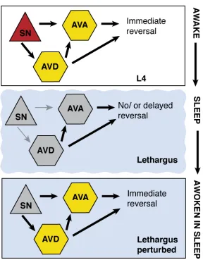
The connections of the C. elegans nervous system are mapped9,10, and functional circuits mediating avoidance defined11. Avoidance behaviors are mediated by mechanosensory and chemosensory neurons that activate downstream circuit components to coordinate motor neuron activity and locomotion. The ASH sensory neuron drives an avoidance circuit and promotes immediate locomotory reversal. When animals are presented with ASH-specific stimuli, ASH stimulates interneurons AVA and AVD, which in turn induces backward locomotion through stimulation of excitatory cholinergic motor neurons in the ventral cord (Figure 1a).12,13,14 Evidence of changing arousal as measured by response delay to ASH-specific stimuli suggests modulation in the avoidance circuit during sleep behavior.
31
measurement and activation of specific components of pertinent circuits. Here we use genetically encoded calcium sensors and light-driven channels to investigate the ASH circuit during sleep and waking. We find that multiple steps in the circuit are dampened during sleep.
4.3 ASH SENSORY NEURON EXHIBITS DECREASED SENSORY RESPONSE
The amphid sensory neuron, ASH, senses multiple aversive stimuli, including mechanical stimulation at the tip of the head, and noxious chemical cues, such as copper, 1-octanol, or high osmolarity.13 We used a chemical stimulus to characterize ASH activity during a “sleep cycle” because it is more consistent and controllable than mechanical stimuli. We fabricated a modified version of a microfluidic olfactory chip17 to accommodate and immobilize fourth larval stage (L4), lethargus, and young adult animals (Figure 1B). These devices permitted temporally controlled delivery of chemical stimuli and simultaneous fluorescence imaging from the ASH. Individual animals were assayed for a six-hour period, during which they were subjected to a brief stimulus every 30 minutes. ASH response was measured during these intervals using the calcium indicator GCaMP3.18 Each animal was imaged before, during and after lethargus. Chemosensory neurons use ligand-binding receptors to open ion channels11, and ASH responds to the addition of 1mM Cu2+ with a robust influx of calcium in the L4 stage. During lethargus, the magnitude of ASH response decreases significantly to copper or glycerol, but full responsivity is recovered upon exit from lethargus (Figure 1C, S1A).
32
glycerol and suggest that modulation in lethargus may affect general excitability or synaptic activity of the ASH neuron.
4.4 BASAL ACTIVITY OF AVA IS SUPPRESSED IN LETHARGUS
GCaMP3 findings were confirmed with Cameleon measurements at the interneuron level. Calcium levels in AVA oscillate and increasing levels correspond with reversals.20 We observed that oscillation of AVA activity is not regular and disappears in lethargus, but reappears in the young adult animals (Figure S3A-B). Therefore, the basal activity and the context in which AVA receives input from ASH changes during lethargus.
4.5 SYNCHRONY BETWEEN AVD AND AVA IS LOST IN LETHARGUS
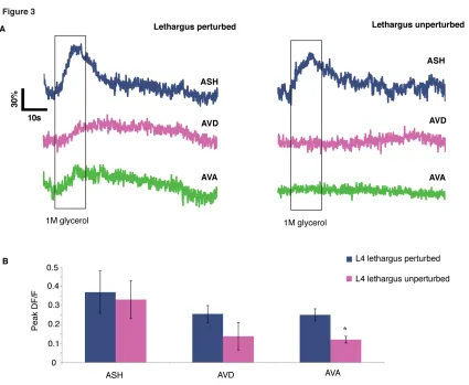
To assess activity across the top two layers of the circuit, we used animals expressing GCaMP3 in ASH, AVD, and AVA. To examine stimulus-driven interneuron activity, we measured responses to ASH-specific cue (1M glycerol)12, which has less variable dynamics (Figure S1A). Each 60-second trial consisted of a 10-second pulse of control buffer or glycerol. Trials with discernable ASH responses were chosen, and instantaneous slopes were calculated over the imaging interval to represent the magnitude of the calcium influxes. In young adult animals without stimulation, the timing of the calcium influxes showed no association with the pulse of the control buffer and did not occur during the stimulus interval (Figure S2B). However, after a glycerol stimulus, the influx of calcium in the AVD and AVA was associated with the stimulation interval (Figure 2B-C). AVD and AVA showed both decreased responsivity during lethargus as well as a loss of coordinated calcium activity. Animals in lethargus exhibited very little activity in the AVD and AVA, and fluctuating calcium levels during the glycerol stimulus were not significantly different from the period preceding the stimulus (Figure 2B). Because the magnitude of activity is considerably smaller in lethargus compared to the young adult, we had to normalize these measurements as binary values. Positive changes in fluorescence were thus counted as individual influx events. There were significantly more influxes during the glycerol stimulus in the young adult, but often little activity was seen in AVA during lethargus even when AVD activity was noted (Figure 2C-E).
33
correlation analysis between each of the neurons imaged. Comparing da