J. exp. Biol. (1982), 99, 331-338 3 3 1
With 3 figures
WPrinted in Great Britain
BLOOD GAS TRANSPORT IN THE CEPHALOPOD,
SEPIA OFFICINALIS
BY KJELL JOHANSEN, OLE BRIX AND GUNNAR LYKKEBOE Department of Zoophysiology, University of Aarhus,
DK-8000 Aarhus C, Denmark
(Received 5 January 1982 - Accepted 3 March 1982)
SUMMARY
Blood gas transport was studied in unrestrained free-swimming cuttle-fish, Sepia officinalis, following cannulations of an efferent branchial (arterial) vessel and the vena cava cephalica with indwelling catheters.
In well-aerated water the arterial pOi averaged about ioo-oo mmHg and was fully saturated with O2. Mixed venous pOi varied between 17 and 40 mmHg, typically corresponding to blood O2 utilizations of 80% or
higher. Some blood samples showed venous pH to exceed arterial, a tendency becoming more distinct during exposure to hypoxic water. The resulting higher O2 affinity of venous compared to arterial blood discourages O2
unloading in the tissues, while promoting efficient O2 loading in the gills.
A high n-value of Sepia blood (« = 4-7) is important for maintaining a large arteriovenous Oa content difference and a high utilization of circulating O2.
INTRODUCTION
The high aerobic metabolism of most cephalopods exceeds that for other aquatic invertebrates of similar size and also surpasses that of many fast-swimming fishes (Altman & Ditmer, 1971).
Since cephalopod blood has a low O2 capacity, typically not exceeding 5 vol%,
the high tissue O2 requirements must be satisfied by high perfusion rates (cardiac
outputs) and by maximum utilization of the blood-borne, circulating O2 pool. The
utilization of O2 transported by the blood and the physiological significance of blood
respiratory properties studied in vitro can never be fully assessed unless information about in vivo circulating levels of blood gases and pH are available from arterial and venous blood. Such information is extremely rare for cephalopods, due to the obvious technical difficulties associated with implanting chronically indwelling intravascular catheters in these animals. Studying the active squid, Loligo, Redfield & Goodkind (1929) measured near-maximal arteriovenous O2 content differences by direct blood
sampling from pinned-down, cut open and artificially ventilated specimens. Based on these measurements blood O2 utilization in Loligo exceeded 90%. The authors
implied that values of blood pH (/>co2) measured concurrently established that nearly
shifts in Loligo. The Bohr factor <p (A log P50/ApH) was reported to be —1-8 forJ
Loligo (Redfield, Coolidge & Hurd, 1926). " Studies of the large Octopus dofleini at rest, based on blood samplings from chroni-cally cannulated animals, showed O2 utilizations averaging 72 % with a maximum of
85 % (Johansen & Lenfant, 1966). O. dofleini also has a large Bohr factor (<j> = — o-8o) (Lenfant & Johansen, 1965).
Blood O2 transport in the phylogenetically primitive Nautilus pompilius showed a
much lower value for O2 utilization of 35%. Nautilus is sluggish and has a small
Bohr factor (0 = - 0 2 0 ) (Johansen, Redmond & Bourne, 1978).
The study to be reported concerns an evaluation of blood gas transport in the common North-Atlantic cuttlefish, Sepia officinalis.
MATERIALS, METHODS AND SURGICAL PROCEDURES
Seven specimens of Sepia officinalis, ranging in weight from 1500 to 1600 g, were used in the study. The animals were obtained by trawling in the English Channel near Plymouth. Before experiments, the animals had been kept for several days in well aerated running sea water at 17 °C. They were fed live crabs at intervals, but food was withheld for at least 24 h prior to anaesthesia.
Anaesthesia was induced by slow addition of ethyl alcohol to an aerated seawater bath to a final concentration of about 3 % in the sea water. Muscle relaxation occurred after about 30 min exposure to the alcohol-water mixture. The surgical procedures lasted about 20 min. The large vena cava cephalica, cannulated for mixed venous blood, is accessible slightly posterior to the anus when the mantle musculature is relaxed. A PE 60 polythene catheter was passed downstream through a hole made in the vessel wall with a cutting-edge surgical suture needle. The catheter was advanced 6-8 cm downstream and tied to the vessel wall by a pursestring ligature.
The cannulation presented no significant obstruction to venous flow since the vena cava cephalica is a large vessel of 8-10 mm diameter. Arterial blood was obtained by cannulation of the efferent branchial artery accessible at the lateral aspect of the gill when the mantle was relaxed. After tying a holding thread around the tip of the gill, a hole was cut in the efferent branchial vessel about 10-15 m m from the holding thread.
Polyethylene catheters (PE 50) were used except in one specimen where the highly contractile gill tissue precluded insertion of a catheter larger than PE 10. The catheters had a standard length of 40 cm and each blood sampling procedure lasted 20-30 s. The catheters were advanced 3-5 cm upstream from the point of insertion. A ligature was tied around the vessel immediately proximal to the point of cannulation.
Cephalopod blood gas transport 333
^ During experiments, arterial and venous blood were drawn simultaneously. The Ramples were kept in stoppered syringes in iced water until analysed for p0%, pCOt and pH using Radiometer equipment. Total blood CO2 content was analysed on a modifiedVan Slyke apparatus (Brix, 1981). During hypoxia experiments, water pOi was brought down to, and kept at, the desired level by nitrogen bubbling. Information on blood respiratory properties used in calculations was taken from Brix, Lykkeboe & Johansen (1981) and unpublished information. O2 capacity was calculated as half
[image:3.451.50.413.255.421.2]of the blood copper content (Ghiretti, 1966) measured on a Perkin Elmer 503 atomic absorption spectrophotometer. All experiments were performed at 17-19 °C.
Table 1. In vivo values for oxygen tension and pH of arterial and venom blood in
normoxic {top panel) and hypoxic water
Also shown are derived values for utilization (Ut) and perfusion requirement Q/Vot> the latter calculated as ((a — v)Ol)~1x ioo.)
Animal no. 2 2 2 3 4 S 5 7 4 4 6 6 7 Pi.o, (mmHg)
1 3 5 I S O 1 5 0 147 143 153 153 133 5° 67 55 51 (mmHg) 85-0 II3-5 IOO-O 63-0 1080 100-5 125-4 1 1 5 - 0
37-S 24-0 29-5 16-0 33-3 •P5.O2 (mmHg)
2 7 8 26-0 2 5 1 2 9 9 1 8 0 17-0 1 7 0 40-0 7-5 I4-S 19-0 14-0 2O-O pHa 7-43 7 6 4 7 6 4 7 4 9 7-55 7 6 9 7 6 5 7-55 7 6 3 7.70 7 6 7 7-83 7 6 1
p H . 7-40 7-57 7-67 7-45 7 6 6 7-73 7 7 3 7-58 7-7O 7 6 8 7 7 2 7-82 7-70
capacity (vol.%) 3 6 1 3 3 2 3-00 3 7 9 3 2 3 3 7 0 3-49 3 2 3 3 2 3 3-O7 3-14 3-02 3-67
s
a Oi(%)'
84-6 9 9 9 9 9 8 8 1 9 9 9 4 9 9 9 9 9 9 9 9 581-6
6 4 7 74-1 71-9 64-2 Sv.O, (%) 2-O 22-1 57'2 6 4 19-3 37-8 37-8 7 1 8
o-8
io-8
4 6 4 53-4 43-7
Ut
(%) 9 7 6 77-9 42-7 92-2
8o-6
6 2 2 6 2 2 27-8 9 9 0 8 3 3 37'4 35-S 31-9
(a - 0 ) 0 ,
(vol.%)
3 1 6 2-85 1-51 2-96 2 8 7 2 5 6 2 5 1 I-I2 2-7O 1 6 8 0-90 o-57 0-79
Q/Vo, 3 1 6 3S'i 66-2 33-8 34-8 39-1 39-8 n 8 9 3 37-o 59-5 I I I - I 175-4 126-6
RESULTS
Table 1 gives values for arterial and venous/>o> and pH from steady-state conditions in
normoxic (133 mmHg < pOt < 153 mmHg) and hypoxic water (30 < />Oj < 8ommHg).
In normoxic water the arterial pOi was very high, exceeding 100 mmHg for 4 out of 5 animals. In two animals arterial pOi was higher than n o mmHg, in one case 125 mmHg, when ambient water pOi was 135 mmHg. This implies near equilibrium between arterial blood and inspired water with respect to 02 tension.
Arterial blood was nearly fully saturated with 02 (Table 1). Mixed venous pOi in normoxic animals ranged between 17 and 40 mmHg. Table 1 shows the venous 02
saturations to vary considerably, because of variable blood pH and the very large Bohr factor. The utilization or turnover of 02 in arterial blood to the tissue also
varied and could be as high as 99%. The blood 02 capacity averaged 3-1 vol%. In
4 animals, total blood C02 content was measured in arterial and venous samples at
normoxic conditions. The increases in blood C02 content (in mM), comparing
arterial and venous blood for the 4 specimens, were from 2-7 to 4-5, from 3-3 to 50, from 3-0 to 4-6, and from 4-7 to 5-4.
During hypoxic exposure the arterial 02 tension fell markedly (Table 1, Fig. 2),
334
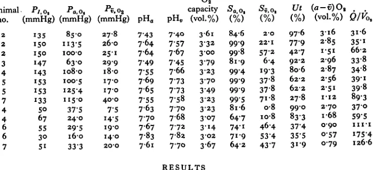
[image:4.451.122.325.47.380.2]60 90 120 Time (min)
Fig. i. Time course of blood gases, pH and percentage utilization of blood O2 in Sepia officinalis
associated with exposure to hypoxic water. w, Water; a, arterial; v, venous; 5, % Oa
satu-ration; Ut, utilization. The data are from one animal.
pre-hypoxic values. Blood pH rose markedly during hypoxia, also correlated with a state of hyperventilation. In some animals under hypoxic conditions, pH of venous blood was higher than that of arterial blood, although this finding was not consistent in all sample sets (Table i). Hypoxia also caused the total CO2 content of arterial
blood to rise (Fig. i). The O2 saturation fell in both arterial and venous blood, but
the relative decline of the two, and thus of the O2 utilization, varied between animals
and experiments.
Fig. i (lower panel) also shows that O2 utilization from circulating blood initially
rises during hypoxic exposure (at P7 ~ 8o mmHg), but declines at more severe O2
deficiency in the water.
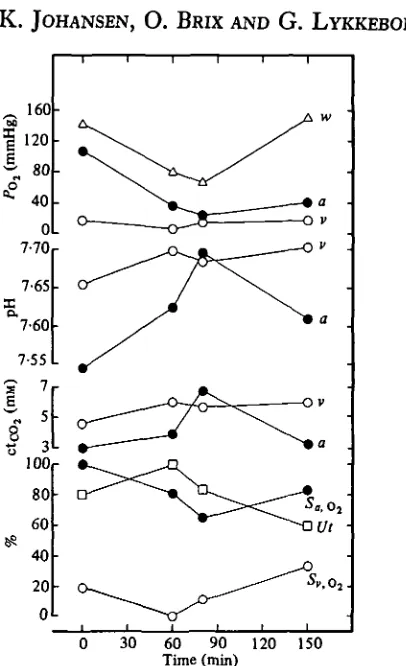
Fig. 2 shows how the arterial and venous O2 tensions relate to ambient pOt during hypoxic exposure. The decline in arterial O2 tension with reduced ambient pOt was associated with a reduced gradient from arterial blood to inspired water. This trend was correlated with a marked hyperventilation. The venous O2 tensions stayed rather
unchanged, after an initial drop in moderately hypoxic water. In combination with a marked alkalosis, this implies an increase in venous O2 saturation and hence a reduced
Cephalopod blood gas transport
335
c o
•a
o •a
60
4 0 -20
20 40 60 80 100 120 160
[image:5.451.73.346.50.265.2]Ambient O2 tension (mmHg)
Fig. 2. Arterial and venous pOt in relation to ambient pOt for Sepia officinalis at 17 °C. # ,
arterial; O, venous.
Fig. 3 shows the relationship between blood O2 affinity (P&0), HcO2 saturation and
blood pOa, based on Oa-binding curves determined for Sepia in vitro (Brix et al.
1981). The relationship between pH and O2 affinity is expressed by the equation:
log P50 = — i-6 pH+ 13-644 at a temperature of 17 °C. The empirically determined n-value for the O2 binding curves was 4 7 . The broken lines show calculated pOi isopleths based on an n-value of 2-5. The pairs of arterial and venous sample sets plotted on to the nomogram (larger data points) express the in vivo blood gas levels from unrestrained Sepia during normoxia (data set I) and during progressive hypoxia (set II, pQ% 80 mmHg; set III, pOt 67 mmHg; and set IV, pOt 30 mmHg). The smaller data points connected by arrows to those experimentally determined show what effect a reduction in n-value would have on arterial and venous O2 saturations.
It is clear that, for some data sets, the venous values are aligned with or displaced to the left of the arterial, implying that venous blood had a higher O2 affinity, i.e.
lower P50 values than arterial. This result reflects the fact that circulating blood pH in some cases was higher in venous compared to arterial blood (Table 1). At a water pOi of 67 and 30 mmHg, arterial and venous blood pH, and hence the corresponding
O2 affinity, were nearly similar.
Fig. 3 also reveals that a reduction in n-value from the very high level present in Sepia blood, results in a marked reduction in arterial saturation and a concurrent increase in the venous saturation, causing a drastic fall in the arteriovenous O2
content difference and hence in utilization of the circulating blood O2.
DISCUSSION
The combination of high tissue O2 requirement and low blood O2 carrying capacity
336
[image:6.451.97.348.57.377.2]70
Fig. 3. The table in the top panel gives the numerical changes in arteriovenous O2 content
difference, O2 utilization and the perfusion requirement (Q/Pi,) »n relation to ambient
water p0, for Sepia officinalis. The nomogram (bottom panel) is constructed from the O2
affinity values, Bohr shift, and «-value published by Brix et al. (1981), for the same species. The larger data symbols in the nomogram are based on in vivo blood sampling from animal 4 at a water pOt of 140 mmHg (symbols I), 80 mmHg (symbols II), 67 mmHg (symbols III) and animal 6 at 30 mmHg water pOl (symbols IV). • , Arterial; O, venous blood. The smaller symbols express the in vivo data points transformed to a nomogram based on an n-value of 2-5.
in turn will depend on the respiratory properties of blood and the ventilatory and circulatory pumps, as well as the diffusion barriers in the gills and tissues. A high blood O2 utilization expressed by a near-maximal difference in O2 content between
arterial and venous blood has long been advocated as a unique trait of cephalopod blood and alleged to depend on an exceptionally high pH sensitivity (Bohr effect) of the binding of O2 to most cephalopod haemocyanins. Recently, however, Lykkeboe,
Brix & Johansen (1980), Brix et al. (1981) and Lykkeboe & Johansen (1982) have pointed out that a Bohr factor (A logPs0/ApH) numerically exceeding i-o, which is
tanta-Cephalopod blood gas transport 337
>
mount to the release of the same mol fraction protons, or, conversely, during O2unloading one mol protons will be bound to the O2 carrier when one mol O2 is
unloaded. This relationship formally expressed in the linkage equation (Wyman, 1964) has the important consequence that if the pH sensitivity of the blood numerically exceeds i-o, there will not be an excess of free protons produced in aerobic metabolism to shift the O2-binding equilibrium of haemocyanin towards that of unbound O2,
i.e. O2 unloading in the tissues will consequently not be promoted by the aerobic
metabolism. Rather, as predicted by Lykkeboe et al. (1980), the pH of venous blood may exceed that of arterial blood, handicapping O2 unloading rather than promoting
it as has been traditionally advocated (Redfield & Goodkind, 1929).
The present study on Sepia is one of very few allowing an evaluation of the import-ance of the uniquely high Bohr shifts in cephalopods for blood gas transport in vivo based on blood samples drawn from unrestrained specimens.
The high arterial O2 tensions in Sepia (Table 1, Fig. 1) at normoxic ambient
conditions, suggest a highly efficient gas exchange in the gills. An arterial p0% of 100 mmHg implies that an O2 extraction from the ventilatory current of 28-5% or
higher will suffice to produce a negative pOt gradient between expired water and arterial blood, characteristic of a counter-current exchange process. Hazelhoff (1939) reported a range for O2 extraction between 50% and 80% in cephalopods, while the
large Octopus dofieini showed an average extraction of 26-8% (Johansen & Lenfant, 1966).
Arterial blood for Sepia in well-aerated water will therefore typically be fully saturated with O2 (Table 1). The more variable venous p0% corresponds to venous O2
saturations that are about 20% or lower, implying O2 utilizations 80% or higher. At
conditions of increased O2 requirement during swimming, or at reduced water pOi, the cephalopod solution to increased or maintained O2 delivery (hypoxic water) can
thus only modestly depend on an increased unloading of O2, but must instead depend
on maximizing the arterial O2 saturation and an increase in cardiac output.
Our data testify that in Sepia venous pH may exceed arterial pH, in both normoxic and hypoxic water (Table 1). This in vivo situation accords with the predictions of Lykkeboe et al. (1980), explainable as a result of the interaction between proton binding and the state of oxygenation of haemocyanins with very high Bohr shifts. As is apparent from Fig. 3, this tendency gives venous blood a higher O2 affinity
than arterial blood, a fact which will actually impede O2 unloading from haemocyanin.
In addition, the O2 affinity of all circulating blood will increase during hypoxia
as a result of a general alkalosis, a finding recently also reported for blood of Octopus vulgaris (Houlihan et al. 1982). This tendency will further reduce Oa unloading,
while at the same time favouring O2 loading. The possibility that anaerobic
produc-tion of protons could aid O2 unloading, for example during exercise, is small due to
the relatively undissociated state of octopine, which is the principal product of anaerobic metabolism in cephalopods (Zammit, 1978).
The very high O2 utilization of Sepia blood in normoxic and moderately hypoxic
water (P7 Oj ~ 80 mmHg) is clearly related to the highly sigmoid shape (high n-value)
of the O2Hc binding curves (Fig. 3). Calculated data points show that arterial
smaller n-value. The high n-value can hence be regarded as a counterbalance for the apparent adverse influence of the blood acid-base status on Oa unloading. Fig. 3
(top panel) also gives the numerical changes in arteriovenous 02 content difference,
02 utilization and the perfusion requirement (£)/J£>a). The latter is seen to increase
dramatically as the blood loses importance in 02 transport.
Accompanying the alkalosis in Sepia during exposure to hypoxia was a notable increase in total C02 content of the blood (Fig. 1). The increase may possibly be related
to the breakdown of arginine phosphate that occurs during hypoxia (Storey & Storey, 1979), since hydrolysis of this compound leads to a build-up of bicarbonate (Burton, 1978). The phenomenon will act to amplify the tendency towards an alkalotic status during hypoxia and thus further substantiate the 02 loading strategy of compensatory
02 transport.
We are grateful to the Marine Biological Association of the United Kingdom at Plymouth, England, for help with a supply of Sepia, and for space and research facili-ties. This investigation was supported by a NATO grant to Drs P. W. Hochachka and K. Johansen, Dr O. Brix was supported by the Danish Natural Science Research Council, J. nr. 511-20410.
REFERENCES
ALTMAN, P. L. & DITMER, D. S. (1971). Eds. Biol. Handbooks: Respiration and Circulation. Fedn.
Am. Socs. exp. Biol.
BURTON, R. F. (1978). Intracellular Buffering. Resp. Physiol. 33, 51-58.
BRIX, O. (1981). A modified Van Slyke apparatus .7. appl. Physiol. 50, 1093-1097.
BRIX, O., LYKKEBOE, G. & JOHANSEN, K. (1981). The significance of the linkage between the Bohr and Haldane effects in cephalopod bloods. Resp. Physiol. 44, 177-186.
GHIRETTI, F. (1966). Molluscan hemocyanins. In Physiology of Mollusca (ed. K. M. Wilbur and C. M. Yonge), pp. 175-208. New York: Academic Press.
HAZELHOFF, E. H. (1939), t)ber die Ausnutzung des Sauerstoffs bei verschiedenen Wassertieren. Z.
vergl. Physiol. 26, 306-327.
HOULIHAN, D. F., INNES, A. J., WELLS, M. J. & WELLS, J. (1982). Oxygen consumption and blood gases of Octopus vulgaris in hypoxic conditions. J. comp. Physiol. (in the press).
JOHANSEN, K. & LENFANT, C. (1966). Gas exchange in the cephalopod, Octopus dofleini. Am. J. Physiol.
aio, 910-918.
JOHANSEN, K., REDMOND, J. R. & BOURNE, G. B. (1978). Respiratory exchange and transport of oxygen in Nautilus pompilius. J. exp. Zool. 205, 27-36.
JOHANSEN, K., BRIX, O., KORNERUP, S. & LYKKEBOE, G. (1982). Factors affecting O2-uptake in the
cuttlefish, Sepia officinalis. J. mar. biol. Ass.U.K. 62, 187-191.
LENFANT, C. & JOHANSEN, K. (1965). Gas transport by hemocyanin containing blood of the cephalopod,
Octopus dofleini. Am. J. Physiol. 209, 991-998.
LYKKEBOE, G. & JOHANSEN, K. (1982). A cephalopod approach to rethinking about the importance of the Bohr and Haldane effects. Pacific Science (in the press).
LYKKEBOE, G., BRIX, O. & JOHANSEN, K. (1980). Oxygen-linked CO8 binding independent of pH in
cephalopod blood. Nature, Lond. 287, 330-331.
REDFIELD, A. C , COOLIDGE, T. & HURD, A. L. (1926). The transport of oxygen and carbon dioxide by some bloods containing hemocyanin. J. biol. Chem. 69, 475-509.
REDFIELD, A. C. & GOODKIND, R. (1929). The significance of the Bohr effect in the respiration and asphyxiation of the squid, Loligo pealei. J. exp. Biol. 6, 240-349.
STOREY, K. B. & STOREY, J. M. (1979). Octopine metabolism in the cuttlefish, Sepia officinalis: octopine production by muscle and its role as an aerobic substrate for non-muscular tissues. J. comp. Physiol.
I31,3ii-3I9-WYMAN, J. (1964). Linked functions and reciprocal effects in hemoglobin: a second look. In Advances
in Protein Chemistry, vol. xix (ed. C. B. Anfinsen, M. L. Anson, J. T. Edsall and F. M. Richards),
223-286.