The role of the teleost gill epithelium in the acclimation of euryhaline teleost fishes to both freshwater and seawater environments has been studied extensively. Na+/K+-ATPase is one of the most important enzymes associated with ion regulation in the fish gill because it indirectly energizes the branchial excretion of NaCl from marine teleosts (see McCormick, 1995; Marshall and Bryson, 1998; Evans et al., 1999). The role of Na+/K+-ATPase in freshwater ion balance is not well understood, but it may be important for establishing electrochemical gradients necessary for NaCl and/or Ca2+ uptake (see Perry, 1997).
Na+/K+-ATPase is a membrane-bound P-ATPase composed of at least three subunits: α, βand γ(Blanco and Mercer, 1998). This ubiquitous transporter is involved in maintaining routine cell volume and resting membrane potential (Blanco and Mercer, 1998) and is extremely abundant in specialized ion-transporting cells. Two well-studied examples in fishes are the marine teleost chloride cell and the elasmobranch rectal gland tubule cell. In these two cell types, Na+/K+-ATPase is localized along the complex basolateral membrane and/or tubule system (Karnaky et al., 1976; Eveloff et al., 1979), the large surface areas of which amplify the possible sites for Na+/K+-ATPase insertion (Karnaky, 1986; Valentich et al., 1995). Many
researchers have reported a positive correlation between environmental salinity and the biochemical activity of branchial Na+/K+-ATPase in teleost fishes, especially in salmonids and anguillids (Kultz and Jurss, 1993; Madsen et al., 1995; McCormick, 1995; Uchida et al., 1996, 1997; Crockett, 1999; D’Cotta et al., 2000). However, in other species, branchial Na+/K+-ATPase activity in freshwater-acclimated individuals is equivalent to or exceeds that of seawater-acclimated fish (Yoshikawa et al., 1993; Madsen et al., 1994; McCormick, 1995; Jensen et al., 1998; Vonck et al., 1998; Kelly et al., 1999; Marshall et al., 1999).
Recently, there have been further insights into the cellular regulation of fish gill Na+/K+-ATPase using heterologous and homologous antibodies and partial cDNA sequences to the α -subunit. For example, changes in branchial Na+/K+-ATPase activity have been positively correlated with Na+/K+ -ATPase-specific mRNA and protein levels (Kisen et al., 1994; Cutler et al., 1995; Madsen et al., 1995; Hwang et al., 1998; Jensen et al., 1998; Lee et al., 1998; D’Cotta et al., 2000), suggesting that changes in fish gill Na+/K+-ATPase activity are due to changes in the relative abundance of the enzyme. In mammals, activation of latent Na+/K+-ATPase has also been shown to be responsible for changes in the activity of the enzyme (Ewart Printed in Great Britain © The Company of Biologists Limited 2000
JEB2916
Changes in Na+/K+-ATPase activity and abundance
associated with environmental salinity were investigated in the gills and rectal gland of the Atlantic stingray Dasyatis
sabina. Using a ouabain-specific ATPase assay and western
blotting, we found that stingrays from fresh water had the highest activity and highest relative abundance of Na+/K+
-ATPase in the gills. Using immunohistochemistry, we also found that gills from freshwater stingrays had the greatest number of Na+/K+-ATPase-rich cells. When freshwater
stingrays were acclimated to sea water for 1 week, the activity and abundance of Na+/K+-ATPase and the number
of Na+/K+-ATPase-rich cells decreased in the gills. In
seawater stingrays, the branchial activity and abundance of Na+/K+-ATPase and the number of Na+/K+-ATPase-rich
cells were further reduced. In rectal glands, the activity and
abundance of Na+/K+-ATPase were lower in freshwater
animals than in seawater-acclimated and seawater stingrays, both of which had equivalent levels. These findings suggest that salinity-associated changes in gill and rectal gland Na+/K+-ATPase activity are due to changes in
the abundance of Na+/K+-ATPase. We conclude that the
gills may be important for active ion uptake in fresh water, while the rectal gland is important for active NaCl excretion in sea water. The results from this study are the first to demonstrate an effect of environmental salinity on Na+/K+-ATPase expression in the gills and rectal gland of
an elasmobranch.
Key words: Na+/K+-ATPase, gill, rectal gland, elasmobranch,
stingray, Dasyatis sabina, salinity, euryhaline.
Summary
Introduction
EFFECTS OF ENVIRONMENTAL SALINITY ON Na
+/K
+-ATPase IN THE GILLS AND
RECTAL GLAND OF A EURYHALINE ELASMOBRANCH (DASYATIS SABINA)
PETER M. PIERMARINI* ANDDAVID H. EVANS
University of Florida, Department of Zoology, Box 118525, 223 Bartram Hall, Gainesville, FL 32611, USA
*e-mail: [email protected]
and Klip, 1995), but this has not been extensively studied in fish.
Na+/K+-ATPase-rich cells have been immunolocalized on both gill lamellae and interlamellar regions of freshwater chum salmon (Oncorhynchus keta), but the cells are only present on the interlamellar region of seawater-acclimated individuals (Uchida et al., 1996, 1997; Ura et al., 1996; Shikano and Fujio, 1998b). It has been suggested that Na+/K+-ATPase-rich cells found on the lamellae may be involved with ion uptake from fresh water, whereas interlamellar Na+/K+-ATPase-rich cells are associated with NaCl excretion in a marine environment (Laurent and Dunel, 1980; Uchida et al., 1996; Seidelin and Madsen, 1999). The guppy (Poecilia reticulata) is an exception, because Na+/K+-ATPase-rich cells are only found in interlamellar regions, regardless of salinity (Shikano and Fujio, 1998a,c, 1999).
In contrast to teleost fishes, the role of the gill epithelium in elasmobranch ion regulation has not been well studied. It is thought that the gills do not play a large role in NaCl excretion since elasmobranchs have a salt-secreting rectal gland (Shuttleworth, 1988; Karnaky, 1998). However, a few studies have suggested that the gills are a potential site of net salt secretion, since plasma NaCl concentrations were maintained well below seawater levels when rectal glands were surgically removed from spiny dogfish Squalus acanthias (Burger, 1965; Evans et al., 1982). In addition, recent evidence of natriuretic peptide receptors in the gills of two marine elasmobranch species (Donald et al., 1997; Sakaguchi and Takei, 1998) may indicate that the gills are involved in ion regulation. In freshwater elasmobranchs, the gills are presumably the site of ion uptake, but data are lacking.
Mitochondria-rich and Na+/K+-ATPase-rich cells have been described from the gills of a few marine elasmobranch species (Laurent and Dunel, 1980; Laurent, 1984; Conley and Mallatt, 1988). In contrast to chloride cells found in teleost gills, elasmobranch Na+/K+-ATPase-rich cells lack the complex basolateral membrane/tubule system, apical crypt and accessory cells. Branchial Na+/K+-ATPase activity has been measured in seawater-acclimated S. acanthias (Jampol and Epstein, 1970; Morgan et al., 1997) and was found to be considerably lower than that in teleost gills and in the elasmobranch rectal gland. No study has localized or measured branchial Na+/K+-ATPase activity from an elasmobranch acclimated to a salinity lower than full-strength sea water.
The mechanisms and regulation of NaCl secretion have been relatively well studied in the marine elasmobranch rectal gland, where Na+/K+-ATPase plays an important role in energizing active NaCl transport (see Riordan et al., 1994; Valentich et al., 1995; Silva et al., 1997). However, the function of the rectal gland in elasmobranchs from low-salinity environments has not been thoroughly investigated. Oguri (1964) and Thorson et al. (1983) have demonstrated a positive correlation between relative rectal gland size and environmental salinity, suggesting a decreased function in fresh water. Only Gerzeli et al. (1976) have looked at biochemical changes associated with the rectal gland in lower salinities, and they demonstrated
that freshwater bull sharks (Carcharhinus leucas) had rectal glands with a lower total ATPase activity than their marine counterparts. Although a salt-secreting gland may appear to have no ion-regulatory function in fresh water, the rectal gland is still composed of numerous secretory tubules in both euryhaline and stenohaline freshwater species (Oguri, 1964; Thorson et al., 1978).
The Atlantic stingray (Dasyatis sabina) is one of the few elasmobranch species that has established reproducing populations in both freshwater and marine environments. It is, therefore, an excellent model for studying elasmobranch osmoregulatory mechanisms. In a previous study, we demonstrated that freshwater Atlantic stingray populations had a lower concentration of major plasma osmolytes and a smaller rectal gland than marine Atlantic stingrays (Piermarini and Evans, 1998). The freshwater stingrays were capable of acclimating to sea water, where their plasma NaCl and urea concentrations were similar to those of marine stingrays, but the size of their rectal gland did not change appreciably (Piermarini and Evans, 1998).
The goals of this study were to compare the activity, relative amount and localization (gills only) of Na+/K+-ATPase in the gills and rectal gland of Atlantic stingrays from freshwater and seawater environments. We also compared the same variables in freshwater stingrays acclimated to sea water for 1 week. This study is the first to demonstrate an effect of environmental salinity on the activity and expression of Na+/K+-ATPase in the gills and rectal gland of an elasmobranch and to demonstrate the distribution of Na+/K+-ATPase-rich cells in the gills of an elasmobranch by immunohistochemistry.
Materials and methods Animal collection and holding conditions
Ten Atlantic stingrays Dasyatis sabina were captured from the St Johns River, FL, USA (Lake Jesup or Lake George), as described by Piermarini and Evans (1998). Animals were held in two 379 l freshwater, closed-system tanks (five stingrays per tank; <1 ‰ salinity). In addition, five marine Atlantic stingrays were captured via hook and line from Cedar Key, FL, USA, transported to Gainesville, FL, USA, and held in a 379 l seawater, closed-system tank (30–32 ‰).
One of the freshwater groups was left in fresh water, while the other was gradually acclimated to sea water as follows. After 1 week in fresh water, the salinity was raised to 16 ‰ over 2 days (8 ‰ per day). After 2 days in 16 ‰, the salinity was raised to 32 ‰ over 3 days. The animals remained at a salinity of 32 ‰ for 1 week before tissue samples were taken. The marine stingrays remained in sea water (32 ‰) for the entire period. All the animals were fed live grass shrimp (Palaemonetes sp.) every other day and were starved for 48 h prior to tissue collection.
2959
Na
/K
-ATPase expression in stingray gills and rectal gland
Collection of tissues
Animals were anesthetized in 4 l of a 0.01 % MS-222 solution made with tank water. For freshwater stingrays, the solution was buffered with a commercial freshwater pH buffer (Seachem) to prevent acidification by the MS-222. Once anesthetized, animals were placed ventral side up in a slanted water bath with the gills immersed in the anesthetic.
To clear the gills and rectal gland of red blood cells, the animal was perfused with elasmobranch Ringer’s solution (Forster et al., 1972) at 4 °C. For freshwater stingrays, the Ringer’s solution was modified by reducing the NaCl concentration to 200 mmol l−1, the urea concentration to 200 mmol l−1 and the trimethylamine oxide concentration to 41 mmol l−1. The skin ventral to the heart and pericardium was removed, and 0.5–1.0 ml of blood was removed from the ventricle with a heparinized 25 gauge needle attached to a 1 ml syringe. An equal volume of heparinized Ringer’s solution was then injected into the ventricle and allowed to circulate for a few minutes. A cannula, connected to a perfusion bottle (positioned 1 m above the animal), was inserted into the conus arteriosus and held by forceps. Once the perfusion had started, the sinus venosus was cut to relieve back pressure. The perfusion was continued until the gills appeared bleached and the fluid exiting the pericardial cavity was clear of blood (usually 3–5 min).
Immediately after the perfusion, the animal was pithed, and the second left and right gill arches were removed and placed in an elasmobranch Ringer’s solution on ice. Gill filaments were trimmed off the arches and placed in fixative (3 % paraformaldehyde, 0.05 % glutaraldehyde, 0.05 % picric acid in 10 mmol l−1 phosphate-buffered saline, pH 7.3) for 24 h at 4 °C. Additional filaments were snap-frozen in liquid nitrogen for western blot analysis or placed in ice-cold SEI buffer (250 mol l−1 sucrose, 10 mmol Na
2EDTA, 50 mol l−1 imidazole, pH 7.3), which was then frozen on dry ice for Na+/K+-ATPase activity measurements. The rectal gland was then excised, and pieces were taken for western blot and Na+/K+-ATPase activity measurements. All samples for western blots and enzyme assays were stored at −80 °C.
Na+/K+-ATPase activity
Na+/K+-ATPase activity was measured using the technique developed by McCormick (1993). In brief, the tissue frozen in SEI buffer was rapidly thawed and homogenized, and the homogenate was centrifuged at 5000 g for 30 s at 4 °C. The supernatant (10µl) was added to four wells of a 96-well microplate on ice, and an assay mixture (200µl) with or without ouabain (0.5 mmol l−1) was then added. The composition of the assay mixture was identical to that described by McCormick (1993), except that the concentrations of Na+, K+ and Mg2+ were 45, 5 and 10 mmol l−1, respectively. These concentrations were optimized for Atlantic stingray tissue and were similar to those used by Morgan et al. (1997) for spiny dogfish gill and rectal gland tissue. Na+/K+-ATPase activity was determined by subtracting the oxidation rate of NADH to NAD (at 340 nm)
in the presence of ouabain from the rate in the absence of ouabain at 25 °C. Absorbance was measured using a Spectramax 250 microplate reader (Molecular Devices). Total protein was measured using a bicinchoninic acid assay (Pierce). Activity was expressed as µmol ADP h−1mg−1protein.
Western blots
Preparation of tissue for western blots was modified from Claiborne et al. (1999). On a single day, gill filaments or a piece of rectal gland from one animal in each experimental condition were prepared for western blotting. The tissue was placed in ice-cold homogenization buffer [250 mmol l−1 sucrose, 1 mmol l−1Na
2EDTA, 2µg ml−1aprotinin, 2µg ml−1 leupeptin, 100µg ml−1phenyl methylsulfonyl fluoride (PMSF) and 30 mmol l−1Tris] and homogenized on ice using a ‘Tissue-tearor’ in a cold room at 4 °C. Homogenates were filtered through cheesecloth and centrifuged (3000 g) for 5 min at 4 °C to remove nuclei and debris. The supernatant was then filtered through cheesecloth and centrifuged (50 000 g) for 30 min at 4 °C to pellet membrane fractions. The pellet was resuspended with a minimal volume of ice-cold homogenization buffer, and an equal volume of a modified Laemmli sample buffer (Laemmli, 1970), without Bromophenol Blue and β -mercaptoethanol, was then added to solubilize the proteins.
The resulting protein samples were centrifuged for 5–10 s at 16 000 g to pellet any undissolved material. The total protein content of the supernatant was determined using a detergent-compatible assay (Bio-Rad), after which Bromophenol Blue was added to a final concentration of 0.01 % and β -mercaptoethanol to a final concentration of 2 %.
A 20µg sample of total protein for gill (5µg for rectal gland) was loaded in triplicate and run on a 7.5 % Tris-HCl precast polyacrylamide gel (Bio-Rad) for 1 h at 125 V. Note that it was necessary to load four times more total protein in gill samples because of the lower α-subunit abundance per microgram of total protein compared with the rectal gland. Proteins were then transferred to a polyvinylidene difluoride (PVDF; Bio-Rad) membrane using a wet (20 % methanol, Tris-glycine) transfer unit for 2.5 h at 90 V in a cold room at 4 °C with stirring.
The PVDF was blocked with Blotto for 1.5 h at 25 °C and then transferred to the primary antibody solution (monoclonal antibody ‘a5’ culture supernatant diluted 1:1000 in Blotto) overnight at 4 °C. The primary antibody, ‘a5’, developed by Dr Douglas Fambrough, was obtained from the Developmental Studies Hybridoma Bank developed under the auspices of the National Institute of Child Health and Human Development and maintained by The University of Iowa, Department of Biological Sciences, Iowa City, IA 52242, USA. This antibody was raised against the α-subunit of avian Na+/K+-ATPase and is not isoform-specific. It has been used numerous times to study Na+/K+-ATPase in fish gills (see Witters et al., 1996; Choe et al., 1999; Schreiber and Specker, 1999; Dang et al., 2000).
Tween-20 (TTBS), then incubated with an alkaline-phosphatase-conjugated goat anti-mouse IgG secondary antibody (Bio-Rad; diluted 1:3000 in Blotto) for 2 h at 25 °C. The PVDF was then washed three times (15 min each) with TTBS, and a substrate solution (Bio-Rad Immun-Star ECL Kit) was applied to the PVDF for 5 min at 25 °C to initiate a luminescent signal. Binding of antibody was detected by exposing Hyperfilm-ECL imaging film (Amersham) to the PVDF membrane. The film was developed according to the manufacturer’s protocol. The film was exposed for a series of times (0, 15, 30, 45, 60, 75, 90 s) to ensure linearity of the signal. Negatives were digitized into TIFF files, using a UMAX flatbed scanner with transparency adapter, and analyzed using NIH Image version 1.61 (National Institutes of Health, USA).
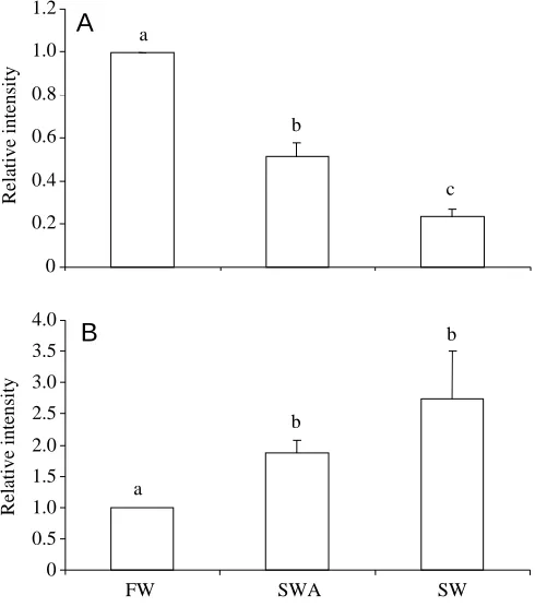
To quantify the relative abundance of the α-subunit, we measured the intensity of the immunopositive bands using densitometry and standardized all measurements to the freshwater condition. Therefore, intensity measurements of freshwater individuals for a particular tissue were 1.0.
Immunohistochemistry
Fixed tissue was stored in two changes of 75 % ethanol at 25 °C, dehydrated in an ethanol series, and embedded in paraffin wax within 2 weeks of fixation. Serial sections of gill tissue, parallel to the long axis of the filament, were cut at 6µm and placed on poly-L-lysine-coated slides. For immunostaining, only slides containing sections from the trailing half of the gill lamellae and filament were chosen. In preliminary trials, we found that Na+/K+-ATPase-rich cells were located exclusively on the trailing half of gill lamellae and filaments (P. M. Piermarini and D. H. Evans, unpublished observation), similar to the observations reported for teleost chloride cells (see Van Der Heijden et al., 1997). The sections were deparaffinized in Hemo-De, hydrated in an ethanol series, and washed in 10 mol l−1phosphate-buffered saline (PBS). A hydrophobic PAP-Pen (Electron Microscopy Suppliers) was used to draw circles around the tissue sections, and 3 % H2O2 was then placed on the sections for 25 min to inhibit endogenous peroxidase activity. Sections were also blocked with a casein solution (Powerblock, Biogenex) for 5 min before application of the primary antibody.
The primary antibody, monoclonal antibody ‘a5’ (diluted 1:175 in PBS), was placed on the sections for 2 h at 25 °C. The antibody was rinsed off, and the sections were washed in PBS for 5 min. After application of the primary antibody, the sections were incubated with a biotinylated anti-mouse IgG secondary antibody (Biogenex) and a horseradish-peroxidase-labeled streptavidin solution (Biogenex) for 20 min each at 25 °C. After washing with PBS for 5 min, antibody binding was visualized by applying a solution of 3,3′-diaminobenzidine tetrahydrochloride (DAB; Biogenex) to the sections for 5 min at 25 °C. No staining was detected when non-immune mouse serum was used instead of primary antibody.
Quantification of the immunohistochemistry was performed by counting the number of immunopositive (Na+/K+ -ATPase-rich) cells per gill lamella and per interlamellar space. For each
animal, three immunostained slides were chosen at random. On a section from each slide, the number of Na+/K+-ATPase-rich cells was counted on 30 randomly selected lamellae and interlamellar regions. The length of the lamella was also measured to standardize cell counts on the lamellae. Results are expressed as the number of cells per lamella and the number of cells per interlamellar region.
Statistical analyses
For mean Na+/K+-ATPase activities and the number of Na+/K+-ATPase-rich cells, differences among groups were detected using a one-way analysis of variance (ANOVA), with a Student–Newman–Keuls post-hoc test. Differences in relative band intensities from western blots were detected using a Kruskal–Wallis non-parametric ANOVA, with a Kruskal–Wallis post-hoc test (Conover, 1980). All tests were two-tailed, and differences were considered significant if P<0.05.
Results Na+/K+-ATPase activity
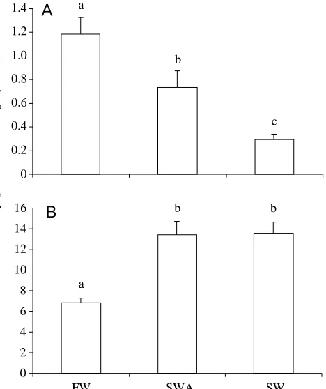
Na+/K+-ATPase activity was measured in the gills and rectal gland of stingrays to compare the overall catalytic activity of the enzyme in the different groups. In gills, Na+/K+-ATPase activity was highest in freshwater, intermediate in seawater-acclimated
0 2 4 6 8 10 12 14 16
FW SWA SW
0 0.2 0.4 0.6 0.8 1.0 1.2 1.4
Na
+/K +-ATPase activity (
µ
mol ADP h
-1 mg -1 protein)
A
B
a
b
c
a
[image:4.612.327.561.72.351.2]b b
Fig. 1. Na+/K+-ATPase activity in the gills (A) and rectal gland (B)
2961
Na
/K
-ATPase expression in stingray gills and rectal gland
and lowest in seawater stingrays (Fig. 1A). In contrast, rectal gland Na+/K+-ATPase activity was lowest in freshwater stingrays and higher in seawater-acclimated and seawater stingrays, in which levels were equivalent (Fig. 1B). If rectal gland mass is taken into account, the total Na+/K+-ATPase activity (rectal gland mass ×Na+/K+-ATPase activity) would be lowest in freshwater stingrays, intermediate in seawater-acclimated stingrays and highest in seawater stingrays.
Western blots
We used western blotting to determine whether the differences in Na+/K+-ATPase activity were correlated with changes in the relative abundance of the enzyme. Antibody ‘a5’ was highly cross-reactive for a band with a molecular mass of 110.9±1.0 kDa in gills and 111.7±1.4 kDa (means ± S.E.M., N=5) in rectal glands (Fig. 2), which is within the expected molecular mass range for the α-subunit (Blanco and Mercer, 1998). In the gills, the relative abundance of the α -subunit followed a similar trend to the Na+/K+-ATPase activities: α-subunit abundance was highest in freshwater, intermediate in seawater-acclimated and lowest in seawater stingrays (Fig. 3A). In the rectal gland, the relative abundance of the α-subunit also followed a similar trend to the Na+/K+ -ATPase activities; α-subunit abundance was lowest in freshwater and higher in seawater-acclimated and seawater stingrays (Fig. 3B).
Immunohistochemistry
Changes in the distribution of gill Na+/K+-ATPase-rich cells associated with salinity were visualized using immunolocalization of the α-subunit on fixed tissue sections (Fig. 4). Staining for Na+/K+-ATPase was restricted to relatively large cells (presumably mitochondra-rich cells) and was not found on respiratory pavement cells (Fig. 4). The number of Na+/K+-ATPase-rich cells per lamella and the sum of the number of Na+/K+-ATPase-rich cells per lamella plus the number of Na+/K+-ATPase-rich cells per interlamellar region followed the same trend as Na+/K+-ATPase activity and relative abundance; freshwater stingrays had the most, seawater-acclimated stingrays had intermediate amounts and seawater stingrays had the fewest (Fig. 5A,C). In the interlamellar region, seawater-acclimated gills had the most Na+/K+-ATPase-rich cells, followed by freshwater and then seawater gills (Fig. 5B).
Although the intensity of the immunostaining was not quantified, there appeared to be qualitative trends. In freshwater stingrays, staining was darkest in Na+/K+-ATPase-rich cells on the ends of the lamellae; the staining appeared to weaken towards the interlamellar region and was weakest in cells from 205
130
77
32
[image:5.612.54.294.68.262.2]MW 1 2 3 4 5 6
Fig. 2. Representative western blot of gill and rectal gland tissue from Atlantic stingrays. Antibody was specific for a 110.9±1.0 kDa protein in gills and a 111.7±1.4 kDa protein in the rectal gland
(means ± S.E.M., N=5), representing the α-subunit of Na+/K+
-ATPase. MW, molecular mass marker (kDa). Lanes 1, 2, 3, gill tissue from freshwater, seawater-acclimated and seawater stingrays
(20µg total protein per lane), respectively. Lanes 4, 5, 6, rectal gland
tissue from freshwater, seawater-acclimated and seawater stingrays
(5µg total protein per lane), respectively. Note the decrease in band
intensity from freshwater to seawater in gills and increase in band intensity from freshwater to seawater in rectal gland. The intensity of gill and rectal gland bands cannot be compared since different amounts of total protein were loaded.
0 0.2 0.4 0.6 0.8 1.0 1.2
0 0.5 1.0 1.5 2.0 2.5 3.0 3.5 4.0
FW SWA SW
A
B
Relative intensity
Relative intensity
a
b
c
a
b
[image:5.612.322.567.75.353.2]b
Fig. 3. Relative intensity of immunopositive bands representing the
α-subunit of Na+/K+-ATPase in the gills (A) and rectal gland (B) of
the interlamellar region (Fig. 4A). In seawater-acclimated stingrays, Na+/K+-ATPase-rich cells stained heavily on both the lamellae and interlamellar region (Fig. 4B). In seawater stingrays, the Na+/K+-ATPase-rich cells were moderately stained in the interlamellar region (Fig. 4C).
Discussion Gills
The results from this study suggest that Na+/K+-ATPase activity associated with the gills of the Atlantic stingray is negatively correlated with salinity. Branchial Na+/K+-ATPase activity was highest in freshwater stingrays, followed by seawater-acclimated and, lastly, seawater animals. In salmonids and anguillids, gill Na+/K+-ATPase activity is positively correlated with environmental salinity, but a negative or no correlation with salinity has been found in other fishes (McCormick, 1995; Jensen et al., 1998; Kelly et al., 1999; Marshall et al., 1999). Differences in stingray gill Na+/K+-ATPase activity were positively correlated with changes in the relative abundance of the α-subunit and in the numbers of Na+/K+-ATPase-rich cells. This suggests that salinity-induced changes in branchial Na+/K+-ATPase activity in the Atlantic stingray are accomplished by regulating the relative amounts of Na+/K+-ATPase protein, probably through the number of Na+/K+-ATPase-rich cells on gill lamellae. If elasmobranch gill Na+/K+-ATPase is regulated like that of
B
[image:6.612.49.291.66.556.2]C
A
Fig. 4. Representative immunostaining for Na+/K+-ATPase-rich cells
in longitudinal sections of gill filaments from freshwater (A), seawater-acclimated (B) and seawater (C) Atlantic stingrays. Scale
bars, 100µm. Na+/K+-ATPase-rich cells occurred on lamellae
(finger-like projections) and/or interlamellar regions (basal to and
between lamellae). Note the differences in Na+/K+-ATPase-rich cell
[image:6.612.328.548.70.379.2]location (lamella versus interlamellar region) and darkness of staining within and between groups.
Fig. 5. Na+/K+-ATPase-rich cell numbers in the gills of freshwater
(FW), seawater-acclimated (SWA) and seawater (SW) Atlantic
stingrays. (A) Number of Na+/K+-ATPase-rich cells per lamella;
(B) number of Na+/K+-ATPase-rich cells per interlamellar region;
(C) ‘sum’ number of Na+/K+-ATPase-rich cells (per lamella plus per
interlamellar region). Values are means +1 S.E.M. Lower case letters indicate statistical categorization of the means as determined by a Student–Newman–Keuls post-hoc test (P<0.05).
0 2 4 6 8 10 12
Number of cells, sum
a
b
c 0
1 2 3 4 5
FW SWA SW
a
b
c
Number of cells
per lamella
a
b
c
A
B
C
0 2 4 6 8
Number of cells
2963
Na
/K
-ATPase expression in stingray gills and rectal gland
teleosts, then the decrease in Na+/K+-ATPase α-subunit protein abundance found in seawater-acclimated and seawater stingrays may be a result of a decrease in α-subunit mRNA levels (Cutler et al., 1995; Madsen et al., 1995; Hwang et al., 1998; Jensen et al., 1998; D’Cotta et al., 2000) brought about by a decrease in transcription or an increase in mRNA degradation. In salmonids, the turnover rate of gill Na+/K+ -ATPase protein does not appear to be affected by salinity (D’Cotta et al., 2000).
Na+/K+-ATPase activity from the gills of a freshwater elasmobranch has not previously been measured. The increased branchial Na+/K+-ATPase activity associated with freshwater stingrays may indicate that the gills have an active transport role in fresh water, possibly for ion uptake. An alternative explanation is that the increase in Na+/K+-ATPase activity is associated with an increased cost of cell volume regulation in freshwater gill cells, but this explanation is unlikely since the immunostaining occurred in specific cell types and not across the entire gill epithelium (see Fig. 4). The levels of branchial Na+/K+-ATPase activity in the seawater-acclimated and seawater stingrays were lower than those in the freshwater stingrays. This was expected since elasmobranchs have a salt-secreting rectal gland that is the primary site of NaCl excretion (Shuttleworth, 1988). Although the branchial Na+/K+-ATPase activity is comparatively low, it may be enough to drive some active NaCl excretion, especially in cases where the rectal gland is removed (see Evans et al., 1982) or reduced in size (see below).
Since Na+/K+-ATPase-rich cells were most prevalent on the lamellae of the freshwater stingrays, it is probable that these cells are involved with active ion uptake. Na+/K+-ATPase-rich, or chloride, cells have been reported on the gill lamellae of several freshwater-acclimated teleost species (Avella et al., 1987; Uchida et al., 1996; Ura et al., 1996; Seidelin and Madsen, 1999; Wong and Chan, 1999) where it has been proposed that they are involved in ion uptake from a freshwater environment (Laurent and Dunel, 1980; Avella et al., 1987; Uchida et al., 1996). The processes of ion uptake and acid–base balance are, however, inextricably linked in the gills of freshwater fish (see Claiborne, 1998; Evans et al., 1999), so the Na+/K+-ATPase-rich cells may also be involved with the excretion of acid (H+) and/or base (HCO
3−).
Since there was an increase in the number and/or staining intensity of Na+/K+-ATPase-rich cells found on the gill filaments of seawater-acclimated and seawater stingrays, it is possible that these cells are involved in active NaCl excretion, a function that has been suggested for the Na+/K+-ATPase-rich cells found in the interlamellar regions of the gills of seawater-acclimated teleosts (see Uchida et al., 1996). However, the presence of Na+/K+-ATPase-rich cells in the gills does not necessarily imply active NaCl excretion. Hagfish (Myxine glutinosa) also possess branchial Na+/K+-ATPase-rich cells (Choe et al., 1999), but are strict ion conformers. Choe et al. (1999) suggested that hagfish Na+/K+-ATPase-rich cells may facilitate acid–base exchange (see Evans, 1984) and/or Ca2+ transport (see Forster and Fenwick, 1994), which is known to
occur across the hagfish gill epithelium. These functions should also be considered for the Na+/K+-ATPase-rich cells found in the gills of seawater-acclimated and seawater stingrays.
Rectal gland
Na+/K+-ATPase activity in rectal glands from seawater-acclimated and seawater stingrays was greater than that from freshwater stingrays. Differences in rectal gland Na+/K+ -ATPase activity were positively correlated with changes in the relative abundance of the α-subunit. This suggests that the salinity-associated regulation of rectal gland Na+/K+-ATPase activity in the Atlantic stingray is due to changes in the relative amounts of Na+/K+-ATPase protein present.
The measurements of freshwater stingray rectal gland Na+/K+-ATPase activity presented in this study are the first to be reported for a freshwater elasmobranch. Even though the freshwater rectal gland Na+/K+-ATPase activity was approximately 50 % lower than that of seawater-acclimated and seawater stingrays, the freshwater activity is still approximately seven times higher than that found in the gills. It is possible that the rectal glands of freshwater stingrays cannot completely inhibit active NaCl secretion, an indication of their relatively recent (in evolutionary time) occurrence in fresh water. Also, maintaining a relatively high Na+/K+ -ATPase activity in lower salinities would probably be adaptive for a species that frequently encounters variations in environmental salinity (see Marshall et al., 1999). An alternative explanation is that some of the Na+/K+-ATPase activity in the freshwater rectal gland may be related to a secondary function, such as the active excretion of xenobiotics that has recently been described in the rectal gland of Squalus acanthias (Miller et al., 1998). Further biochemical studies on the rectal gland of the freshwater stingray should help elucidate its function.
An alternative explanation is that seawater-acclimated stingrays may compensate for the reduced rectal gland mass by increasing the secretion rate and/or NaCl concentration of the secreted rectal gland fluid. In addition, the relative abundance of the cystic-fibrosis transmembrane regulator (CFTR) chloride channel, which is thought to be the rate-limiting step in active NaCl excretion by the rectal gland (Riordan et al., 1994), may increase. Further studies on other transporters involved with active NaCl excretion in the rectal gland (i.e. CFTR, Na+/K+/2Cl− cotransporter, K+ channel) as well as functional studies on isolated, perfused glands would be helpful in comparing the relative salt-secreting capabilities of the rectal glands of seawater-acclimated and seawater stingrays.
In conclusion, we have presented data for different patterns of Na+/K+-ATPase expression associated with environmental salinity in the gills and rectal gland of a euryhaline elasmobranch. The activity and abundance of Na+/K+-ATPase in the gills were negatively correlated with salinity, which suggests that the gills are important for active ion uptake from low-salinity environments. In contrast, Na+/K+-ATPase activity and abundance in the rectal gland were positively correlated with salinity, which supports earlier findings that the rectal gland is the primary site of NaCl excretion in marine elasmobranchs. The evolution of the euryhaline lifestyle found in the Atlantic stingray is probably linked to this plasticity of Na+/K+-ATPase expression found in the gills and rectal gland. It would be interesting to study closely related species with a more restricted salinity tolerance (i.e. Dasyatis say, Dasyatis americana, Potamotrygon sp.) to determine whether their ability to express Na+/K+-ATPase differentially is more limited. In addition, studies on other important ion regulatory transporters (i.e. H+-ATPase, Na+/K+/2Cl− cotransporter, CFTR) would be useful to examine the rare phenomenon of elasmobranch euryhalinity on a biochemical and cellular level.
We would like to thank the following people and organizations for their input and/or assistance with this study: Elena Amesbury, Craig Aubrey, Dr Lauren Chapman, Keith Choe, Dr Linda Green, Dr Louis Guillette Jr, Dr Karl Karnaky Jr, Dr Steve McCormick, Dr Frank Nordlie, Dr Buck Snelson, Dr Colette St. Mary, Dr Jill Verlander-Reed, University of Florida Zoology Department, University of Florida Seahorse Key Marine Laboratory and the Electron Microscopy Core Laboratory, Biotechnology Program, University of Florida. This work was supported by an EPA-STAR Graduate Research Fellowship U-915419-01-0 (P.M.P.), an American Elasmobranch Society Student Research Award (P.M.P.), an Explorer’s Club Exploration Fund (P.M.P.) and NSF Grant IBN-9604824 (D.H.E.).
References
Avella, M., Masoni, A., Bornancin, M. and Mayer, G. N. (1987). Gill morphology and sodium influx in the rainbow trout (Salmo
gairdneri) acclimated to artificial freshwater environments. J. Exp. Zool. 241, 159–170.
Blanco, G. and Mercer, R. W. (1998). Isozymes of the Na–K-ATPase: Heterogeneity in structure, diversity in function. Am. J.
Physiol. 275, F633–F650.
Burger, J. W. (1965). Roles of the rectal gland and kidneys in salt and water excretion in the spiny dogfish. Physiol. Zool. 38, 191–196.
Choe, K. P., Edwards, S., Morrison-Shetlar, A. I., Toop, T. and
Claiborne, J. B. (1999). Immunolocalization of Na+/K+-ATPase in
mitochondrion-rich cells of the Atlantic hagfish (Myxine glutinosa) gill. Comp. Biochem. Physiol. 124A, 161–168.
Claiborne, J. B. (1998). Acid–base regulation. In The Physiology of
Fishes (ed. D. H. Evans), pp. 177–198. Boca Raton, FL: CRC Press.
Claiborne, J. B., Blackston, C. R., Choe, K. P., Dawson, D. C., Harris, S. P., MacKenzie, L. A. and Morrison, S. A. I. (1999). A mechanism for branchial acid excretion in marine fish:
identification of multiple Na+/H+antiporter (NHE) isoforms in gills
of two seawater teleosts. J. Exp. Biol. 202, 315–324.
Conley, D. M. and Mallatt, J. (1988). Histochemical localization of
Na+–K+ATPase and carbonic anhydrase activity in gills of 17 fish
species. Can. J. Zool. 66, 2398–2405.
Conover, W. J. (1980). Practical Nonparametric Statistics. New York: John Wiley & Sons.
Crockett, E. L. (1999). Lipid restructuring does not contribute to
elevated activities of Na+/K+-ATPase in basolateral membranes
from the gill of seawater-acclimated eel (Anguilla rostrata). J. Exp.
Biol. 202, 2385–2392.
Cutler, C. P., Sanders, I. L., Hazon, N. and Cramb, G. (1995).
Primary sequence, tissue specificity and expression of the Na+,K+
-ATPase alpha-1 subunit in the European eel (Anguilla anguilla).
Comp. Biochem. Physiol. 111B, 567–573.
Dang, Z., Lock, R. A. C., Flik, G. and Wendelaar Bonga, S. E.
(2000). Na+/K+-ATPase immunoreactivity in branchial chloride
cells of Oreochromis mossambicus exposed to copper. J. Exp. Biol. 203, 379–387.
D’Cotta, H., Valotaire, C., Le Gac, F. and Prunet, P. (2000).
Synthesis of gill Na+–K+-ATPase in Atlantic salmon smolts:
differences in α-mRNA and α-protein levels. Am. J. Physiol. 278,
R101–R110.
Donald, J. A., Toop, T. and Evans, D. H. (1997). Distribution and characterization of natriuretic peptide receptors in the gills of the spiny dogfish, Squalus acanthias. Gen. Comp. Endocr. 106, 338–347.
Evans, D. H. (1984). Gill Na+/H+and Cl−/HCO3−exchange systems
evolved before the vertebrates entered fresh water. J. Exp. Biol. 113, 465–469.
Evans, D. H., Oikari, A., Kormanik, G. A. and Mansberger, L. (1982). Osmoregulation by the prenatal spiny dogfish Squalus
acanthias. J. Exp. Biol. 101, 295–305.
Evans, D. H., Piermarini, P. M. and Potts, W. T. W. (1999). Ionic transport in the fish gill epithelium. J. Exp. Zool. 283, 641–652. Eveloff, J., Karnaky, K. J., Jr, Silva, P., Epstein, F. H. and Kinter,
W. B. (1979). Elasmobranch rectal gland cell: Autoradiographic localization of tritium-labeled ouabain-sensitive (sodium, potassium)-ATPase in rectal gland of dogfish, Squalus acanthias.
J. Cell Biol. 83, 16–32.
Ewart, H. S. and Klip, A. (1995). Hormonal regulation of the
Na+–K+-ATPase: Mechanisms underlying rapid and sustained
2965
Na
/K
-ATPase expression in stingray gills and rectal gland
efflux from the hagfish, Eptatretus cirrhatus, gill pouch by an extract of corpuscles of Stannius from an eel (Anguilla
dieffencachii): Teleostei. Gen. Comp. Endocr. 94, 92–103.
Forster, R. P., Goldstein, L. and Rosen, J. K. (1972). Intrarenal control of urea reabsorption by renal tubules of the marine elasmobranch, Squalus acanthias. Comp. Biochem. Physiol. 42A, 3–12.
Gerzeli, G., De Stefano, G. F., Bolognani, L., Koenig, K. W., Gervaso, M. V. and Omodeo-Salé, M. F. (1976). The rectal gland in relation to the osmoregulatory mechanisms of marine and freshwater elasmobranchs. In Investigations of the Ichthyofauna of
Nicaraguan Lakes (ed. T. B. Thorson), pp. 619–627. Lincoln:
School of Life Sciences, University of Nebraska.
Hwang, P. P., Fang, M. J., Tsai, J. C., Huang, C. J. and Chen, S.
T. (1998). Expression of mRNA and protein of Na+–K+-ATPase
alpha subunit in gills of tilapia (Oreochromis mossambicus). Fish
Physiol. Biochem. 18, 363–373.
Jampol, L. M. and Epstein, F. H. (1970). Sodium–potassium-activated adenosine triphosphatase and osmotic regulation in fishes.
Am. J. Physiol. 218, 607–611.
Jensen, M. K., Madsen, S. S. and Kristiansen, K. (1998). Osmoregulation and salinity effects on the expression and activity
of Na+,K+-ATPase in the gills of European sea bass, Dicentrarchus
labrax (L.). J. Exp. Zool. 282, 290–300.
Karnaky, K. J. (1986). Structure and function of the chloride cell of
Fundulus heteroclitus and other teleosts. Am. Zool. 26, 209–224.
Karnaky, K. J. (1998). Osmotic and ionic regulation. In The
Physiology of Fishes (ed. D. H. Evans), pp. 157–176. Boca Raton,
FL: CRC Press.
Karnaky, K. J., Jr, Kinter, L. B., Kinter, W. B. and Stirling, C. E. (1976). Teleost chloride cell. II. Autoradiographic localization of gill Na,K-ATPase in killifish (Fundulus heteroclitus) adapted to low and high salinity environments. J. Cell Biol. 70, 157–177. Kelly, S. P., Chow, I. N. K. and Woo, N. Y. S. (1999). Haloplasticity
of black seabream (Mylio macrocephalus): Hypersaline to freshwater acclimation. J. Exp. Zool. 283, 226–241.
Kisen, G., Gallais, C., Auperin, B., Klungland, H., Sandra, O., Prunet, P. and Andersen, O. (1994). Northern blot analysis of the
Na+,K+-ATPase alpha-subunit in salmonids. Comp. Biochem.
Physiol. 107B, 255–259.
Kultz, D. and Jurss, K. (1993). Biochemical characterization of isolated branchial mitochondria-rich cells of Oreochromis
mossambicus acclimated to fresh water or hypersaline sea water. J. Comp. Physiol. 163B, 406–412.
Laemmli, U. K. (1970). Cleavage of structural proteins during assembly of the head of bacteriophage T4. Nature 227, 680–685. Laurent, P. (1984). Gill internal morphology. In Fish Physiology,
vol. XA (ed. W. S. Hoar and D. J. Randall), pp. 73–183. Orlando, FL: Academic Press.
Laurent, P. and Dunel, S. (1980). Morphology of gill epithelia in fish. Am. J. Physiol. 238, R147–R159.
Lee, T. H., Tsai, J. C., Fang, M. J., Yu, M. J. and Hwang, P. P.
(1998). Isoform expression of Na+–K+-ATPase alpha-subunit in
gills of the teleost Oreochromis mossambicus. Am. J. Physiol. 275, R926–R932.
Madsen, S. S., Jensen, M. K., Nohr, J. and Kristiansen, K. (1995).
Expression of Na+–K+-ATPase in the brown trout, Salmo trutta: In
vivo modulation by hormones and seawater. Am. J. Physiol. 269,
R1339–R1345.
Madsen, S. S., McCormick, S. D., Young, G., Endersen, J. S., Nishioka, R. S. and Bern, H. A. (1994). Physiology of seawater
acclimation in the striped bass, Morone saxatilis (Walbaum). Fish
Physiol. Biochem. 13, 1–11.
Marshall, W. S. and Bryson, S. E. (1998). Transport mechanisms of seawater teleost chloride cells: An inclusive model of a multifunctional cell. Comp. Biochem. Physiol. 119A, 97–106. Marshall, W. S., Emberley, T. R., Singer, T. D., Bryson, S. E. and
McCormick, S. D. (1999). Time course of salinity adaptation in a strongly euryhaline estuarine teleost, Fundulus heteroclitus: a multivariable approach. J. Exp. Biol. 202, 1535–1544.
McCormick, S. D. (1993). Methods for nonlethal gill biopsy and
measurement of Na+,K+-ATPase activity. Can. J. Fish. Aquat. Sci.
50, 656–658.
McCormick, S. D. (1995). Hormonal control of gill Na+,K+-ATPase
and chloride cell function. In Cellular and Molecular Approaches
to Fish Ionic Regulation, vol. 14 (ed. C. M. Wood and T. J.
Shuttleworth), pp. 285–315. San Diego: Academic Press. Miller, D. S., Masereeuw, R., Henson, J. and Karnaky, K. J., Jr
(1998). Excretory transport of xenobiotics by dogfish shark rectal gland tubules. Am. J. Physiol. 275, R697–R705.
Morgan, J. D., Wilson, J. M. and Iwama, G. K. (1997). Oxygen
consumption and Na+,K+-ATPase activity of rectal gland and gill
tissue in the spiny dogfish, Squalus acanthias. Can. J. Zool. 75, 820–825.
Oguri, M. (1964). Rectal glands of marine and fresh-water sharks: comparative histology. Science 144, 1151–1152.
Perry, S. F. (1997). The chloride cell: Structure and function in the gills of freshwater fishes. Annu. Rev. Physiol. 59, 325–347. Piermarini, P. M. and Evans, D. H. (1998). Osmoregulation of the
Atlantic stingray (Dasyatis sabina) from the freshwater Lake Jesup of the St. John River, Florida. Physiol. Zool. 71, 553–560. Riordan, J. R., Forbush, B. and Hanrahan, J. W. (1994). The
molecular basis of chloride transport in shark rectal gland. J. Exp.
Biol. 196, 405–418.
Sakaguchi, H. and Takei, Y. (1998). Characterisation of C-type natriuretic peptide receptors in the gill of dogfish Triakis scyllia. J.
Endocr. 156, 127–134.
Schreiber, A. M. and Specker, J. L. (1999). Metamorphosis in the summer flounder Paralichthys dentatus: changes in gill mitochondria-rich cells. J. Exp. Biol. 202, 2475–2484.
Seidelin, M. and Madsen, S. S. (1999). Endocrine control of Na+,K+
-ATPase and chloride cell development in brown trout (Salmo
trutta): Interaction of insulin-like growth factor-I with prolactin and
growth hormone. J. Endocr. 162, 127–135.
Shikano, T. and Fujio, Y. (1998a). Immunolocalization of Na+,K+
-ATPase and morphological changes in two types of chloride cells in the gill epithelium during seawater and freshwater adaptation in a euryhaline teleost, Poecilia reticulata. J. Exp. Zool. 281, 80–89.
Shikano, T. and Fujio, Y. (1998b). Immunolocalization of Na+/K+
-ATPase in branchial epithelium of chum salmon fry during seawater and freshwater acclimation. J. Exp. Biol. 201, 3031–3040. Shikano, T. and Fujio, Y. (1998c). Relationships of salinity
tolerance to immunolocalization of Na+,K+-ATPase in the gill
epithelium during seawater and freshwater adaptation of the guppy,
Poecilia reticulata. Zool. Sci. 15, 35–41.
Shikano, T. and Fujio, Y. (1999). Changes in salinity tolerance and branchial chloride cells of newborn guppy during freshwater and seawater adaptation. J. Exp. Zool. 284, 137–146.
mechanisms that mediate the secretion of chloride by the rectal gland of Squalus acanthias. J. Exp. Zool. 279, 504–508.
Thorson, T. B., Brooks, D. R. and Mayes, M. A. (1983). The evolution of freshwater adaptation in stingrays. Nat. Geogr. Res.
Rep. 15, 663–694.
Thorson, T. B., Wotton, R. M. and Georgi, T. A. (1978). Rectal gland of freshwater stingrays, Potamotrygon spp. (Chondrichthyes: Potamotrygonidae). Biol. Bull. 154, 508–516.
Uchida, K., Kaneko, T., Yamaguchi, A., Ogasawara, T. and Hirano, T. (1997). Reduced hypoosmoregulatory ability and alteration in gill chloride cell distribution in mature chum salmon (Oncorhynchus keta) migrating upstream for spawning. Mar. Biol. 129, 247–253.
Uchida, K., Kaneko, T., Yamauchi, K. and Hirano, T. (1996). Morphometrical analysis of chloride cell activity in the gill
filaments and lamellae and changes in Na+,K+-ATPase activity
during seawater adaptation in chum salmon fry. J. Exp. Zool. 276, 193–200.
Ura, K., Soyano, K., Omoto, N., Adachi, S. and Yamauchi, K.
(1996). Localization of Na+,K+-ATPase in tissues of rabbit and
teleosts using an antiserum directed against a partial sequence of
the α-subunit. Zool. Sci. 13, 219–227.
Valentich, J. D., Karnaky, K. J., Jr and Moran, W. M. (1995). Phenotypic expression and natriuretic peptide-activated chloride
secretion in cultured shark (Squalus acanthias) rectal gland epithelial cells. In Cellular and Molecular Approaches to Fish
Ionic Regulation, vol. 14 (ed. C. M. Wood and T. J. Shuttleworth),
pp. 285–315. San Diego, CA: Academic Press.
Van Der Heijden, A. J. H., Verbost, P. M., Eygensteyn, J., Li, J., Bonga, S. E. W. and Flik, G. (1997). Mitochondria-rich cells in gills of tilapia (Oreochromis mossambicus) adapted to fresh water or sea water: quantification by confocal laser scanning microscopy.
J. Exp. Biol. 200, 55–64.
Vonck, A. P. M. A., Bonga, S. E. W. and Flik, G. (1998). Sodium and calcium balance in Mozambique tilapia, Oreochromis
mossambicus, raised at different salinities. Comp. Biochem. Physiol. 119A, 441–449.
Witters, H., Berckmans, P. and Vangenechten, C. (1996).
Immunolocalization of Na+,K+-ATPase in the gill epithelium of
rainbow trout, Oncorhynchus mykiss. Cell. Tissue Res. 283, 461–468.
Wong, C. K. C. and Chan, D. K. O. (1999). Chloride cell subtypes in the gill epithelium of Japanese eel Anguilla japonica. Am. J.
Physiol. 277, R517–R522.
Yoshikawa, J. S. M., McCormick, S. D., Young, G. and Bern, H.
A. (1993). Effects of salinity of chloride cells and
sodium,potassium-ATPase activity in the teleost Gillichthys