AIDS-Related CNS Cryptococcosis: Radiologic-Pathologic Correlation
Vincent P. Mathews, 1.5 Piero L. Alo,4 Jonathan D. Glass,2 A. J. Kumar, 1 and Justin C. McArthur3 PURPOSE: This study evaluates the effectiveness of cranial CT and MR in detecting autopsy
findings of AIDS-related CNS cryptococcosis. METHODS: Final imaging studies compared with
pathology were CT in eight patients (five with contrast) and MR in five patients (all with Gd-DTPA).
RESULTS: Neither modality effectively identified cryptococcal meningitis. Punctate hyperintensi-ties were seen in all patients with MR and corresponded pathologically to both perivascular spaces
dilated by cryptococcal infection and cryptococcomas. Pathologically, cryptococcomas were more common than dilated perivascular spaces. MR detected more cryptococcomas than did CT, but both modalities underestimated the number of lesions seen at autopsy. Contrast enhancement of cryptococcomas and cryptococcal meningitis was uncommon. CONCLUSIONS: CNS
cryptococ-cosis was more effectively demonstrated by MR than by CT, but both modalities underestimated the pathologic extent of the disease. Cryptococcal lesion contrast enhancement was unusual possibly because of the immunocompromised state of our patients and the unique characteristics
of the organism itself.
Index terms: Acquired immunodeficiency syndrome (AIDS); Brain, infection; Radiologic-pathologic
correlations
AJNR 13:1477-1486, Sep/Oct 1992
Cryptococcus neoformans
is the most
com-mon fungus to involve the central nervous system
(CNS)
.
This ubiquitous organism typically enters
the body via the respiratory tract and then
spreads hematogenously from the lungs to the
CNS ( 1 ). Intracranial cryptococcosis primarily
manifests itself as meningitis, although mass
le-sions can develop (2-11).
CNS cryptococcosis is
a particularly important neurologic problem in
patients with the acquired immunodeficiency
syndrome (AIDS) since
Cryptococcus
neofor-mans
ranks as the third most frequent CNS
pathogen in these patients following only the
Received September 3, 1991; revision requested October 23; revision received January 24, 1992 and accepted January 28.
This work was supported in part by National Institutes of Health Grant NS 26643.
Presented at the annual meeting of the American Society of Neuro-radiology, Washington, DC, June 1991.
From the 1
Departments of Radiology, 2
Pathology (Neuropathology), and 3
Neurology, The Johns Hopkins Medical Institutions, Baltimore, Maryland and the 4
Department of Human Biopathology, University of Rome La Sapienza, Rome, Italy.
5
Address reprint requests to V. P. Mathews, MD, at present address: Department of Radiology, Bowman Gray School of Medicine, Medical Center Blvd., Winston-Salem, NC 27157-1088.
AJNR 13:1477-1486, Sep/Oct 1992 0195-6108/92/1305-1477 © American Society of Neuroradiology
human immunodeficiency virus (HIV) and
Toxo-plasma gondi (
12, 13).
AIDS patients are not only
at increased risk to develop cryptococcal
infec-tion
,
but they also tend to present with more
disseminated forms of the infection (14)
.
Several reports have been written describing
computed tomography
(CT)
and magnetic
reso-nance
imaging
(MR) findings in patients with CNS
cryptococcosis (6, 7
,
9,
15-18). Two recent large
series have presented CT ( 1 0) and CT and MR
(11) features in
35
and 29 patients, respectively.
However, only two reports of two patients each
have directly compared the imaging and
patho-logical findings (7, 17). This
study
compares
cranial CT and MR with autopsy
findings
in
13
patients with AIDS-related CNS
cryptococcosis,
with particu
l
ar
emphasis
on
determining
the
ef-fectiveness of CT and MR
in
detecting the
path-ologic changes.
Materials and Methods
The medical records of 66 patients with AIDS-related CNS cryptococcosis who had received consultation from
the Department of Neurology at our institution from Jan-uary 1, 1986 to December 31, 1990 were retrospectively
evaluated. Of the 66 patients 13 patients who died near
the time of their final brain imaging study and were
1478 MATHEWS
sequently autopsied are the focus of this study. One of the patients was included in a previous report (19). The interval between the last imaging study and death ranged from 4 hours to 28 days, with a mean duration of 8.5 days and median duration of 6 days. The study group consisted of 12 men and one woman, with ages ranging between 28
years and 56 years (mean age
=
38 years). Risk factors for AIDS included homosexuality or bisexuality in eight pa-tients, intravenous drug abuse in three patients, and het-erosexual contact with an HIV-infected partner in one patient. No risk factor was identified in one patient. Seven of 13 patients had acute episodes of cryptococcalmenin-gitis that directly contributed to their demise. Three patients
experienced relapses of previously controlled cryptococcal meningitis that contributed to their deaths. Three patients had previous episodes of cryptococcal meningitis that clin-ically appeared to be adequately suppressed at the time of death.
Eight patients had CT scans as their last brain imaging
study and five of these were given intravenous iodinated
contrast. Scans were performed with 4-mm section thick-ness through the posterior fossa, 8-mm section thickness through the rest of the brain, 20-cm field of view, and 512 X 512 matrix. In addition, two of these eight patients had recent MR scans without contrast administration. The five remaining patients had MR studies with Gd-DTPA admin-istration as their last imaging study of the brain. Two of these five patients also had recent noncontrast-enhanced CT scans. MR was performed on a 1.5-T instrument using routine spin-echo imaging to obtain T1-weighted (600/20/ 1 TR/TE/excitations) and spin-density and T2-weighted (3000/30, 100/1) images. Images were acquired with 5-mm
section thickness, 2-mm intersection gap, 24-cm field of view, and 256 X 256 matrix. The CT and MR studies were
evaluated for abnormalities that might suggest
cryptococ-cal infection such as leptomeningeal thickening, punctate lesions representing dilated perivascular spaces, or
intra-cranial masses. Presence or absence of abnormal contrast
enhancement of any type was also noted. For each patient, the parenchymal lesions were counted and categorized as to whether they were less than or greater than 3 mm in
size to correlate with pathologic specimens. Tien et al [11) did not state a specific measurement but assumed that in patients with CNS cryptococcosis tiny hyperintense foci on
T2-weighted images represented dilated perivascular
spaces, as originally reported by Wehn et al (17). While there has been, to our knowledge, no MR study of brain
specimens affected by cryptococcosis, one group has found etat crible in four brain specimens imaged with MR. The perivascular spaces were usually 3 mm or less, al-though, in one specimen, 4-mm spaces were observed (20). We attempted to evaluate punctate hyperintense foci on MR by choosing a lesion size (<3 mm) based on material presented in the literature describing dilated perivascular spaces and hypothesized that small lesions less than 3 mm in size would be due to dilated perivascular spaces.
Evalu-ation of the relative performance of CT and MR was done by examining how well each modality detected autopsy
findings overall and not how effectively the two modalities
AJNR: 13, September /October 1992
performed in the same cases, since only four of the 13 patients had both studies near death.
Autopsies were performed on all patients within 36 hours after death. All brains were fixed in buffered formalin (10%) for at least 2 weeks. Brains were cut in coronal or horizontal sections and evaluated grossly. Histologic eval-uation was performed on 5-micron thick sections stained with hematoxylineosin or with mucicarmine. Definitions of pathologic entities pertinent to this study were modified from Vinters and Anders (21). A
perivascular space
is actually a potential space that surrounds a parenchymal vessel and represents an extension of the subarachnoid space. Adilated perivascular space was defined
histologi-cally as a well-marginated circular space within the brain tissue with a vessel located inside the space (Fig. 1 D). The diameter of a dilated perivascular space can vary from a few times to several times the diameter of the vessel it
surrounds. When infected with cryptococci, the space con-tained mucinous material, inflammatory cells and
orga-nisms, and was associated with compression, but not
invasion or destruction of the surrounding parenchyma. In this study, the term "dilated perivascular space" refers to one infected with cryptococci, unless specified otherwise. Parenchymal invasion by cryptococcal organisms, either by extension out of the perivascular space or by direct involvement unrelated to a vessel, was termed a
crypto-coccoma.
Histologically, cryptococcomas were irregularly marginated lesions which contained mucinous material, inflammatory cells, and organisms, and replaced brain parenchyma. They frequently were confluent (Fig. 2D). Cryptococcomas were counted and categorized as to whether they were less than or greater than 3 mm in size, to test the hypothesis stated above that tiny lesions on MR correspond to dilated perivascular spaces.Results
Twelve of 13 patients had abnormal final im
-aging studies as described in Table 1. The most
common abnormalities noted were punctate
hy-perintensities (<3 mm in size) seen on
T2-weighted MR images in five patients. These
le-sions were primarily seen in the basal ganglia and
midbrain, but could also be found throughout the
brain. Pathologically, these imaging findings cor
-responded to either dilated perivascular spaces or
small cryptococcomas. Cryptococcomas and di
-lated perivascular spaces were usually both
pres-ent in the same areas within the specimens
.
Overall, cryptococcomas were seen more
fre-quently than dilated perivascular spaces,
al-though the more frequent lesion varied from one
area to another.
AJNR: 13, September /October 1992
as their final brain imaging study, but in no patient
who had CT as their final brain imaging study.
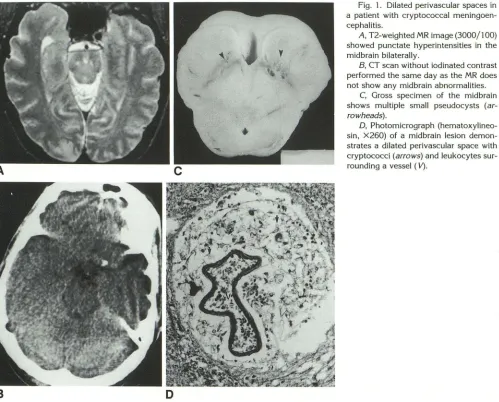
Grossly
,
there are usually several dilated spaces
that are not individually resolved on MR, but
instead are seen as single larger lesions (Fig
.
1)
.
Histologically, the dilated perivascular spaces
were as described above, with the space around
the vessel being several times the size of the
vessel diameter. The vast majority of these
le-sions were less than 3 mm in size and were not
detected by imaging
.
In one case, a 56-year-old
man, some of the enlarged perivascular spaces
seen with MR were not filled with organisms, but
presumably were dilated in association with
age-and/ or HIV -related atrophy.
At autopsy
,
12 of 13 brains had
cryptococco-mas within brain parenchyma. In six of these 12
,
the cryptococcomas measured greater than 3
mm and presumably could have been seen on
imaging studies. The number of cryptococcomas
AIDS-RELATED CRYPTOCOCCOSIS 1479
Fig. 1. Dilated perivascular spaces in
a patient with cryptococcal meningoen
-cephalitis.
A, T2-weighted MR image (3000/100) showed punctate hyperintensities in the
midbrain bilaterally.
8, CT scan without iodinated contrast performed the same day as the MR does
not show any midbrain abnormalities. C, Gross specimen of the midbrain
shows multiple small pseudocysts (ar-rowheads).
D, Photomicrograph (hematoxylin eo-sin, X260) of a midbrain lesion demon -strates a dilated perivascular space with
cryptococci (arrows) and leukocytes s ur-rounding a vessel ( V).
greater than 3 mm in these six specimens ranged
from one to 14 or more (in some cases the
confluent cryptococcomas were difficult to
iden-tify separately). The basal ganglia
,
thalamus
,
and
midbrain were the most frequent locations of the
larger cryptococcomas, although lesions greater
than 3 mm were also present in the cerebellum
,
pons
,
corpus callosum
,
and cortex. In all six of
these pa
t
ients
,
the final imaging studies (three
MRs
,
three CTs) showed abnormalities consistent
with cryptococcomas (hyperintense on long TR
images, hypointense on short TR images
,
hypo-dense or isohypo-dense mass on CT
;
Figs
.
2 and 3)
.
[image:3.612.60.559.78.480.2]1480 MATHEWS AJNR: 13, September /October 1992
A
B
c
D
E
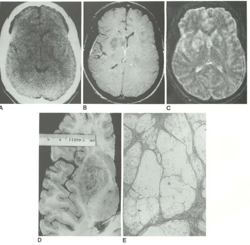
Fig. 2. Right basal ganglia cryptococcomas in a patient with CNS cryptococcosis.
A, CT scan without iodinated contrast shows mass effect on the frontal horn of the right lateral ventricle without a discrete abnormality of attenuation.
B, Gd-DTPA-enhanced T1-weighted MR image (600/20) demonstrates nonenhancing hypointense masses within the right caudate and lentiform nuclei.
C, T2-weighted MR image (3000/1 00) (degraded by motion artifact) shows hyperintense lesions in both lentiform nuclei and in the right caudate nucleus.
D, Horizontally sectioned autopsy specimen of the right cerebral hemosphere reveals that the right lentiform nucleus mass is composed of many confluent pseudocysts. The caudate nucleus lesion is of similar composition as seen on an adjacent section.
£, Photomicrograph (hematoxylineosin, X40) demonstrates multiple confluent cryptococcomas in the right basal ganglia.
more lesions than CT in each case. MR
detected
lesions in the basal ganglia, caudate, cerebellum,
and brain stem better than CT, but both
modali-ties
were equally poor
in detecting cortical
cryp-tococcomas
present
at autopsy
.
None of the
[image:4.612.58.567.79.577.2]con-AJNR: 13, September /October 1992
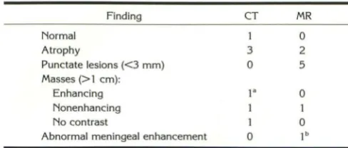
TABLE 1: Findings on final imaging study before autopsy in 13 patients with CNS cryptococcosis
Finding CT MR
Normal 1 0
Atrophy 3 2
Punctate lesions (<3 mm) 0 5
Masses (> 1 em):
Enhancing 1. 0
Nonenhancing 1
No contrast 1 0
Abnormal meningeal enhancement 0 1b
• Pathologically found to be primary CNS lymphoma.
b This patient had focal cerebritis and adjacent vascular congestion as discussed in the text.
trast enhancement on the three Gd
-
DTPA-en-hanced MR scans or the three iodinated
contrast-enhanced CT studies performed on patients with
these lesions. (Note discussion of mass lesions
greater than 1 em below
.) The brain parenchyma
adjacent to the cryptococcomas did not
demon-strate inflammatory infiltration, necrosis, or
hem-orrhage in any of the cases.
Small intraparenchymal cryptococcomas (<3
mm) were quite common in our histologic
speci-mens. In all cases in which there were larger
cryptococcomas, smaller cryptococcomas were
also noted pathologically. The smaller
cryptococ-comas were not seen with CT despite being quite
numerous (range, one to 1 04) in some cases. MR
detected some of these lesions in every case, but
most of these small cryptococcomas went
un-detected.
Imaging studies demonstrated mass lesions
greater than 1 em in four patients. One of these
patients showed contrast enhancement on CT.
The enhancing mass was found to be primary
CNS lymphoma at autopsy and not a
cryptococ-coma
.
One patient had a contrast-enhanced CT
and another had a Gd-DTPA-enhanced MR (Fig
.
2)
showing
no contrast enhancement of their
masses
.
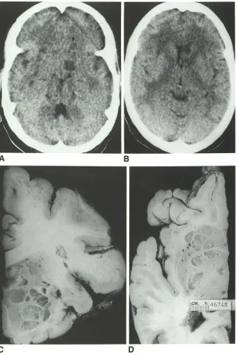
One patient with multiple masses seen
on CT (Fig
.
3) was not given
contrast.
Patholog-ically
,
the masses in these latter three patients
were found to be multiple confluent
cryptococ-comas in the basal ganglia.
All 13 patients had cryptococcal meningitis as
manifested grossly by opacification of the
men-inges and the presence of mucoid material
pro-duced by the cryptococcal organisms within the
subarachnoid space. Cryptococcal organisms and
inflammatory cells (lymphocytes and occasional
histiocytes) were seen histologically within the
subarachnoid spaces and leptomeninges
.
The
meningeal inflammatory reaction was usually
AIDS-RELATED CRYPTOCOCCOSIS 1481
mild. Abnormal meningeal contrast
enhancement
was seen
in
one of five
enhanced MR
studies and
none of the five
enhanced CT scans.
The patient
who demonstrated
gyriform
enhancement
(Fig.
4)
pathologically was
found
to have
a focal
bac-terial cerebritis related
to
Nocardia
septicemia
,
with severe
vascular
congestion
near
the area of
enhancement.
The
cryptococcBI meningitis in this
patient
was
no more
severe pathologically than
that seen in the
other
nonenhancing cases and
probably did not
account for the contrast
en-hancement
.
Atrophy was
a common finding
both
on
im-aging
studies
and at
autopsy.
Five of 13 patients
(three on CT
,
two
on MR)
exhibited subjective
imaging evidence
of cerebral atrophy. Since the
presence of atrophy was not
a
primary
focus of
this
study
,
no quantitative
evaluation of
brain
volume was performed.
Of the seven patients who died during
their
first
episode of CNS cryptococcosis
,
three
had
both small
(<3
mm)
and
large
(>3
mm)
crypto-coccomas pathologically
,
while four had only
small cryptococcomas. All three of the patients
who died during relapses of previously
controlled
cryptococcosis had both
small and
large
crypto-coccomas. One of the
three
patients who died
with clinically suppressed CNS cryptococcosis
had no cryptococcomas
, while the other two had
only small cryptococcomas.
.
Discussion
CNS cryptococcosis
is
an important
clinical
problem in patients with AIDS ranking as the
third most frequent cause of CNS
infection
in
AIDS (12
,
13)
and
presenting more often
as a
disseminated
infection in these
patients than in
other patients
(14).
The diagnosis of
cryptococcal
meningitis in AIDS patients
is complicated
by
the
fact
that
cerebrospinal
fluid cell
count,
protein
,
and glucose may be normal or only mildly
ab-normal in
comparison
to
the
nearly always
ab-normal laboratory values in HIV
-negative pati
ents
with cryptococcal meningitis
.
The presence
of
fever
and headache in
a
patient with AIDS
or at
risk for AIDS should prompt an investigation
for
CNS cryptococcal
disease.
CT
and
MR
are
im-portant diagnostic
techniques in any
patient
with
HIV
infection
or AIDS
and neurologic
dysfunction
since intracranial mass lesions
are frequent (22).
The most
common mass
lesions detected by
CT
are due to toxoplasmosis
,
although lymphoma
[image:5.614.52.296.102.206.2]1482 MATHEWS
Fig. 3. CT demonstration of both lu -cent and isodense cryptococcomas in a
patient with CNS cryptococcosis. A and B, CT scans without iodinated
contrast show hypodense lesions
(arrow-heads) in the base of the left cerebral hemisphere (A) and mass effect on the
left lateral ventricle due to an ill-defined
slightly hypodense lesion (B). Note the
absence of attenuation abnormalities on the right.
C, Coronal autopsy specimen of the
left cerebral hemisphere shows multiple
pseudocysts in the basal ganglia co
rre-sponding to the largest lucent lesion in A and the ill-defined mass in B.
D, Horizontal autopsy specimen of the
right cerebral hemisphere demonstrates
multiple pseudocysts in the basal ganglia
that were not detected by CT.
A
c
cryptococcosis
must be
considered
(23-25).
Of
concern
in patients with cryptococcosis is the
report that 20
%
of patients with mass lesions and
meningitis
and
10
%
of patients with mass lesions
without
meningitis had no signs or symptoms
suggesting
CNS disease (5).
The typical pathologic findings of CNS
cryp-tococcosis in our series were meningitis, dilated
perivascular
spaces
,
and
cryptococcomas.
Cryp-toco
cca
l meningitis
is
the most common
neuro-logic manifestation of
cryptococcosis and
often
AJNR: 13, September /October 1992
B
D
has an insidious chronic course
(1,
22).
In
re-sponse to the host immune system
'
s attack on
the organisms
,
the cryptococci will produce a
mucoid material (26)
.
When the meningeal
dis-ease involves the extensions of the subarachnoid
space that accompany perforating arteries into
the parenchyma
,
the production of voluminous
mucoid material may enlarge these perivascular
[image:6.612.220.561.80.590.2]AJNR: 13, September /October 1992
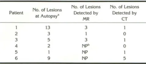
TABLE 2: Detection of cryptococcomas >3 mm in size
No. of Lesions No. of Lesions No. of Lesions
Patient
at Autopsy• Detected by Detected by
MR CT
13 3 1
2 3 1 0
3 5 3 1
4 2 NPb 0
5 1 NP 1
6 9 NP 5
• Some lesions that may have arisen from multiple smaller lesions that had become confluent were counted as a single lesion.
b NP, not performed.
Note.- The final imaging study was MR in patients 1-3 and CT in patients 4-6.
within brain parenchyma, can develop when the
organisms have extended directly from the
peri-vascular spaces into the parenchyma (26) or,
possibly, when they have invaded the paren
-chyma from other meningeal or ependymal
sur-faces
.
In the detection of meningeal disease due to
cryptococcosis, imaging techniques were not
sen-sitive, since only one of 10 studies with contrast
administration was positive. The meningeal
con-trast enhancement in this one MR case was more
likely due to a superimposed bacterial cerebritis
and its associated vascular congestion than due
to cryptococcosis
,
since the severity of
crypto-coccal meningitis present was not significantly
different than that seen in the cases without
contrast enhancement. Gd-DTPA-enhanced MR
·
has been shown to be more effective than
con-trast-enhanced CT in detecting meningeal
en-hancement in bacterial and tuberculous
menin-gitis (27, 28). Cryptococcal meninmenin-gitis was
asso-ciated with meningeal enhancement on
Gd-DTPA-enhanced MR in one of four cases (11) and
neither of two cases (28) in two other clinical
studies. Gyriform enhancement on CT
examina-tions of cryptococcal meningitis has been
re-ported (6, 9), although it is not commonly seen
(10, 11)
.
An obvious pitfall in these clinical studies
w
i
thout pathologic correlation is that other con
-ditions
,
in particular, infarction, can cause gyri
-form cortical and meningeal enhancement (29).
Our results indicate that AIDS-related cryptococ
-cal meningitis does not typi-cally produce
menin-geal enhancement on CT or MR.
MR was more effective than CT in demonstrat
-ing abnormalities due to dilated perivascular
spaces both in our series and in previous studies
(11, 17)
.
Not stated in these other works is our
observation that the punctate hyperintensities on
AIDS-RELATED CRYPTOCOCCOSIS 1483
MR often do not represent a single dilated
peri-vascular space
,
but
,
instead
,
represent s
e
veral
spaces in close proximity to each other. We also
found that these punctate MR abnormalities were
more often due to small intraparenchymal
cryp-tococcomas that likely develop
e
d as a progr
e
s-sion of cryptococcosis involving p
e
riva
s
cular
spaces and
,
therefore
,
are located in the same
areas as dilated perivascular spaces. However
,
dilated perivascular spaces and small
cryptococ-comas can result in an id
e
ntical imaging
appear-ance. In one of our patients
,
enlarged perivascular
spaces were not uniformly a consequence of
cryptococcosis but, instead
,
were probably at
least partially due to age- or HIV -related atrophy
.
Generalized atrophy has been reported to be
common in AIDS patients (30
,
31) and
,
specifi-cally, those with cryptococcosis (1 0). HIV
infec-tion itself may be the cause of this parenchymal
loss
,
although no correlation has been found
between the degree of atrophy and the severity
of HIV infection (31 ). Since atrophy may enlarge
the perivascular spaces to the extent that they
are visible on MR, the observation of punctate
hyperintensities is not specific for CNS
crypto-coccosis in patients with AIDS. However
,
this
finding should raise the suspicion of cryptococcal
disease, especially if other signs of diffuse atrophy
such as ventricular and sulcal enlargement are
not present or the patient has clinical signs or
symptoms of meningitis.
MR was more effective than CT not only in
identifying pathologic changes due to dilated
per-ivascular spaces but also in demonstrating more
cryptococcomas in each of the patients that had
both studies performed near the time of death. In
the detection of cryptococcomas greater than 3
mm
,
imaging techniques were sensitive in
de-tecting abnormal cases, since every patient with
these lesions had an abnormal imaging study
.
Howeve
r,
imaging was not sensitive in detecting
individual lesions
,
since the actual number of
lesions present pathologically was underesti
-mated by more than 50
%.
Volume averaging of
small lesions with normal brain or lack of
radio-graphic contrast between some cryptococcomas
and surrounding parenchyma are two possible
reasons that many lesions cannot be seen
retro-spectively on imaging studies.
Some authors have called the larger lesions
composed of organisms and mucoid material
"
gelatinous pseudocysts
"
(7
,
1 0) which is an
[image:7.615.58.297.95.200.2]con-1484 MATHEWS AJNR: 13, September /October 1992
B
c
D
E
Fig. 4. Superficial gyriform enhancement in a patient with cryptococcal meningitis.
A and B, T1-weighted MR images (600/20) before (A) and after (B) injection of Gd-DTPA shows superficial gyriform enhancement
(arrowheads) in the right occipital and temporal lobes.
C, T2-weighted MR image (3000/1 00) demonstrates abnormal hyperintensity in the right occipital lobe (solid arrow). Note that the
right thalamic lesion (open arrow) and two of the smaller punctate hyperintensities in the basal ganglia (arrowheads) corresponded
pathologically to intraparenchymal cryptococcomas. Other punctate hyperintensities seen corresponded to dilated perivascular spaces.
D, Horizontal gross specimen of the occipital lobes shows bilateral leptomeningeal thickening (arrowheads) and right occipital lobe
discoloration in the area of signal abnormality in C. This discoloration proved histologically to be severe vascular congestion secondary to bacterial cerebritis.
£, Photomicrograph (hematoxylineosin, X 150) of cryptococcal meningitis shows interstitial debris with a moderate inflammatory response as evident by the number of dark staining leukocytes (arrowheads).
fusion regarding cryptococcal disease. In our
ex-perience, "gelatinous pseudocysts
"
are the typical
lesions that occur pathologically when the
cryp-tococcal organisms cause a mass lesion in the
brain
.
In other words
,
gelatinous pseudocysts and
cryptococcomas are equivalent terms. In some
cases, the relative amounts of mucoid material,
inflammatory cells
,
and organisms result in x-ray
attenuation that causes the appearance of the
low-density gelatinous pseudocysts described by
others (Fig
.
3A)
.
In other cases
,
the constituents
are such that greater x-ray attenuation occurs
and the lesions are nearly isodense to brain tissue
(Figs. 2A and 36). However, on gross pathological
evaluation in both situations, the lesions have the
same appearance of pseudocystic areas filled with
mucoid material.
Contrast enhancement of cryptococcomas,
similar to meningitis as mentioned above
,
is
AJNR: 13, September /October 1992
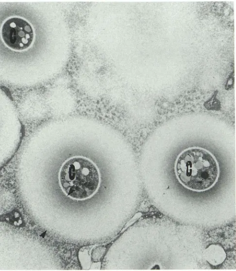
Fig. 5. Electron microscopy (X5300) shows three cryptococci
(C). Note the thick polysaccharide capsule (between arrowheads)
surrounding the microorganisms. (Courtesy of Dr Alo.)
inflammation was present in our specimens. AIDS
patients are unable to mount effective
inflam-matory responses, unlike immunocompetent
pa-tients in
other
studies (2, 3,
5)
.
This may account
for the observation of enhancement of
parenchy-mal lesions in other studies. Three patients with
AIDS-related cryptococcosis studied with
Gd-DTPA-
enhanced
MR in one study (11) showed
enhancement of parenchymal lesions, but none
of these patients had a tissue diagnosis via biopsy
or autopsy to exclude another etiology. The
cryp-tococcal organism is unusual in that it produces
a polysaccharide coat (Fig. 5) that may protect
the organism from inflammation even in
immu-nologically normal patients (26)
.
In addition
,
the
cryptococcal polysaccharide may have
immuno-suppressive properties (32)
.
This may account for
the lower incidence of inflammatory blood-brain
barrier breakdown and
consequent
contrast
en-hancement in cryptococcosis when compared to
bacterial meningitis (27, 28) or brain abscess (33,
34).
In summary, the typical pathologic
findings
in
AIDS-related CNS cryptococcosis are meningitis,
perivascular spaces dilated by
cryptococci, and
intraparenchymal cryptococcomas
.
MR
was
more effective than CT
in
detecting dilated
peri-AIDS-RELATED CRYPTOCOCCOSIS 1485
vascular spaces and cryptococcomas. Both
im
-aging modalities,
however
,
und
e
restimat
ed
the
actual number of
lesion
s
identified pathologically
and failed
to
detect
cryptococcal meningitis.
Con-trast
enhancement of cryptococcomas or
cryp-tococcal
meningitis
was uncommon
probably
be-cause
of
the immunocompromised
state
of these
patients and the uniqu
e
characteristics of the
organism itself
.
Acknowledgments
We thank the medical and nursing staff of the Johns
Hopkins Hospial AIDS service and the Johns Hopkins
Hospital neuroradiology and MR technologists for their roles
in caring for our patients.
References
1. Enzmann DR. Imaging of infections and inflammations of the central
nervous system: computed tomography, ultrasound, and nuclear magnetic resonance. New York: Raven Press, 1984:176-177 2. Everett BA, Kusske JA, Rush JL, Pribram HW. Cryptococcal infection
of the central nervous system. Surg f'leurol1978;9: 157-163 3. Harper CG, Wright DM, Parry G, O'Connor MJ. Cryptococcal granu
-loma presenting as an intracranial mass. Surg f'leurol 1979; 11 :425-429
4. Long JA, Herdt JB, DiChiro G, Cramer HR. Cerebral mass lesions in Torulosis demonstrated by computed tomography. J Comput Assist
Tomogr 1980;4:766-769
5. Fujita NK, Reynard M, Sapico FL, Guze LB, Edwards JE. Cryptococcal
intracerebral mass lesions: the role of computed tomography and
nonsurgical management. Ann Intern Med 1981 ;94:382-388
6. Cornell SH, Jacoby CG. The varied computed tomographic appear -ance of intracranial cryptococcosis. Radiology 1982; 143:703-707 7. Garcia CA, Weisberg AL, Lacorte WSJ. Cryptococcal intracerebral
mass lesions: CT-pathologic considerations. Neurology 1985;35:731-734
8. Waterson JA, Gilligan BS. Cryptococcal infections of the central nervous system: a ten year experience. Clin Exp f'leurol1987;23: 1
27-137
9. Tan CT, Kuan BB. Cryptococcus meningitis, clinicai-CT scan consid -erations. f'leuroradiology 1987;29:43-46
10. Popovich MJ, Arthur RH, Helmer E. CT of intracranial cryptococcosis. AJf'IR 1990; 11:139-142
11. Tien RD, Chu PK, Hesselink JR, Duberg A, Wiley C. Intracranial cryptococcosis in immunocompromised patients: CT and MR findings in 29 cases. AJf'IR 1991;12:283-289
12. Levy RM, Bredesen DE, Rosenblum ML. Neurologic manifestations of the acquired immunodeficiency syndrome (AIDS): experience at
UCSF and review of the literature. J f'leurosurg 1985;62:475-495 13. Gabuzda DH, Hirsch MS. Neurologic manifestations of infection with
human immunodeficiency virus: clinical features and pathogenesis.
Ann Intern Med 1987; 107:383-391
14. Eng RHK, Bishburg E, Smith SM, Kapila R. Cryptococcal infections in patients with acquired immune deficiency syndrome. Am J Med 1986;81: 19-23
15. Tress B, Davis B. Computed tomography of intracerebral toruloma.
f'leuroradiology 1979; 17:223-226
[image:9.612.55.298.78.356.2]1486 MATHEWS
17. Wehn SM, Heinz ER, Burger PC, Boyko OB. Dilated Virchow-Robin
spaces in cryptococcal meningitis associated with AIDS: CT and MR findings. J Comput Assist Tomogr 1989; 13:756-762
18. Riccio T J, Hesselink JR. Gd-DTPA enhanced MR of multiple
crypto-coccal brain abscesses. AJNR 1989; 1 O:S65-S66
19. Balakrishnan J, Becker PS, Kumar AJ, Zinreich SJ, McArthur JC,
Bryan RN. Acquired immunodeficiency syndrome: correlation of
radiologic and pathologic findings in the brain. Radiographies
1990;10:201-215
20. Braffman BH, Zimmerman RA, Trojanowski JQ, Gonatas NK, Hickey
WF, Schlaepfer WW. Brain MR: pathologic correlation with gross and
histopathology. I. Lacunar infarction and Virchow-Robin spaces.
AJNR 1988;9:621-628
21. Vinters HV, Anders KH. Neuropathology of AIDS. Boca Raton, FL:
CRC Press, 1990:61-65
22. Dismukes WE. Cryptococcal meningitis in patients with AIDS. J Infect Dis 1988; 157:624-628
23. Kelly WM, Brant-Zawadzki M. Acquired immunodeficiency syndrome: neuroradiologic findings. Radiology 1983; 149:485-491
24. Whelan MA, Kricheff II, Handler M, et al. Acquired immunodeficiency
syndrome: cerebral computed tomographic manifestations. Radiology
1983;149:477-484
25. Elkin CM, Leon E, Grenell SL, Leeds NE. Intracranial lesions in the
acquired immunodeficiency syndrome: radiological (computed to-mographic) features. JAMA 1985;253:393-396
AJNR: 13, September /October 1992
26. Hassin GB. Torulosis of the central nervous system. J Neuropathol
Exp Neurol1947;6:44-60
27. Mathews VP, Kuharik MA, Edwards MK·. D'Amour PG, Azzarelli B, Dreesen RG. Gd-DTPA-enhanced MR imaging of experimental bac-terial meningitis: evaluation and comparison with CT. AJNR 1988;9: 1 045-1 050
28. Chang KH, Han MH, Roh JK, Kim 10, Han MC, Kim C-W.
Gd-DTPA-enhanced MR imaging of the brain in patients with meningitis:
Comparison with CT. AJNR 1990; 11 :69-76
29. Elster AD, Moody DM. Early cerebral infarction: gadopentetate di-meglumine enhancement. Radiology 1990;177:627-632
30. Levy RM, Rosenbloom S, Perrett LV. Neuroradiologic findings in AIDS: a review of 200 cases. AJNR 1986;7:833-839
31. Chrysakopoulous HS, Press GA, Grafe MR, Hesselink JR, Wiley CA.
Encephalitis caused by human immunodeficiency virus: CT and MR imaging manifestations with clinical and pathologic correlation. Ra-diology 1990;175:185-191
32. Diamond RD. Cryptococcus neoformans. In: Mandell GL, Douglas RG Jr, Bennet JE, eds. Principles and practice of infectious diseases. 3rd
ed. New York: Churchill Livingstone, 1990:1980-1989
33. Enzmann DR, Britt RH, Yeager AS. Experimental brain abscess
evolution: computed tomographic and neuropathologic correlation.
Radiology 1979; 133:113-122
34. Runge VM, Clanton JA, Price AC, et al. Evaluation of contrast-enhanced MR imaging in a brain abscess model. AJNR 1985;6: