Clinical Ophthalmology
Dovepress
R e v i e w
open access to scientific and medical research
Open Access Full Text Article
Perioperative visual loss in ocular and nonocular
surgery
Kathleen T Berg Andrew R Harrison Michael S Lee
Department of Ophthalmology, University of Minnesota, Minneapolis, MN, USA
Correspondence: Michael S Lee 420 Delaware St Se, MMC 493, Minneapolis, MN 55455, USA Tel +1 612 625 3553 Fax +1 612-626-3119 email mikelee@umn.edu
Abstract: Incidence estimates for perioperative vision loss (POVL) after nonocular surgery
range from 0.013% for all surgeries up to 0.2% following spine surgery. The most common neuro-ophthalmologic causes of POVL are the ischemic optic neuropathies (ION), either ante-rior (AION) or posteante-rior (PION). We identified 111 case reports of AION following nonocular surgery in the literature, with most occurring after cardiac surgery, and 165 case reports of PION following nonocular surgery, with most occurring after spine surgery or radical neck dissection. There were an additional 526 cases of ION that did not specify if the diagnosis was AION or PION. We also identified 933 case reports of central retinal artery occlusion (CRAO), 33 cases of pituitary apoplexy, and 245 cases of cortical blindness following nonocular surgery. The incidence of POVL following ocular surgery appears to be much lower than that seen follow-ing nonocular surgery. We identified five cases in the literature of direct optic nerve trauma, 47 cases of AION, and five cases of PION following ocular surgery. The specific pathogenesis and risk factors underlying these neuro-ophthalmic complications remain unknown, and physicians should be alert to the potential for loss of vision in the postoperative period.
Keywords: perioperative, postoperative, vision loss, ocular surgery, nonocular surgery
Introduction
Vision loss is a rare, and devastating, complication that can be seen following both ocular and nonocular surgeries. The specific pathogenesis of perioperative vision loss (POVL) remains elusive in most cases, with much controversy surrounding patient and surgical risk factors. Many publications have speculated on causation. Unfortunately, given the rarity of POVL, these are based solely on retrospective data. We present a detailed review of the literature on the subject of vision loss following ocular and nonocular surgical procedures.
Method of literature review
We searched the National Library of Medicine’s PubMed database with a subsequent review of the accompanying references (last accessed February 8, 2010). The major search words and word combinations included: posterior ischemic optic neuropathy; anterior ischemic optic neuropathy; central retinal artery occlusion; pituitary apoplexy; cortical blindness; optic nerve trauma; postoperative vision loss; postoperative blind-ness; perioperative vision loss; perioperative blindblind-ness; and ocular surgery. In addition, the citations from the above searches were also included. Cases from the non-English literature and cases prior to 1970 were not included. Cases with documented direct surgical trauma to orbital structures other than the optic nerve were not included.
Clinical Ophthalmology downloaded from https://www.dovepress.com/ by 118.70.13.36 on 21-Aug-2020
For personal use only.
Number of times this article has been viewed
This article was published in the following Dove Press journal: Clinical Ophthalmology
Dovepress
Berg et al
Cases attributed to steroid injection into orbital structures were also not included.
Neuro-ophthalmologic causes
of vision loss after nonocular
surgery
incidence and prevalence of POvL
Vision loss occurring after nonocular surgery with general anesthesia typically results from: anterior ischemic optic neuropathy (AION); posterior ischemic optic neuropathy (PION); central retinal artery occlusion (CRAO); pituitary apoplexy; or cortical blindness. While studies of 400,0001
and 61,0002 patients undergoing general anesthesia have
demonstrated a low incidence of POVL in noncardiac, nonspinal fusion procedures, the actual incidence, overall, remains unknown. However, the risk of POVL is believed to be highest following cardiac or spine surgeries. A recent review of 5.6 million patients from the National Inpatient Sample (NIS) found that the incidence of POVL to be 8.64/10,000 after cardiac procedures and 3.09/10,000 after spinal fusion, while the incidence was only 0.66/10,000 after cholecystectomy, and just 0.12/10,000 after appendectomy.3
In a retrospective study of 501,342 noncardiac surgeries carried out at one institution, only four patients (0.0008%) developed persistent vision loss not attributed to direct surgical trauma to optic or cerebral tissues.1 In a separate
study at the same institution, 17 patients were found to have perioperative ION out of a total of 27,915 coronary artery bypass graft (CABG) procedures (0.06%).4 Similar studies
of POVL following cardiac surgery have shown comparable findings, with the incidence rates ranging from 0.09%5 to
0.113%.6 Other studies suggest that the rate of POVL may
be even higher after spine surgery. A study of over 225,000 surgeries at a single institution over a 15-year period found three cases of POVL out of 3,351 spine surgeries (0.09%), a 50-fold higher rate compared with other surgeries in the study.7 Additional large-scale studies of POVL after spine
surgery have demonstrated incidence rates varying from 0.094%8 to 0.2%,9 though the higher incidence seen in
the latter study may be because it was carried out at three tertiary care specialty hospitals, which may have attracted more complex cases.
ischemic optic neuropathies
The two most common types of POVL are AION and PION.10,11 In a recent study of 126,666 surgeries from a single
institution, there were 17 cases of ischemic optic neuropathy (ION) (0.013%): 8 AION and 9 PION.12 The type of ION
varies depending on the type of surgery performed, with AION occurring most often after cardiac surgery45 and PION
occurring most often after prone-positioned spine surgery or radical neck dissection.10,13
In a recent analysis of 4,728,815 spine surgeries from the NIS registry, the overall incidence of postoperative ION was found to be 0.006%.8 Other incidence estimates for ION
following spine surgery range from 0.028%14 to 0.12%.9
A review of 27,915 cardiac surgeries found the overall incidence of ION to be 0.06%, with 12 cases of AION and five cases of PION,4 while another study of 9,701 CABG
procedures found an ION incidence of 0.11%, with eight cases of AION and three cases of PION.6 A review of 602
CABG procedures found the incidence of AION to be 1.3%,15
while a similar study of 7,685 CABG procedures found an ION incidence of 0.09%.5
When the incidence values from these large-scale reviews are compiled, the overall incidence of AION is 0.00079% (1/126,666) after spine surgery and 0.024% (42/172,569) following cardiac surgery. There were no cases of AION following noncardiac, nonspine surgery in any of the large-scale reviews. When the incidence values for PION are compiled, the overall incidence is 0.005% (7/140,768) after spine surgery, 0.0061% (10/164,282) following cardiac surgery, and 0.0032% (4/126,666) after nonspinal, noncardiac surgery.
Diagnosis
AION is likely caused by occlusion or relative hypoperfu-sion of the anterior optic nerve head by the posterior ciliary arteries.16 Typical presentation includes sudden painless
vision loss and a visual field defect, most commonly inferior altitudinal in nature. Diffuse or segmental disc edema is observed on funduscopy at symptom onset, with the devel-opment of optic nerve atrophy after 4–8 weeks (Figure 1).17
Three forms of AION are recognized: perioperative, arteritic, and nonarteritic.18 The arteritic form of AION, associated
with giant cell arteritis (GCA), often presents in the elderly with an elevated erythrocyte sedimentation rate (ESR) and C-reactive protein (CRP), and systemic symptoms such as weight loss and jaw claudication. It is important to note that inflammatory markers are often extremely elevated after general surgery. Because of this, in the absence of other features of GCA such as systemic symptoms or abnormal temporal arteries, it is not advisable to check ESR and CRP in a patient who awakes from surgery with visual loss. If these are checked, an elevated ESR alone is not enough to diagnose arteritic AION in the postoperative period.19 The
Clinical Ophthalmology downloaded from https://www.dovepress.com/ by 118.70.13.36 on 21-Aug-2020
Dovepress Perioperative vision loss
gold standard for diagnosing arteritic AION is the temporal artery biopsy. Giant cell arteritis is considered an ophthalmic emergency, requiring the urgent administration of systemic corticosteroids to prevent the development of permanent, bilateral blindness.18
PION is believed to result from infarction to the intraor-bital optic nerve, supplied by pial blood vessels. PION is much less common than AION. There are three recognized types of PION: perioperative, arteritic, and nonarteritic; the perioperative type is the most common.20 PION presents
with similar signs and symptoms as AION, and is dif-ferentiated from AION based on the presence of a normal optic disc and fundoscopic examination at the onset of visual symptoms (Figure 2).13 Gadolinium enhancement21
and/or restricted diffusion22 may be seen on orbital magnetic
resonance imaging (MRI), while the MRI is typically unre-markable in AION.
Pathogenesis, risk factors, and outcomes
The pathogenesis of postoperative ION remains undefined. AION may occur in the setting of substantial blood loss or hypotension, such as that seen following gastrointestinal bleeding or hemodialysis. It is for this reason that some have suggested that intraoperative hypotension and anemia underlie the development of ION.23 Of 83 cases of ION
following spine surgery listed in the American Society of Anesthesiologists (ASA) Postoperative Visual Loss Registry, 96% had 1 L or more of blood loss or 6 hours or more of total anesthesia time, (with a mean anesthetic time of 9.6 hours).10
Blood loss, with or without arterial hypotension, has been shown to cause the release of endogenous vasoconstrictors due to the activation of the sympathetic nervous system, which can produce choroidal and optic nerve ischemia.24
The use of vasoconstricting agents to correct intraoperative hypotension has also been suggested to promote optic nerve ischemia.5,11,24 Analysis of the NIS registry found that patients
who developed ION were more likely to have hypotension, peripheral vascular disease, or anemia.8 However, there have
been cases of ION development in patients with blood pres-sure readings .90 mmHg24 and in those with minimal (less
than 500 mL) blood loss.10,25,26
Postoperative AION occurs most often after cardiac sur-gery, especially CABG. The mild to moderate hypothermia used during bypass could cause increased blood viscosity, which may lead to watershed infarction of the optic nerve.5
Indeed, it has been shown that cerebral blood flow decreases by 6%–7% for every degree Centigrade drop in body tem-perature.27 Cardiopulmonary bypass has also been shown to
promote complement activation, causing a five-fold increase in C3a levels, a known smooth muscle spasminogen.28
A recent time-matched, case-control study found that patients who developed AION after CABG had lower postoperative hemoglobin values, a higher rate of atherosclerotic disease, and were more likely to have had a coronary angiogram within 48 hours of surgery when compared with control CABG patients.4 A similar study found that CABG patients
who developed postoperative AION had prolonged bypass times, lower hematocrit levels, marked perioperative weight gain from large fluid infusions required to support intraop-erative blood pressure, and lower cardiac outputs requiring higher doses of inotropic medications.15
CABG could also contribute to the development of AION through elevations in intraocular pressure (IOP). It has been noted that AION has occurred with elevations in IOP,29,30 and
there is experimental evidence that elevated IOP can cause optic nerve ischemia by reducing blood flow to the eye.11
Figure 2 Normal appearing optic disc is seen in all cases of acute posterior ischemic optic neuropathy.
Figure 1 Optic disc edema observed in all cases of acute anterior ischemic optic neuropathy.
Clinical Ophthalmology downloaded from https://www.dovepress.com/ by 118.70.13.36 on 21-Aug-2020
Dovepress
Berg et al
The perfusion pressure of the eye is equal to the mean arterial pressure (MAP) minus the IOP, and dramatic changes in per-fusion pressure may overwhelm the autoregulation of blood flow to the eye.18 It has been shown that there is a rapid rise
in IOP lasting about 30 minutes when bypass pump circula-tion begins,31 and IOP may remain elevated for as long as
three days after bypass.32 The IOP elevation seen in CABG
is believed to be due in part to hemodilution, which causes an increase in intraocular blood flow along with a decrease in colloidal osmotic pressure.31 Autoregulation to the eye may
also be disrupted by other systemic processes often seen in patients who are candidates for CABG, such as aging and diabetes mellitus, as well as marked arterial hypotension,33
which often occurs during bypass procedures. Heart disease patients also often have chronic hypertension, which may reduce tolerance to periods of hypotension by shifting the autoregulatory curve of optic nerve blood flow.34
A variety of hemodynamic derangements could conceiv-ably cause PION including: anemia, hypotension, increased venous pressure, prone positioning during surgery, increased cerebrospinal fluid pressure, direct ocular compression, and embolism. Alteration is usually seen in more than one of these parameters in a given patient, suggesting that the mechanism of infarction may be multifactorial. Anemia and hypotension are nearly always seen in patients who develop postoperative PION.13 The pial vessels that supply the posterior optic nerve
lack an autoregulatory mechanism, and are therefore prone to ischemia during periods of systemic hypotension and when the blood oxygen carrying capacity is decreased.35 However,
studies that compared patients with POVL after spine surgery to controls found no difference in perioperative hematocrit and blood pressure, suggesting that hypotension and anemia alone are not enough to precipitate PION.12,36
Postoperative PION is most commonly seen follow-ing spine surgery in the prone position. Prone positionfollow-ing may lead to increased orbital venous pressure through an increase in abdominal venous pressure, especially when patients are obese.37 Spine surgery patients are often placed
in the Trendelenburg position, which also results in greater orbital venous pressure (Figure 3). Prone positioning may exert excessive pressure on the globe, and has been shown to raise the IOP in both anesthetized38 and awake39 patients.
The use of a Mayfield headrest can also exert excess pres-sure on the globe, especially in patients with exophthalmos or a flattened nasal bridge (Figure 4).40 However, an elevated
IOP would be expected to decrease perfusion pressure to the optic nerve head and central retinal artery, resulting in AION or CRAO, rather than PION.13 However, avoidance of the
prone position and Mayfield headrests alone appears to be insufficient in preventing postoperative PION, as PION has been documented following surgery in the supine position, with the use of head pins instead of foam cushions in the prone position,10,41 and in the lateral decubitus position.42
A study of 37 patients with POVL (including 14 PION and 8 AION patients) following spine surgery found that patients were more likely to have surgical times greater than 6.5 hours and substantial blood loss (3.5 L or more) when compared to age- and surgery-matched controls. Periopera-tive hematocrit and blood pressure did not differ between cases and controls. This study has been recognized as one of the few comparing hemodynamic factors between cases and controls. However, this study was poorly designed. It included
Figure 4 Mayfield headrest used in spine surgery. The edge of the headrest may compress the globe and lead to central retinal artery occlusion.
Figure 3 Positioning in spine surgery typically involves the patient lying prone in the Trendelenburg position. The level of the head is below the level of the feet, which leads to facial edema.
Clinical Ophthalmology downloaded from https://www.dovepress.com/ by 118.70.13.36 on 21-Aug-2020
Dovepress Perioperative vision loss
10 patients from pre-existing case reports and the rest were cases from the authors’ institution or data received from a solicitation to a spine society. The controls were taken from the authors’ institution rather than matched to where the surgery took place. Finally, all of the hemodynamic variables were not available for all of the cases.36 Meanwhile, Holy
and colleagues identified 17 patients with ION from a single institution. Each case was assigned two control patients, matched for age and gender, who underwent similar surger-ies at the same institution with a similar date of surgery. All preoperative, intraoperative, and postoperative hemodynamic variables were available for cases and controls. There was no significant difference between cases and controls in any single hemodynamic variable, suggesting that the cause of ION following nonocular surgery is multifactorial. Almost all patients who develop postoperative ION experience some degree of blood loss, anemia, and hypotension, though not to a greater degree than that seen in control patients who did not suffer ION after surgery.12
There have been many documented cases of PION fol-lowing radical neck dissection. Distension of ophthalmic veins, with accompanying facial edema, due to internal jugular vein ligation has been proposed as a potential mecha-nism for PION development. The venous distention could cause compression of the orbital apex, resulting in reduced perfusion and further anoxia of the posterior aspect of the optic nerve.23,43
While there is no established treatment for postopera-tive AION or PION, some have documented an improve-ment in visual acuity after the correction of anemia with blood transfusion,9,44 the correction of hypotension with
vasopressors,45 or the use of high-dose corticosteroids.9
However, some patients recover full acuity without any treatment,46 suggesting that the improvements seen following
the proposed interventions may be merely coincidental.
Anterior ischemic optic neuropathy
literature review
Through our review of the literature, we encountered 111 reports of AION following nonocular surgery, as well as an additional 526 reports defined only as “ischemic optic neuropathy” (Table 1).8,10,47,48 While AION was most
com-monly seen after: cardiac procedures;4–6,12,15,19,47,49–57 it also
occurred after spine surgery;9,10,12,36,58–63 shoulder surgery;64,65
radical neck dissection;66–70 total parathyroidectomy;55
peripheral vascular surgery;71 emergent Cesarian section;44
transurethral prostatectomy;72 radical prostatectomy;73 hip
replacement;74 osteosynthesis;74 bilateral knee replacement;74
Table 1 ischemic optic neuropathy cases following nonocular surgery
Number of patients, reference
Age; gender Surgery performed
Diagnosis
a2718 NA; 51% male Spine surgery iON, not
further specified
a2453 NA Spinal, orthopedic,
Cardiac, and general Surgery
iON, not further specified 810 36–64; 72% male Spine surgery iON, not
further specified
247 NA CABG iON, not
further specified 1910 36–64; 72% male Spine surgery AiON a836 12–68; gender NA Spine surgery AiON
162 65 F Spine surgery AiON
162 60 F Spine surgery AiON
162 41 M Spine surgery AiON
158 57 M Spine surgery AiON
159 50 F Spine surgery AiON
160 16 F Spine surgery AiON
161 44 M Spine surgery AiON
19 56 F Spine surgery AiON
112 71 M Spine surgery AiON
124 57–73; 71% male CABG AiON
815 58–70; 63% male CABG AiON
86 39–81; 82% male CABG AiON
75 44–61; all male CABG AiON
363 NA CABG AiON
112 60 M CABG AiON
112 54 M CABG AiON
112 64 M CABG AiON
112 55 M CABG AiON
112 72 M CABG AiON
112 60 M CABG AiON
112 54 M CABG AiON
119 54 M CABG AiON
119 54 M CABG AiON
119 63 M CABG AiON
131 46 M CABG AiON
131 63 M CABG AiON
130 55 M CABG AiON
130 68 M CABG AiON
171 68 M CABG AiON
171 74 M CABG AiON
155 56 M CABG AiON
155 44 M CABG AiON
156 71 F CABG AiON
156 58 M CABG AiON
157 63 M CABG AiON
151 71 F CABG AiON
153 70 M CABG53 AiON
154 63 M CABG AiON
167 47 M Neck dissection AiON
169 48 M Neck dissection AiON
(Continued)
Clinical Ophthalmology downloaded from https://www.dovepress.com/ by 118.70.13.36 on 21-Aug-2020
Dovepress
Berg et al
Table 1 (Continued) Number of patients, reference
Age; gender Surgery performed
Diagnosis
170 60 M Neck dissection AiON
155 16 F Total
parathyroidectomy AiON
149 40 M Dynamic
cardiomyoplasty
AiON
152 64 F Coronary
catheterization
AiON
171 62 F Peripheral vascular
surgery
AiON
164 45 M Shoulder surgery AiON
165 55 M Shoulder surgery AiON
174 52 M Hip replacement AiON
174 75 M Hip replacement AiON
174 80 F Hip replacement AiON
174 55 M Osteosynthesis AiON
174 66 M Bilateral knee
replacement
AiON
144 29 F Cesarian section AiON
172 66 M Transurethral
prostatectomy
AiON
173 50 M Radical
prostatectomy
AiON
175 47 F Liposuction AiON
5610 36–64; 72% male Spine surgery PiON
1420 mean age 43.3;
gender NA
Spine surgery PiON
a1436 12–68; gender NA Spine surgery PiON
137 66 M Spine surgery PiON
137 43 M Spine surgery PiON
137 12 M Spine surgery PiON
137 57 M Spine surgery PiON
137 44 M Spine surgery PiON
114 13; gender NA Spine surgery PiON
114 13; gender NA Spine surgery PiON
114 43; gender NA Spine surgery PiON
114 29; gender NA Spine surgery PiON
112 63 M Spine surgery PiON
112 77 M Spine surgery PiON
112 66 M Spine surgery PiON
162 49 F Spine surgery PiON
19 79 F Spine surgery PiON
19 27 F Spine surgery PiON
19 37 M Spine surgery PiON
177 24 M Spine surgery PiON
177 44 M Spine surgery PiON
178 33 F Spine surgery PiON
179 24 M Spine surgery PiON
171 13 M Spine surgery PiON
180 51 M Spine surgery PiON
142 59 M Spine surgery PiON
181 48 M Spine surgery PiON
146 58 M Spine surgery PiON
182 68 F Spine surgery PiON
160 10 M Spine surgery PiON
183 60 M Spine surgery PiON
(Continued)
Table 1 (Continued) Number of patients, reference
Age, gender Surgery performed
Diagnosis
143,181 67 M Neck dissection PiON
184 71 M Neck dissection PiON
185 48 M Neck dissection PiON
186 49 M Neck dissection PiON
168 64 M Neck dissection PiON
1182 73 M Neck dissection PiON
187 37 M Neck dissection PiON
11 77 M Neck dissection PiON
11 67 F Neck dissection PiON
54 57–73; 71% male CABG PiON
36 39–81; 82% male CABG PiON
112 61 M CABG PiON
112 68 F CABG PiON
121 57 F CABG PiON
188 46 F CABG PiON
171 81 F CABG PiON
391 59 M Rhinoplasty PiON
191 40 M Rhinoplasty PiON
191 23 F Rhinoplasty PiON
188 62 M Drainage of infected
hip prosthesis
PiON
189 64 M Shoulder surgery PiON
137 65 F Knee surgery PiON
112 66 M Knee surgery PiON
112 62 M Osteosynthesis PiON
174 51 M Osteosynthesis PiON
123 59 F Gastroduodenotomy PiON
171 51 F Abdominal explorationPiON
11 52 M Thoracotomy PiON
112 88 M Thoracotomy and
segmentectomy
PiON
125 43 F Breast augmentation
and abdominal liposuction
PiON
190 41 M Axillary vein grafting PiON
126 42 M Laparoscopic
nephrectomy
PiON
112 70 M Femoral aneurysm
repair
PiON
1420 mean age 58.3;
gender NA NA PiON
Notes: aStudy contains information compiled from data submitted to a registry, or
compiled case reports, which may be redundant with that seen in the other papers listed.
Abbreviations: iON, ischemic optic neuropathy; AiON, anterior ischemic optic neuropathy; PiON, posterior ischemic optic neuropathy; NA, information not available; M, male; F, female; CABG, cardiopulmonary bypass grafting.
and liposuction.75,76 Patients ranged in age from 1655,60 to
80 years.74 Of the 75 cases that provided individual patient
information, 43 cases were bilateral (57%). Of the 32 cases in which the information was provided for individual patients, 27 (84%) reported perioperative “anemia”, which we defined as a hemoglobin of less than 10 gm/dL and/or a hematocrit less than 30%. Of the 38 cases that provided
Clinical Ophthalmology downloaded from https://www.dovepress.com/ by 118.70.13.36 on 21-Aug-2020
Dovepress Perioperative vision loss
perioperative blood pressure measurements for individual patients, 18 (48%) reported perioperative systolic blood pressure (SBP) measurements of less than 90 mmHg and/ or MAP measurements less than 70.
Posterior ischemic optic neuropathy
literature review
We are aware of 165 cases of postoperative PION in the literature, as well as an additional 526 reports defined only as “ischemic optic neuropathy” (Table 1).8,10,47,48
The most common procedure that resulted in PION was spine surgery.9,10,12,14,20,36,37,42,46,60,62,71,77–83 PION has also been
documented following: radical neck dissection;1,43,68,84–87
CABG;4,6,21,71,88 knee surgery;12,37 osteosynthesis;12,74
drainage of an infected hip prosthesis;88 shoulder
surgery;89 gastroduodenotomy;23 abdominal exploration;71
thoracotomy;1,12 axillary vein grafting;90 femoral aneurysm
repair;12 laparoscopic nephrectomy;26 rhinoplasty;91 and breast
augmentation with abdominal liposuction.25 Patients ranged in
age from 1060 to 81.71 Of the 80 cases that provided individual
patient data, 47 (59%) were bilateral. Of the 42 cases in which the information was provided for individual patients, 28 (67%) reported perioperative “anemia”, which we defined as hemo-globin of less than 10 gm/dL and/or hematocrit less than 30%. Of the 42 cases that provided blood pressure measurements, 28 (67%) reported perioperative SBP readings of less than 90 mmHg and/or MAP measurements less than 70.
Central retinal artery occlusion
Central retinal artery occlusion (CRAO) has been well-documented in children and adults following trauma,92 and
embolic,93 thrombotic, or vasospastic episodes.94 Of the
93 spine surgery cases submitted to the ASA Postoperative Visual Loss Registry, there were 10 cases of CRAO, 7 of which resulted in acuity readings of no light perception.10
In a recent analysis of 4,728,815 spine surgeries from the NIS registry, the overall incidence of postoperative CRAO was found to be 0.001%.8
Diagnosis
CRAO presents with a cherry-red spot on the macula, a white ground-glass appearance of the retina, attenuated arterioles with preserved choriocapillaries, and an afferent pupillary defect (Figure 5).10
Pathogenesis, risk factors, and outcomes
CRAO occurring after general surgery is typically observed following external ocular compression. When compared with
cases of ION after spine surgery, patients who developed CRAO were found to have significantly lower mean anesthetic durations and median estimated blood losses. Periocular trauma was documented in the majority (70%) of cases, evidenced by ipsilateral decreased supraorbital sensation, ophthalmoplegia, corneal abrasion, ptosis, or unilateral erythema.10 The ophthalmic artery gives rise to the central
retinal artery, as well as the posterior ciliary artery, which supplies the choroid. Extensive pressure on the globe may raise IOP above the SBP leading to retinal ischemia.95
Experiments in rhesus monkeys have shown that the retina can tolerate pressure-induced ischemia for approximately 95 minutes and still recover, although it suffers permanent ischemic damage after 105 minutes.96 The majority of
post-operative CRAO cases occur in prone-positioned patients, or in those positioned supine with excessive ocular pressure secondary to an anesthetic mask.95
No Class I data on the effective treatment for CRAO exists, and the accompanying blindness is often permanent. It has been suggested that the use of a foam headrest with orbital cutouts may prevent postoperative CRAO development.95
CRAO literature review
We are aware of 933 documented cases of postoperative CRAO (Table 2), with the overwhelming majority seen following spinal surgery.8,10,40,48,95,97–104 Cases were also
documented after hip/femur treatment,33 knee replacement,3
cholecystectomy,3 appendectomy,3 colorectal resection,3
and cardiac surgery.3 There was also one case following hip
replacement in the lateral decubitus position without direct pressure on the orbit. The authors speculate that the CRAO
Figure 5 Funduscopic appearance of central retinal artery occlusion demonstrates a cherry-red spot on the macula, attenuated arterioles, and retinal whitening from edema.
Clinical Ophthalmology downloaded from https://www.dovepress.com/ by 118.70.13.36 on 21-Aug-2020
Dovepress
Berg et al
in this case may have been due to venous hypertension from a chest strap, which could have resulted in IOP elevation.105
All cases were unilateral, and most authors reported postop-erative periocular swelling and/or erythema. Patients ranged in age from 1295,97,102 to 73 years.103
Pituitary apoplexy
Diagnosis
Pituitary apoplexy is a rare clinical syndrome caused by infarction or hemorrhage of an existing pituitary adenoma. There is typically rapid enlargement of the adenoma, leading to compression of parasellar structures and the abrupt onset of signs and symptoms such as headache, meningismus, vomiting, visual field defects, unilateral or bilateral vision loss, ophthalmoplegia, and/or stupor.106 MRI is the imaging
modality of choice in the diagnosis of pituitary apoplexy, with a sensitivity of 88% (Figure 6).107
Pathogenesis, risk factors, and outcomes
Autopsy studies have suggested that the overall incidence of pituitary adenomas in adults is 1.4%, with the vast majority being completely asymptomatic.108 There are many
fac-tors implicated in the development of pituitary apoplexy, including major surgery.109 During surgery, fluctuations in
blood pressure, hypotension, blood dilution with crystalloid, anticoagulation, excessive steroid secretion, and transient increases in intracranial pressure may all play a role in pituitary apoplexy pathogenesis.110 Postoperative pituitary
apoplexy occurs most often after cardiac surgery,110
espe-cially CABG.111 In fact, an autopsy study of patients who
died within 10 days of CABG found that 15% had evidence
of pituitary necrosis, suggesting that pituitary apoplexy following CABG may be even more common than its clini-cal recognition.108 Factors specifically associated with bypass
that may lead to pituitary apoplexy include reduced tissue oxygenation, embolism, anticoagulation, edema, and/or positive pressure ventilation.111
The recommended management for pituitary apoplexy is urgent transsphenoidal decompression surgery and high-dose corticosteroid administration.112 However, a more
Table 2 Central retinal artery occlusion case reports
Number of patients, reference Age, gender Surgery performed
a8643 NA Spinal, orthopedic, cardiac, and general surgery
b478 NA Spine surgery
b1010 33–59; gender NA Spine surgery
197 12 F Spine surgery
140 28 F Spine surgery
198 30 M Spine surgery
199 23 M Spine surgery
1100 NA Spine surgery
1101 29 M Spine surgery
1102 12 F Spine surgery
1103 73 M Spine surgery
1104 38 M Spine surgery
195 12 F Spine surgery
1105 58 M Hip replacement
Notes: aStudy results include all causes of retinal vascular occlusion, not exclusively CRAO; bStudy contains information compiled from data submitted to registry, or
compiled case reports, which may be redundant with that seen in other papers listed. Abbreviations: NA, information not available; M, male; F, female.
Figure 6 Head computed tomography of a patient with pituitary apoplexy. The hyperdense region represents hemorrhage within a pre-existing pituitary adenoma.
Clinical Ophthalmology downloaded from https://www.dovepress.com/ by 118.70.13.36 on 21-Aug-2020
Dovepress Perioperative vision loss
conservative medical approach can be considered when vision is unaffected, or when the visual deficit is stable or improving, because spontaneous recovery may ensue.113
Pituitary apoplexy literature review
We are aware of 33 cases in the literature of postoperative pituitary apoplexy (Table 3). The majority of cases occurred following cardiac surgery, especially CABG,111,114–124 as well
as mitral valve repair,125 and aortic valve replacement.126
Pituitary apoplexy was also seen following: abdominal aortic aneurysm repair;127 liposuction;128 knee arthroplasty;129,130
hip arthroplasty;114,130 spine surgery;131 cholecystectomy;132
orchiectomy;114 and endoscopic sinus surgery.110 Patients
ranged in age from 45131 to 79 years.123
Cortical blindness
Diagnosis
Cortical blindness results from the destruction or denervation of the primary visual cortex. The parieto-occipital region of the brain is a watershed zone for the middle cerebral and posterior cerebral arteries, and may undergo infarction during periods of systemic hypotension. Stroke after general surgery is very rare, although the risk appears to be increased after cardiac or vascular surgery, especially CABG. The majority of postoperative strokes are ischemic or embolic in nature.133
Bilateral lesions cause a syndrome with signs and symptoms ranging from bilateral lower visual field defects, difficulty in the visual judgment of size, distance, and movement, disrupted smooth oculomotor pursuit, restricted visual atten-tion, and optic ataxia.134 Patients have reduced vision with
normal pupil reaction, intact corneal reflexes, and normal eye movement. Bilateral complete vision loss is rare, and is usually seen in association with other neurologic symptoms associated with vertebrobasilar strokes, such as ataxia, hemi-syndrome, diplopia, and nausea.135 Patient evaluation should
include a detailed neurologic examination and brain com-puted tomography (CT) or MRI to evaluate the lesion.133
Pathogenesis, risk factors, and outcomes
Postoperative cortical blindness results from embolism, generalized cerebral underperfusion, or both.133 Symptoms
may be immediate, identified in the first postoperative day, or delayed, presenting from the second postoperative day onward.136 Early embolism leading to stroke in CABG is
thought to result from manipulations of the heart and aorta, or from the release of particulate matter from the bypass pump. Delayed embolism is generally due to postoperative atrial fibrillation, myocardial infarction, and hypercoagula-bility due to surgical trauma and associated tissue injury. Most strokes due to hypoperfusion are diagnosed in the first postoperative day; delayed hypoperfusion strokes are usually due to postoperative dehydration or blood loss.133 It has been
suggested that repeated short episodes of extreme hypoten-sion are more likely to result in cortical blindness than a longer episode of moderate hypotension.137 Risk factors for
developing postoperative stroke include: hypertension, dia-betes mellitus, renal insufficiency, smoking history, COPD, peripheral vascular disease, cardiac disease, and/or systolic dysfunction. Patients with symptomatic carotid stenosis may be at increased risk of postoperative stroke, including cortical
Table 3 Pituitary apoplexy case reports
Number of patients, reference
Age, gender Surgery performed
1114 69 F CABG
1114 65 M CABG
1116 57 M CABG
1116 55 M CABG
1118 58 M CABG
1118 65 M CABG
1118 68 M CABG
1122 72 M CABG
1122 64 M CABG
1123 79 M CABG
1123 64 M CABG
1183 77 M CABG
1183 59 M CABG
1117 70 M CABG
1111 60 F CABG
1119 61 M CABG
1120 71 M CABG
1121 60 M CABG
1115 63 M CABG
1115 62 M CABG
1115 55 M CABG
1125 56 M Mitral valve
replacement
1126 68; gender NA Aortic valve
replacement
1127 73 M Abdominal aortic
aneurysm repair
1129 65 F Knee arthroplasty
1130 61 M Knee arthroplasty
1114 72 M Hip arthroplasty
1130 76 M Hip arthroplasty
1131 45 M Spine surgery
1128 50 F Liposuction
1114 52 M Orchiectomy
1132 47 M Cholecystectomy
1110 67 M endoscopic sinus
surgery
Abbreviations: NA, information not available; M, male; F, female; CABG, cardio-pulmonary bypass grafting.
Clinical Ophthalmology downloaded from https://www.dovepress.com/ by 118.70.13.36 on 21-Aug-2020
Dovepress
Berg et al
blindness, and may benefit from carotid revascularization prior to undergoing surgery.133
The prognosis of postoperative parieto-occipital stroke ranges from total permanent blindness to brief periods of transient ischemic attacks with full recovery of visual acuity.136 Cortical blindness is generally seen as permanent
after the window for spontaneous recovery has passed, which is thought to be several months post-lesion.138
Cortical blindness literature review
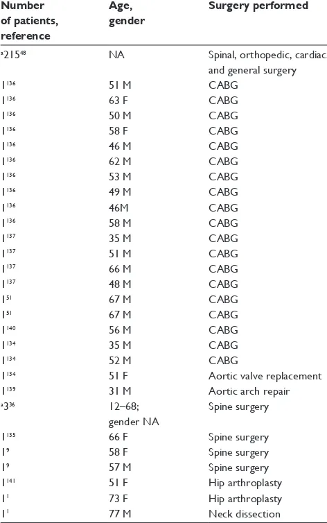
We are aware of 245 cases of postoperative cortical blindness in the literature (Table 4). Most cases were seen following CABG.3,51,134,136,137,139,140 Cases were also seen following:
aortic arch repair;139 aortic valve replacement;134 radical neck
dissection;1 total hip arthroplasty;1,3,141 knee replacement;3
cholecystectomy;3 appendectomy;3 colorectal resection;3
and spine surgery.3,9,36,135 Patients ranged in age from 31139
to 77 years.1
Neuro-ophthalmologic causes of vision
loss after ocular surgery
Neuro-ophthalmologic complications of ocular surgery include optic neuropathies such as AION, PION, and direct trauma to the optic nerve. Vision loss following ocular sur-gery due to CRAO, central retinal vein occlusion (CRVO), globe rupture, retinal detachment, and retrobulbar hemor-rhage occur much more commonly and will not be discussed here. The majority of large-scale incidence data for vision loss is for orbital surgery, such as lateral decompressions and cavernous hemangioma removal, rather than for ocular surgery. The incidence of POVL appears to be much lower following ocular surgery when compared to orbital surgery. In a review of 67 lateral decompression surgeries, there were seven cases of unilateral POVL (2.99%).142 Another
review found 14 cases of POVL out of 2500 orbital sur-geries (0.56%), with all 14 occurring during 1150 orbital exploration surgeries (1.22%).143 We are not aware of any
large-scale reviews providing incidence measurements for POVL following ocular surgery.
Traumatic optic neuropathy
Diagnosis
Periocular injection of anesthetic can result in direct trauma to the optic nerve, especially following retrobulbar injec-tion, though they may also occur following peribulbar or sub-Tenon’s injection. Other potential complications of retrobulbar injection include retrobulbar hemorrhage, central retinal artery or vein occlusion, brain stem anesthesia,144 or
globe rupture with retinal detachment,145 and will not be
discussed here.
Postoperative vision loss from trauma to the optic nerve occurs immediately. An orbital MRI is important for the proper diagnosis in patients presenting with symptoms of optic neuropathy in the first 24 hours following periocular injection. Orbital MRI may show focal enlargement of the optic nerve or enhancement of the optic nerve sheath with gadolinium. The bright ring of signal from cerebrospinal fluid may be absent around the damaged optic nerve on T2-weighted images.146
The MRI may also demonstrate normal results.
Prevention, treatment, and outcomes
The sub-Tenon’s “blind” insertion technique has been shown to be safer than either retrobulbar or peribulbar injections,
Table 4 Cortical blindness case reports
Number of patients, reference
Age, gender
Surgery performed
a21548 NA Spinal, orthopedic, cardiac,
and general surgery
1136 51 M CABG
1136 63 F CABG
1136 50 M CABG
1136 58 F CABG
1136 46 M CABG
1136 62 M CABG
1136 53 M CABG
1136 49 M CABG
1136 46M CABG
1136 58 M CABG
1137 35 M CABG
1137 51 M CABG
1137 66 M CABG
1137 48 M CABG
151 67 M CABG
151 67 M CABG
1140 56 M CABG
1134 35 M CABG
1134 52 M CABG
1134 51 F Aortic valve replacement
1139 31 M Aortic arch repair
a336 12–68;
gender NA
Spine surgery
1135 66 F Spine surgery
19 58 F Spine surgery
19 57 M Spine surgery
1141 51 F Hip arthroplasty
11 73 F Hip arthroplasty
11 77 M Neck dissection
Notes: aStudy contains information compiled from data submitted to registry, or
compiled case reports, which may be redundant with that seen in other papers listed.
Abbreviations: NA, information not available; M, male; F, female; CABG, cardio-pulmonary bypass grafting.
Clinical Ophthalmology downloaded from https://www.dovepress.com/ by 118.70.13.36 on 21-Aug-2020
Dovepress Perioperative vision loss
while still providing equally effective anesthesia and akinesia. This is believed to be due to the cannula’s relatively short length and blunt tip, which limits damage to the optic nerve.147–149 There have been rare reports of optic nerve
trauma following sub-Tenon’s injection; therefore changing the mode of injection alone is not enough to prevent the occurrence of optic nerve trauma.150 Corticosteroid
admin-istration may relieve optic nerve swelling and restore visual acuity, and a trial of corticosteroids should be considered in patients presenting with evidence of direct needle trauma postoperatively.146
Direct optic nerve trauma
literature review
We are aware of five reported cases of optic nerve trauma following ocular surgery. Most cases were following retrob-ulbar injection,151–154 although cases were also seen following
peribulbar155 and sub-Tenon’s injection.150 One patient showed
partial recovery of visual acuity after receiving corticoster-oid therapy.153 Three other patients showed no improvement
despite receiving corticosteroids152,154 (follow-up information
is unknown for one patient).150
ischemic optic neuropathies
While rare, there have been a number of documented cases of AION or PION developing after ocular surgery with the use of periocular or general anesthesia. A retrospec-tive review of 5787 cataract extractions found two cases of AION within six weeks of surgery. However, one patient had a history of spontaneous AION in the contralateral eye 21 months prior.156
Pathogenesis, risk factors, and outcomes
The pathogenesis of AION after ocular surgery remains controversial. It has been postulated that elevated intraocular pressure, in eyes with vulnerable optic nerve circulation, may promote ischemia of the optic nerve head follow-ing cataract extraction.157 However, there have been many
cases of ION development in eyes with normal IOP.158–160
Elevated IOP has also been postulated to play a role in the development of AION following laser in-situ keratomileusis (LASIK).162 In order to create a lamellar flap during LASIK,
the IOP must be raised to .65 mmHg by a suction ring on the anterior segment of the eye.162 However, AION has also
been documented following the use of a femtosecond laser flap with low suction ring, a technique that maintains the IOP at or below 30–40 mmHg,163 and in another patient whose
IOP was maintained at 25 mmHg through the administration
of glaucoma medication.164 It is worth noting that both of
these patients had small cup-to-disc ratios in the fellow eye, a proposed risk factor for AION development.165 Some
have suggested that AION occurring after ocular surgery is coincidental in nature. However, one study compared the likelihood of developing AION 0–6 months or 6–12 months after cataract extraction found that all 18 cases of AION were within the first 6 postoperative months, which differed significantly (P , 0.001) from the uniform distribution expected if there were no temporal relationship to surgery.166
Approximately 50% of patients who develop AION following cataract extraction who undergo contralateral extraction at a later date developed AION in the fellow eye, compared to 19% in a control group who did not have cataract surgery.167
This suggests that some patients may be predisposed to developing AION after ocular surgery.
No treatment has been proven effective in the manage-ment of postoperative ischemic optic neuropathies. Many patients report spontaneous improvement in visual acuity, although none enjoy complete improvement.146 Improvements
in microkeratome design with shorter suction times and faster cutting have caused a decrease in the incidence of optic nerve damage after LASIK.168 Contralateral cataract extraction
should be approached with caution in patients who develop ION postoperatively, as these patients are at increased risk of developing ION in the fellow eye.
Anterior ischemic optic neuropathy
literature review
We are aware of 43 cases in the literature of AION occurring after ocular surgery. The most common surgery was cataract extraction,156,157,169–171 followed by LASIK.164,172,173 Cases
were also seen following vitrectomy,174 scleral buckle,175
pneumoplethysmography,176 and strabismus surgery.177
Patients ranged in age from 26177 to 94 years old.174
Posterior ischemic optic neuropathy
literature review
We are aware of five cases in the literature of PION occurring after ocular surgery. The most common surgery was LASIK.162,164 There was also one case following
bleph-aroplasty178 and one case following cataract extraction.179
Patients ranged in age from 29164 to 78 years.179
Conclusions
Potential neuro-ophthalmologic complications following nonocular surgery include anterior ischemic optic neuropathy, posterior ischemic optic neuropathy, central retinal artery
Clinical Ophthalmology downloaded from https://www.dovepress.com/ by 118.70.13.36 on 21-Aug-2020
Dovepress
Berg et al
occlusion, pituitary apoplexy, and cortical blindness. Reports have attempted to connect anemia, hypotension, blood loss, and other hemodynamic variables to the pathophysiology of AION and PION. These variables occur in nearly all cases of cardiac and spine surgery and yet, AION and PION occur rarely. There may be an individual predisposition of certain patients, or multiple factors that lead to a “perfect storm” of events resulting in AION or PION. Meanwhile CRAO almost always occurs following spinal surgery in the prone position, where ocular compression by the headrest prevents ocular perfusion.
Cortical blindness occurs most frequently after cardiac or vascular surgery, and may be due to embolism or general cerebral underperfusion. Pituitary apoplexy is seen most often following cardiac surgery in patients with pre-existing pituitary adenomas. Causes likely include fluctuations in blood pressure, hypotension, blood dilution with crystalloid, anticoagulation, excessive steroid secretion, and transient increases in intracranial pressure. While there are currently no established treatments available for AION, PION, CRAO, or cortical blindness, patients with pituitary apoplexy may benefit from urgent transsphenoidal decompression surgery and corticosteroid administration.
Potential neuro-ophthalmologic complications following ocular surgery include traumatic optic neuropathy, anterior ischemic optic neuropathy, and posterior ischemic optic neu-ropathy. Traumatic optic neuropathy is seen most frequently following retrobulbar injection, although it may also occur after peribulbar, or even sub-Tenon’s, injection. Diagnosis can often be confirmed with orbital MRI, and visual acu-ity may improve with corticosteroid administration. AION is seen most often after cataract extraction or laser in-situ keratomileusis, and may be linked to elevated intraocular pressure during surgery. PION is seen most often following LASIK. There is no established treatment available for AION or PION. Contralateral cataract extraction requires careful consideration and patient counseling in patients who develop AION after unilateral extraction, because of the increased risk of developing of AION in the fellow eye.
Disclosure
The authors report no conflicts of interest in this work.
References
1. Warner ME, Warner MA, Garrity JA, MacKenzie RA, Warner DO. The frequency of perioperative vision loss. Anesth Analg. 2001;93(6): 1417–1421.
2. Roth S, Thisted RA, Erickson JP, Black S, Schreider BD. Eye injuries after nonocular surgery. A study of 60,965 anesthetics from 1988 to 1992.
Anesthesiology. 1996;85(5):1020–1027.
3. Shen Y, Drum M, Roth S. The prevalence of perioperative visual loss in the United States: a 10-year study from 1996 to 2005 of spinal, orthopedic, cardiac, and general surgery. Anesth Analg. 2009;109(5): 1534–1545.
4. Nuttall GA, Garrity JA, Dearani JA, Abel MD, Schroeder DR, Mullany CJ. Risk factors for ischemic optic neuropathy after car-diopulmonary bypass: a matched case/control study. Anesth Analg. 2001;93(6):1410–1416.
5. Sweeney PJ, Breuer AC, Selhorst JB, et al. Ischemic optic neuropa-thy: a complication of cardiopulmonary bypass surgery. Neurology. 1982;32(5):560–562.
6. Kalyani SD, Miller NR, Dong LM, Baumgartner WA, Alejo DE, Gilbert TB. Incidence of and risk factors for perioperative optic neuropathy after cardiac surgery. Ann Thorac Surg. 2004;78(1): 34–37.
7. Roth S, Barach P. Postoperative visual loss: still no answers – yet.
Anesthesiology. 2001;95(3):575–577.
8. Patil CG, Lad EM, Lad SP, Ho C, Boakye M. Visual loss after spine surgery: a population-based study. Spine (Phila Pa 1976). 2008;33(13):1491–1496.
9. Stevens WR, Glazer PA, Kelley SD, Lietman TM, Bradford DS. Ophthalmic complications after spinal surgery. Spine (Phila Pa 1976). 1997;22(12):1319–1324.
10. Lee LA, Roth S, Posner KL, et al. The American Society of Anesthesiologists Postoperative Visual Loss Registry: analysis of 93 spine surgery cases with postoperative visual loss. Anesthesiology. 2006;105(4):652–659.
11. Williams EL, Hart WM Jr, Tempelhoff R. Postoperative ischemic optic neuropathy. Anesth Analg. 1995;80(5):1018–1029.
12. Holy SE, Tsai JH, McAllister RK, Smith KH. Perioperative ischemic optic neuropathy: a case control analysis of 126,666 surgical procedures at a single institution. Anesthesiology. 2009;110(2):246–253. 13. Buono LM, Foroozan R. Perioperative posterior ischemic optic
neuropa-thy: review of the literature. Surv Ophthalmol. 2005;50(1):15–26. 14. Chang SH, Miller NR. The incidence of vision loss due to
periop-erative ischemic optic neuropathy associated with spine surgery: the Johns Hopkins Hospital Experience. Spine (Phila Pa 1976). 2005;30(11):1299–1302.
15. Shapira OM, Kimmel WA, Lindsey PS, Shahian DM. Anterior isch-emic optic neuropathy after open heart operations. Ann Thorac Surg. 1996;61(2):660–666.
16. Lessell S. Nonarteritic anterior ischemic optic neuropathy: enigma variations. Arch Ophthalmol. 1999;117(3):386–388.
17. Miller NR. Anterior ischemic optic neuropathy: diagnosis and manage-ment. Bull N Y Acad Med. 1980;56(7):643–654.
18. Hayreh SS. Anterior ischemic optic neuropathy. Clin Neurosci. 1997;4(5):251–263.
19. Alpert JN, Pena Y, Leachman DR. Anterior ischemic optic neuropathy after coronary bypass surgery. Tex Med. 1987;83(8):45–47.
20. Sadda SR, Nee M, Miller NR, Biousse V, Newman NJ, Kouzis A. Clinical spectrum of posterior ischemic optic neuropathy. Am J
Ophthalmol. 2001;132(5):743–750.
21. Vaphiades MS. Optic nerve enhancement in hypotensive ischemic optic neuropathy. J Neuroophthalmol. 2004;24(3):235–236.
22. Purvin V, Kuzma B. Intraorbital optic nerve signal hyperintensity on magnetic resonance imaging sequences in perioperative hypotensive ischemic optic neuropathy. J Neuroophthalmol. 2005;25(3):202–204. 23. Johnson MW, Kincaid MC, Trobe JD. Bilateral retrobulbar optic nerve
infarctions after blood loss and hypotension. A clinicopathologic case study. Ophthalmology. 1987;94(12):1577–1584.
24. Hayreh SS. Anterior ischemic optic neuropathy. VIII. Clinical features and pathogenesis of post-hemorrhagic amaurosis. Ophthalmology. 1987;94(11):1488–1502.
25. Rath EZ, Falick Y, Rumelt S. Posterior ischemic optic neuropathy follow-ing breast augmentation and abdominal liposuction. Can J Ophthalmol. 2009;44(3):346–347.
26. Metwalli AR, Davis RG, Donovan JF. Visual impairment after laparo-scopic donor nephrectomy. J Endourol. 2004;18(9):888–890.
Clinical Ophthalmology downloaded from https://www.dovepress.com/ by 118.70.13.36 on 21-Aug-2020
Dovepress Perioperative vision loss 27. Reuler JB. Hypothermia: pathophysiology, clinical settings, and
man-agement. Ann Intern Med. 1978;89(4):519–527.
28. Chenoweth DE, Cooper SW, Hugli TE, Stewart RW, Blackstone EH, Kirklin JW. Complement activation during cardiopulmonary bypass: evidence for generation of C3a and C5a anaphylatoxins. N Engl J Med. 1981;304(9):497–503.
29. Katz B. Anterior ischemic optic neuropathy and intraocular pressure.
Arch Ophthalmol. 1992;110(5):596–597.
30. Tomsak RL, Remler BF. Anterior ischemic optic neuropathy and increased intraocular pressure. J Clin Neuroophthalmol. 1989;9(2):116–118.
31. Larkin DF, Connolly P, Magner JB, Wood AE, Eustace P. Intraocu-lar pressure during cardiopulmonary bypass. Br J Ophthalmol. 1987;71(3):177–180.
32. Deutch D, Lewis RA. Intraocular pressure after cardiopulmonary bypass surgery. Am J Ophthalmol. 1989;107(1):18–22.
33. Hayreh SS. Factors influencing blood flow in the optic nerve head.
J Glaucoma. 1997;6(6):412–425.
34. Haefliger IO, Meyer P, Flammer J, Luscher TF. The vascular endothelium as a regulator of the ocular circulation: a new concept in ophthalmology?
Surv Ophthalmol. 1994;39(2):123–132.
35. Isayama Y, Takahashi T, Inoue M, Jimura T. Posterior ischemic optic neuropathy. III. Clinical diagnosis. Ophthalmologica. 1983;187(3): 141–147.
36. Myers MA, Hamilton SR, Bogosian AJ, Smith CH, Wagner TA. Visual loss as a complication of spine surgery. A review of 37 cases. Spine
(Phila Pa 1976). 1997;22(12):1325–1329.
37. Dunker S, Hsu HY, Sebag J, Sadun AA. Perioperative risk fac-tors for posterior ischemic optic neuropathy. J Am Coll Surg. 2002;194(6):705–710.
38. Hunt K, Bajekal R, Calder I, Meacher R, Eliahoo J, Acheson JF. Changes in intraocular pressure in anesthetized prone patients.
J Neurosurg Anesthesiol. 2004;16(4):287–290.
39. Lam AK, Douthwaite WA. Does the change of anterior chamber depth or/and episcleral venous pressure cause intraocular pressure change in postural variation? Optom Vis Sci. 1997;74(8):664–667.
40. Wolfe SW, Lospinuso MF, Burke SW. Unilateral blindness as a com-plication of patient positioning for spinal surgery. A case report. Spine
(Phila Pa 1976). 1992;17(5):600–605.
41. Cheng MA, Sigurdson W, Tempelhoff R, Lauryssen C. Visual loss after spine surgery: a survey. Neurosurgery. 2000;46(3):625–631. 42. Heitz JW, Audu PB. Asymmetric postoperative visual loss after
spine surgery in the lateral decubitus position. Br J Anaesth. 2008;101(3):380–382.
43. Marks SC, Jaques DA, Hirata RM, Saunders JR Jr. Blindness following bilateral radical neck dissection. Head Neck. 1990;12(4):342–345. 44. Kawasaki A, Purvin V. Recovery of postoperative visual loss
fol-lowing treatment of severe anaemia. Clin Experiment Ophthalmol. 2006;34(5):497–499.
45. Connolly SE, Gordon KB, Horton JC. Salvage of vision after hypotension-induced ischemic optic neuropathy. Am J Ophthalmol. 1994;117(2):235–242.
46. Lee LA, Lam AM. Unilateral blindness after prone lumbar spine surgery.
Anesthesiology. 2001;95(3):793–795.
47. Breuer AC, Furlan AJ, Hanson MR, et al. Central nervous system com-plications of coronary artery bypass graft surgery: prospective analysis of 421 patients. Stroke. 1983;14(5):682–687.
48. Shen Y, Drum M, Roth S. The prevalence of perioperative visual loss in the United States: a 10-year study from 1996 to 2005 of spinal, orthopedic, cardiac, and general surgery. Anesth Analg. 2009;109(5):1534–1545.
49. Robinson RJ, Cecere R, Chiu RC. Binocular blindness following dynamic cardiomyoplasty. J Card Surg. 1996;11(1):75–78.
50. Larkin DF, Wood AE, Neligan M, Eustace P. Ischaemic optic neu-ropathy complicating cardiopulmonary bypass. Br J Ophthalmol. 1987;71(5):344–347.
51. Shahian DM, Speert PK. Symptomatic visual deficits after open heart operations. Ann Thorac Surg. 1989;48(2):275–279.
52. Tomsak RL. Ischemic optic neuropathy associated with retinal embolism. Am J Ophthalmol. 1985;99(5):590–592.
53. Moster ML. Visual loss after coronary artery bypass surgery. Surv
Ophthalmol. 1998;42(5):453–457.
54. Sha’aban RI, Asfour WM. Visual loss after coronary artery bypass surgery. Saudi Med J. 2000;21(1):90–92.
55. Jaben SL, Glaser JS, Daily M. Ischemic optic neuropathy fol-lowing general surgical procedures. J Clin Neuroophthalmol. 1983;3(4):239–244.
56. Busch T, Sirbu H, Aleksic I, Stamm C, Zenker D, Dalichau H. Anterior ischemic optic neuropathy: a complication after extracorporal circula-tion. Ann Thorac Cardiovasc Surg. 1998;4(6):354–358.
57. Tidow-Kebritchi S, Jay WM. Anterior ischemic optic neuropathy following off-pump cardiac bypass surgery. Semin Ophthalmol. 2003;18(4):166–168.
58. Katzman SS, Moschonas CG, Dzioba RB. Amaurosis secondary to massive blood loss after lumbar spine surgery. Spine (Phila Pa 1976). 1994;19(4):468–469.
59. West J, Askin G, Clarke M, Vernon SA. Loss of vision in one eye following scoliosis surgery. Br J Ophthalmol. 1990;74(4): 243–244.
60. Kim JW, Hills WL, Rizzo JF, Egan RA, Lessell S. Ischemic optic neuropathy following spine surgery in a 16-year-old patient and a ten-year-old patient. J Neuroophthalmol. 2006;26(1):30–33.
61. Dilger JA, Tetzlaff JE, Bell GR, Kosmorsky GS, Agnor RC, O’Hara JF Jr. Ischaemic optic neuropathy after spinal fusion. Can J Anaesth. 1998;45(1):63–66.
62. Katz DM, Trobe JD, Cornblath WT, Kline LB. Ischemic optic neuropathy after lumbar spine surgery. Arch Ophthalmol. 1994; 112(7):925–931. 63. Tice DA. Ischemic optic neuropathy and cardiac surgery. Ann Thorac
Surg. 1987;44(6):677.
64. Fournier JH, Velis E. Visual loss after shoulder surgery under general anesthesia diagnosed as caused by ocular compression with elec-troretinography testing: case report and review of the literature. Int
Surg. 2004;89(4):209–211.
65. Gilbert ME, Savino PJ, Sergott RC. Anterior ischaemic optic neuropathy after rotator cuff surgery. Br J Ophthalmol. 2006;90(2):248–249. 66. Aydin O, Memisoglu I, Ozturk M, Altintas O. Anterior ischemic optic
neuropathy after unilateral radical neck dissection: case report and review. Auris Nasus Larynx. 2008;35(2):308–312.
67. Gotte K, Riedel F, Knorz MC, Hormann K. Delayed anterior ischemic optic neuropathy after neck dissection. Arch Otolaryngol Head Neck
Surg. 2000;126(2):220–223.
68. Chutkow JG, Sharbrough FW, Riley FC Jr. Blindness follow-ing simultaneous bilateral neck dissection. Mayo Clin Proc. 1973;48(10):713–717.
69. Wilson JF, Freeman SB, Breene DP. Anterior ischemic optic neuropathy causing blindness in the head and neck surgery patient. Arch Otolaryngol
Head Neck Surg. 1991;117(11):1304–1306.
70. Strome SE, Hill JS, Burnstine MA, Beck J, Chepeha DB, Esclamado RM. Anterior ischemic optic neuropathy following neck dissection.
Head Neck. 1997;19(2):148–152.
71. Brown RH, Schauble JF, Miller NR. Anemia and hypotension as contributors to perioperative loss of vision. Anesthesiology. 1994;80(1):222–226.
72. Sadaba LM, Garcia-Layana A, Maldonado MJ, Berian JM. Bilateral ischemic optic neuropathy after transurethral prostatic resection: a case report. BMC Ophthalmol. 2006;6:32.
73. Williams GC, Lee AG, Adler HL, et al. Bilateral anterior ischemic optic neuropathy and branch retinal artery occlusion after radical prostatec-tomy. J Urol. 1999;162(4):1384–1385.
74. Kaeser PF, Borruat FX. Visual Loss After Orthopedic Procedures.
J Arthroplasty. 2010 Feb 8. [Epub ahead of print].
75. Minagar A, Schatz NJ, Glaser JS. Liposuction and ischemic optic neuropathy. Case report and review of literature. J Neurol Sci. 2000;181(1–2):132–136.
76. Foroozan R, Varon J. Bilateral anterior ischemic optic neuropathy after liposuction. J Neuroophthalmol. 2004;24(3):211–213.
Clinical Ophthalmology downloaded from https://www.dovepress.com/ by 118.70.13.36 on 21-Aug-2020