_____________________________________________________________________________________________________
*Corresponding author: E-mail: docbutchi@gmail.com;
www.sciencedomain.org
Smile Correction Using an Esthetic Crown
Lengthening with Either Diode Laser or Scalpel – A
Comparative Clinical Patient Perspective Study
Pradeep Koppolu
1, Butchibabu Kalakonda
1*, Ashank Mishra
2,
Amara Swapna Lingam
1, Essam Elkhatat
1, Almzru AbdulAziz Ibrahim
1,
Faisal A. Alhegbani
1, Falah Saeed Falah Alshahrani
1, AlNassar Ibrahim Nassar
1and Alothman Abdulrahman Abdulaziz
11
Al-Farabi Colleges, Riyadh, KSA. 2
Sri Sai College of Dental Surgery, Vikarabad, India.
Authors’ contributions
This work was carried out in collaboration between all authors. Authors PK and BK designed the study, wrote the protocol and wrote the first draft of the manuscript. Authors AM, ASL and EE managed the literature searches. All authors read and approved the final manuscript.
Article Information
DOI: 10.9734/BJMMR/2017/30656 Editor(s): (1) Ibrahim El-Sayed M. El-Hakim, Ain Shams University, Egypt and Riyadh College of Dentistry and Pharmacy, Riyadh,
Saudi Arabia. Reviewers: (1) Anil Patil, Bharati Vidyapeedth Unviersity, India. (2)Angelo Troedhan, General Hospital of Vienna “Hietzing”, Austria. Complete Peer review History:http://www.sciencedomain.org/review-history/17299
Received 23rd November 2016 Accepted 11th December 2016 Published 21st December 2016
ABSTRACT
Background and Objectives: A “gummy smile” which creates disharmony in a perfect smile is
indeed a matter of concern and patient-related esthetic considerations should always be given a higher emphasis in the selection of the surgical techniques. The aim of this clinical study was to compare the patient perceptions of smile correction with an esthetic crown lengthening either with the laser or the scalpel.
Methods: Fourteen patients who presented with a “gummy smile” or an excessive gingival display
associated with an altered passive eruption were recruited for the study. They were randomly assigned to either the laser or the scalpel group. Visual analog scores for pain and patient perceptions related to the esthetic change and expectations from the treatment were evaluated.
Results: The visual analog scores for pain and discomfort were significantly lower intra operatively
and after the first day for the laser group compared to the scalpel. But there was no significant difference from the 3rd to the 7th day between both groups. There was a significant perception of the esthetic change after a week in the laser group but comparing the esthetic outcomes achieved by both groups after four weeks, there was no significant difference. Comparative patient perceptions related to expectations of treatment between the groups was not significant.
Conclusion: The outcome of this study demonstrated that laser can be used effectively as an
alternative treatment to scalpel in smile correction associated with an altered passive eruption.
Keywords: Gummy smile; excessive gingival display; esthetic crown lengthening; gingivectomy; altered passive eruption; diode lasers.
1. INTRODUCTION
Smile is an important component of facial expression that enhances the esthetics and self-esteem of an individual.
The current concept of smile correction involves three components; the teeth, lip framework and the gingival scaffold. Hence, the current scenario of correction of a person’s smile not only involves the work of an esthetic dentist in modifying or reshaping the color or contour of the teeth, but also the esthetic perception of a Periodontist in shaping up of the gingival architecture, which is a crucial part of the smile correction [1].
The trademark of a pleasant smile as envisioned by most dentists is full length exposure of the maxillary teeth with a 1mm visibility of the mid facial gingiva, which is considered favorable. However, a gingival display of more than 3-4 mm is unpleasant, paving way to the term “gummy smile” or “excessive gingival display” [2].
There are multiple reasons which contribute to a gummy smile; ranging from extra oral causes such as vertical maxillary excess, short upper lip, labial muscular hyper mobility and numerous other intra oral causes such as short clinical tooth length and altered passive eruption (APE) [1,3].
Most often, APE is the culprit for a gummy smile as evident in most young female patients with a reported 14% as compared to men at 7% [4]. APE has been further sub classified into two types. An excessive amount of keratinized gingiva with short crowns is the hallmark characteristic of type 1 APE where as excessive gingival display or gummy smile associated with an overgrowth of the maxillary process with normal gingival dimensions is the characteristic of type 2 APE. A further sub classification was based on the relative distance between the existing osseous crest and the CEJ [5].
Literature is replete with various treatment modalities for the management of “gummy smile” caused by type 1 APE such as gingivoplasty/ gingivectomy with the use of scalpel or electrocautery [6,7]. But with the advent of new technology, lasers have become the preferential mode of treatment by providing painless, minimally invasive and precise surgical procedures in esthetic dentistry. Lasers have thus become an integral part of smile rehabilitations [8].
Due to paucity of studies comparing lasers against scalpels for smile correction procedures, our present study was intended to compare the patient’s perceptions related to smile correction by an esthetic crown lengthening and evaluate the clinical efficacy of lasers against the scalpel for the treatment of gummy smile. We have hypothesized that patients in the laser group might have less morbidity and a better appreciation of the esthetic change as compared with the scalpel. The aim of this clinical study was to compare the patient perceptions of smile correction with an esthetic crown lengthening either with the laser or the scalpel.
2. MATERIALS AND METHODS
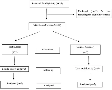
A total of 16 patients attending a private dental hospital presenting with a complaint of esthetic concerns related to their smile were initially screened and finally 14 patients (8 women, 6 men) aged between 18 to 30 years, falling into the scope of the study, were elected (Fig. 1). Patients who met the following criteria were included in the study:
1. Absence of any systemic disease, 2. Good oral hygiene routine, 3. No attachment loss,
Fig. 1. Consort flow chart of the study
Smokers, noncompliant patients, patients that underwent any periodontal treatment in the area and those with delayed wound healing capabilities attributed to their systemic conditions (diabetes etc) as well as pregnant and lactating mothers were excluded from the study.
Pre treatment assessment comprised of clinical evaluation of the selected cases by an UNC-15 periodontal probe to check whether they met the criteria of altered passive eruption. The protocol was adapted from Rebeiro et al.[9] the cemento-enamel junction (CEJ) location was first investigated employing a probe. Cases with an overlap of the gingival margin of the enamel coronal to the CEJ, with presence of sufficient distance between the osseous crest and the CEJ assessed by radiographs, were considered as altered passive eruption by definition.
The study protocol was properly explained to the patients and a signed informed consent was taken from all the patients. The patients were randomly assigned to either the Test (Laser) or Control (Scalpel) groups by a computer- generated randomization.
After a thorough oral prophylaxis, the patients were assessed for their willingness in
maintenance of a proper oral hygiene before being recalled for the correction of
gummy smile. A proper assessment of the subject’s smile was done prior to the anesthesia as any form of anesthesia alters the smile pattern.
For the laser group, a topical anesthetic agent, Precaine® B (20% benzocaine) was sufficient. A diode laser (Sunny™, MSI, Bengaluru) with an 808-nm wavelength was used.
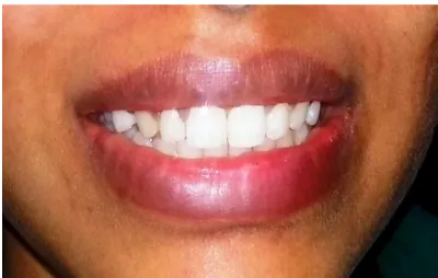
Fig. 2. Preoperative photograph showing gummy smile
Fig. 3. Intraoperative photograph during the procedure with laser
Fig. 4. Postoperative photograph after gummy smile correction with laser
In the scalpel group, after sufficient anesthesia was achieved using 2% lignocaine with 1:80,000 adrenaline, a periodontal probe was used to delineate a proper biologic width and demarcation of the excess gingival tissue to be removed. Crown lengthening with an external bevel-gingivectomy was planned using the sulcular and external bevel incisions and the excess gingival tissue was removed using Gracey curettes for recontouring the gingival (Figs. 5-7). The required 3 mm of distance for
establishment of a proper biologic width between the CEJ and the bone crest was properly checked in both groups.
Fig. 5. Preoperative photograph showing gummy smile of case 2
Fig. 6. Intraoperative photograph during the procedure with scalpel
Fig. 7. Postoperative photograph after gummy smile correction with scalpel
prescribed to be used twice a day for 2 weeks to enhance a proper plaque control for both groups.
The observation period was one month. The perceptions of the patient’s pain and discomfort related to the procedure were evaluated through a visual analog scale (VAS) intraoperatively and at the 1st, 3rd and the 7th day.
The VAS comprised of 10 cm scale with 0 indicated as “no” pain and 10 indicated as “worst pain imaginable”. Patients were instructed to mark their pain levels on this scale. To get the patients acquainted with the VAS scale, a preliminary pilot trial was done explaining the VAS protocol to the patients.
The esthetic satisfaction perceived by the patients after the treatment was analyzed by a questionnaire without a statistical analysis considering the small sample. The patients were asked to fill a questionnaire comprising of questions related to the esthetic change after a week and the expected outcomes related to both the treatment protocols after one month. The evaluation of the treatment outcome by a professional photographer was implemented through a questionnaire in this study.
2.1 Statistical Analysis
Statistical analysis was performed using Statistical Package for Social Sciences SPSS Version 20.0 (SPSS Inc, Chicago Illinois, USA). The statistical significance of data for patient perceptions related to pain and discomfort within
the groups was determined by one way analysis of variance & Post Hoc test and between the groups was determined by the paired t-test. Changes were considered significant at the P < 0.05 levels.
3. RESULTS
None of the 14 patients included in the study complained of any adverse events throughout the study period. Fig. 1 depicts the consort flow chart of the study.
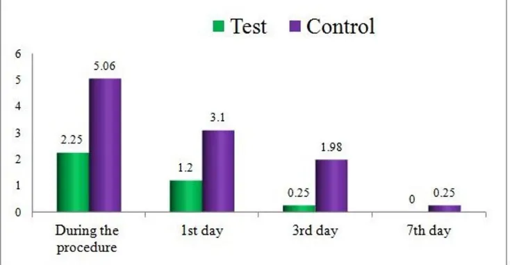
Comparison of the mean VAS scores of the levels of pain for both groups, observed intra operatively, 1st, 3rd and the 7th day of the study are summarized in Table 1 and Graph 1.
Study analysis revealed that there was a significant difference in VAS scores of pain (P < 0.05), in the laser group displaying significantly lower VAS scores intraoperatively and on the first day. No relevant significance was found on the 3rd and the 7th days.
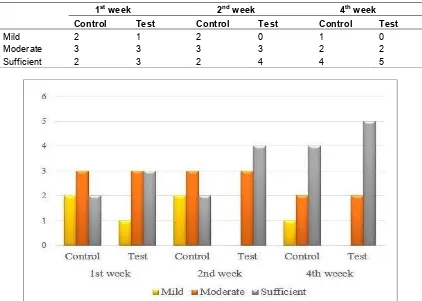
Table 2 and Graph 2 summarize the patient’s perception of the cosmetic change within a period of 4 weeks between both groups. 5 out of 7 patients in test group reported the result to be satisfactory after a week. However, during the same period, only 2 out of 7 patients in the control group were satisfied with the cosmetic change. After a four week observation period, both groups expressed almost similar cosmetic change perceptions (5 from laser and 4 from scalpel groups).
Table 1. Comparison of VAS scores between test and control groups
Vas
Group VAS score comparison
Test (laser) Control (scalpel) P value
During the procedure 2.55 ± 0.85 5.06 ± 0.51 0.041* 1st day 1.29 ± 0.57 3.1 ± 0.21 0.048* 3rd day 0.25 ± 0.43 1.98 ± 0.33 0.064# 7th day 0 0.25 ± 0.23 0.178#
*Statistically significant (P<0.05), #statistically not significant (P>0.05), VAS=Visual Analog Scale
Table 2. Patient evaluation for perception of cosmetic change at end of 1st, 2nd and 4th week
after treatment
1st week 2nd week 4th week
Control Test Control Test Control Test
Mild 2 1 2 0 1 0
Moderate 3 3 3 3 2 2
Sufficient 2 3 2 4 4 5
Graph 2. Perception of cosmetic change at end of 1st, 2nd and 4th week after treatment
Table 3 summarizes the perception regarding
preferred expectation of treatment after 4 weeks. 3 out of 7 patients in the control
group and 5 out of 7 in the test group were totally satisfied with the expected result. Two patients were dissatisfied in the control group which was not observed in the test
group.
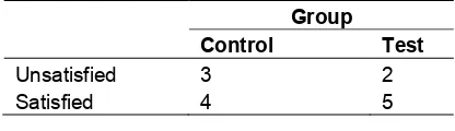
Table 4 summarizes the perception of the result evaluated by a professional photographer. He was dissatisfied with smile in two patients in the
control group and with one patient in the test group after the smile correction.
Table 3. Patient evaluation for perception regarding expected outcome from the
treatment after 4th week
Group
Control Test
Table 4. Patient evaluation for perception of the result by a professional photographer
Group
Control Test
Unsatisfied 3 2 Satisfied 4 5
4. DISCUSSION
This study is the first of its kind, comparing the patient perceptions related to pain and esthetic outcomes for a smile correction using either the laser or scalpel. Our results stand by our claim and fully support our hypothesis that the laser group would result in better outcomes compared to the scalpel group in terms of patient perceptions of pain and discomfort in the initial period of the treatment. This initial perception is crucial for the patient’s acceptability of the treatment modality.
Gummy smile associated with an APE type 1A corresponds to a gingival position placed above the CEJ sufficient enough over the alveolar bone, in which osseous recontouring can be avoided
and the procedure can be accomplished by a gingivectomy / gingivoplasty. Treatment of
excessive gingival display associated with APE type 1A includes conventional crown lengthening employing scalpel, electrocautery and lasers [6-8].Lasers have been utilized in our study as they had a proven advantage of being painless, providing excellent hemostasis and delivering precise treatment.
Infiltration anesthesia is a major concern for certain apprehensive patients. Comparing the anesthesia patterns in our study, almost 90.4% of the subjects in the laser group underwent gingival recontouring with topical anesthesia alone whereas most of the patients (99%) of the scalpel group had to bear the pain of infiltration anesthesia. Our results are in accordance with studies by Panagiotis et al. [10] and Fornaini et al. [11] as they have used only topical anesthesia in all their laser soft tissue procedures without the need of any infiltration anesthesia.
Comparing the laser group with the scalpel group, the results of the present study have revealed that the duration of the procedure and the simplicity accompanied with the precise handling of the tissues had better outcomes for the laser group. There was minimal bleeding as
the laser can seal vessels with a diameter of 0.5 mm enhancing hemostasis in the surgical area which made the procedure gain high acceptability and reshaping of the gingiva was done with proper control as the laser tip can precisely remove the gingival epithelium without causing any damage to the underlying bone [12].
Assessment of pain is cumbersome considering the physiological and psychological factors involved. We have used a VAS scale which has been reported to be sensitive and reliable [13]. When the patient perceptions for pain were compared between both groups, there was a reduction in the VAS scores intra operatively and on the 1stday in the laser group which might have been attributed to the fact that laser aids in the deposition of a protein coagulum which acts as a biological dressing sealing the sensory nerves and making the wound sterile with less inflammation, reduced bacteremia and pain
according to the studies in the literature [14-16].
The higher VAS scores post operatively in the
scalpel group can be attributed to the more invasive surgical procedure, accompanied
by an open wound which heals at secondary intention.
When comparing the perceptions of the subjects related to the esthetic outcomes, subjects in the laser group have responded positively to the esthetic change seen after a week compared with those of the scalpel group. This might have been attributed to a better visualization of the surgical field immediate post operatively as the lasers coagulate the tissue, leading to a better visualization, hence increasing the patient’s acceptance. Both groups reported equal levels of satisfaction towards the treatment procedure after 1 month.
This was the first study of its kind incorporating an evaluation of the treatment outcome by a professional photographer implemented through a questionnaire. We were convinced that he would be apt in visualizing the smile change pre and post treatment and to compare the differences in opinion if any. Though there were no major scoring differences for both groups, the laser group had a slight advantage over the scalpel group.
the use of analgesics among both groups through a small observational study and it was evident that none of the subjects in the test group had used an analgesic whereas a total of four subjects of the control group had used the analgesics.
One major limitation of our study was the sample size. Considering the treatment protocol aimed at the correction of gummy smile associated with APE type 1A category, it was indeed difficult to include patients fulfilling the above mentioned criteria coupled with the follow up in a private dental office. There were certain other limitations concerning the correlation of VAS score between the gender and the sexes and analysis of esthetic change and outcomes assessed by a scoring system without statistics considering the sample size.
5. CONCLUSION
Providing an esthetic blueprint of a smile correction incorporating team work and satisfying patient’s concerns is indeed a challenge. Considering the results within the limitations of our study; lasers have indeed taken that challenge in part and have shown that they have some clinical relevance and can provide a precise, patient-friendly and safe treatment modality enhancing a superior esthetic outcome satisfying patient’s esthetic demands in smile corrections comprising of esthetic crown lengthening for the treatment of altered passive eruption.
Further studies with a larger sample size incorporating a wide array of parameters would throw light on the esthetic outcomes of such procedures.
ETHICAL APPROVAL
All authors hereby declare that all experiments have been examined and approved by the appropriate ethics committee and have therefore been performed in accordance with the ethical standards laid down in the 1964 Declaration of Helsinki.
COMPETING INTERESTS
Authors have declared that no competing interests exist.
REFERENCES
1. Garber DA, Salama MA. The aesthetic smile: Diagnosis and treatment. Periodontol 2000. 1996;11:18-28.
2. Allen EP. Use of mucogingival surgical procedures to enhance esthetics. Dent Clinic North Am. 1988;32:307-330.
3. Silberberg N, Goldstein M, Smidt A. Excessive gingival display-etiology, diagnosis, and treatment modalities. Quintessence Int. 2009;40:809-818. 4. Tjan AH, Miller GD, The JG. Some esthetic
factors in a smile. J Prosthet Dent. 1984; 51:24-28.
5. Coslet JG, Vanarsdall R, Weisgold A. Diagnosis and classification of delayed passive eruption of the dentogingival junction in the adult. Alpha Omegan. 1977;70:24–28.
6. Lee EA. Aesthetic crown lengthening: Classification, biologic rationale, and treatment planning considerations. Pract Proced Aesthet Dent. 2004;16:769-778. 7. Chu SJ, Karabin S, Mistry S. Short tooth
syndrome: Diagnosis, etiology and treatment management. J Calif Dent Assoc. 2004;32:143-152.
8. Adams TC, Pang PK. Lasers in aesthetic dentistry. Dent Clin North Am. 2004;48: 833-860.
9. Ribeiro FV, Hirata DY, Reis AF, Santos VR, Miranda TS, Faveri M, Duarte PM. Open-flap versus flapless esthetic crown lengthening: 12-month clinical outcomes of a randomized controlled clinical trial. J Periodontol. 2014;85:536-544.
10. Panagiotis K, Christos S, WaseemJ, Tahwinder U, Michael V, Marios T, Irene S. Upper lip laser frenectomy without infiltrated anaesthesia in a paediatric patient: A case report. Cases J. 2009;2: 713-718.
11. Fornaini C, Rocca JP, Bertrand MF, Merigo E, Nammour S, Vescovi P. Nd: YAG and diode laser in the surgical management of soft tissues related to orthodontic treatment. Photomed Laser Surg. 2007;25:381-92.
12. Pick RM, Colvard MD. Current status of lasers in soft tissue dental surgery. J Periodontol. 1993;64:589-602.
13. Scott J, Huskisson EC. Graphic representation of pain. Pain. 1976;2:175– 84.
wound healing following CO2 laser and
conventional surgical excision of canine buccal mucosa. Arch Oral Biol. 1983;28: 287-291.
15. Fenner J, Martin W, Moseley H, Wheatley DJ. Shear strength of tissue bonds as a function of bonding temperature: A
proposed mechanism for laser-assisted tissue welding. Laser Med Sci. 1992;7:39-43.
16. Wigdor H, Walsh J, Featherstone JDB, Visuri SR, Fried D, Waldvogel JL. Lasers in dentistry. Lasers Surg Med. 1995;16: 103-133.
_________________________________________________________________________________
© 2017 Koppolu et al.; This is an Open Access article distributed under the terms of the Creative Commons Attribution License
(http://creativecommons.org/licenses/by/4.0), which permits unrestricted use, distribution, and reproduction in any medium,
provided the original work is properly cited.
Peer-review history: