_____________________________________________________________________________________________________ *Corresponding author: E-mail: [email protected];
www.sciencedomain.org
Comparison of Shear Bond Strength of Different
Orthodontic Metal Bracket-bases Bonded on Enamel
Surface – an In vitro Study
Nirav Dholakiya
1*, Hina Desai
1, Mohammed Shahid Dal
1, Nikunj Patel
1,
Rahul Aghera
1and Nishil Agrawal
11
Department of Orthodontics and Dentofacial Orthopaedics, Manubhai Patel Dental College, Hospital and Oral Research Institute, Vadodara, India.
Authors’ contributions
This work was carried out in collaboration between all authors. Author ND designed the study, performed the statistical analysis, wrote the protocol and wrote the first draft of the manuscript. Authors HD, MSD and NP managed the analyses of the study. Authors RA and NA managed the
literature searches. All authors read and approved the final manuscript.
Article Information
DOI: 10.9734/BJMMR/2016/29260 Editor(s): (1) James Anthony Giglio, Adjunct Clinical Professor of Oral and Maxillofacial Surgery, School of Dentistry, Virginia Commonwealth University, Virginia, USA. Reviewers: (1) Andrea Scribante, University of Pavia, Italy. (2)Devinder Preet Singh, Dr. Harvansh Singh Judge Institute Of Dental Sciences & Hospital, Panjab University, Chandigarh, India. Complete Peer review History:http://www.sciencedomain.org/review-history/16596
Received 31st August 2016 Accepted 2nd October 2016 Published 18th October 2016
ABSTRACT
Aim: To compare the shear bond strength of different metal bracket bases bonded on enamel
surface and further to evaluate the Adhesive Remnant Index score to localize the sites of adhesive fracture.
Materials and Methods: Four types of premolar metal brackets were selected according to their
different mesh designs: (G1, Gemini Series,3M Unitek; G2,Micro Sprint, Forestadent; G3,Equilibrium 2,Dentaurum and G4,Mini-Master Series, American Orthodontics). One hundred brackets for each type were used and bonded on enamel surfaces of extracted human premolars (Transbond XT, 3M Unitek) and were tested to evaluate shear bond strength with an Instron Universal Testing Machine (Star Testing Systems, India). All data were analysed with ANOVA, Tukey’s HSD Post hoc test, and with descriptive statistics. The adhesive fracture site was also evaluated and classified with ARI score.
Results: G2 showed greater shear bond strength when compared with other samples (P < 0.001).
G2 and G3 showed statistically significant differences in comparison with other groups with respect to shear bond strength. There was no statistically significant difference between G1 and G4. The ARI index demonstrated a large variability. G2 showed 70% of the adhesive fracture at cement-enamel interface.
Conclusion: G2 showed the highest shear bond strength in comparison with other groups. ARI
score showed that G2 resulted in 70% adhesive fractures at the cement-enamel interface. An increased size of the bracket-base enhances adhesion but affects the adaptability to surface morphology of the enamel, increasing the risk of fracture at the interface with the bracket.
Keywords: Bracket-base; shear bond strength; adhesion; ARI score.
1. INTRODUCTION
The acid etching technique, introduced by Buonocore, has allowed the replacement of metal bands with directly cemented brackets [1]. The use of metal bracket with a retentive base was first reported by Mitchell in 1967 [2].
The bond strength of bracket is influenced by various factors, including the size and design of bracket base [3-12]. The material and design of the bracket must be having ability to deliver orthodontic forces and masticatory loads. It should also be an aesthetic and at the end of treatment, there should not be any damage to enamel surface during removal of the bracket [6]. The mechanical interlock between base-adhesive and resin-enamel plays an important role for the adhesion of metal brackets.
There are so many types of metal brackets with different types of bracket bases available in the market. They can be classified into two principle groups: Brackets having soldered base and brackets having integral bases. In the first group, the metal bases are soldered to the bracket bodies. In this group, various types of bases are used such as perforated bases, mesh foils, and photo-etched bases. In the second group, the base and the remaining parts of the bracket are a single casted. Four types of bases are categorised under this group: retention groove bases, mesh bases, waffle bases, and laser-structured bases.
There is a mechanical undercut which provides a room for the orthodontic adhesives to extend before polymerization. Most of the metal brackets with fine brazed mesh provide good retention [7]. Metal bracket bases have two types of designs: a single-piece casting formed with a retention groove on the base, and a mesh or a concave, circular form that is welded by laser with silver directly to the bracket body.
The laser-structured bases have many hole-shaped cavities on the base of the brackets that are realized by a laser beam scanned over the base surface. This type of design provides the retention. Other bracket bases are sandblasted, chemically etched, or sintered with porous metal powder [6-12].
Several studies [13,14] have reported that bond failure in metal brackets bonded to enamel surface occurs with 15 seconds of acid etching time in three possiblr areas: between the resin and enamel, within the resin itself, or at the resin-bracket base interface. However, they also found that there were relatively greater chances of bond failure between the resin and bracket because of concentration of stress and defects in the resin film. So to overcome all these problems, a bracket with sufficient retentive bonding between the resin and metal bracket base is needed.
Among all these factors the base retentive system is only one of the factors that influence the shear bond strength of metal brackets effectively. Furthermore, the entire bonding procedure and moisture contamination of an etched enamel can also modify the retention of metal brackets as well as affect the shear bond strength [15,16].
Many studies have compared the shear bond strength of metal brackets with different types of retentive bases [11,15,17-19].
The aim of this study was to evaluate and compare the shear bond strength of different types of retentive bases. Furthermore, the specimens were examined by using an adhesive remnant index (ARI) to localize the sites of adhesive fracture.
2. MATERIALS AND METHODS
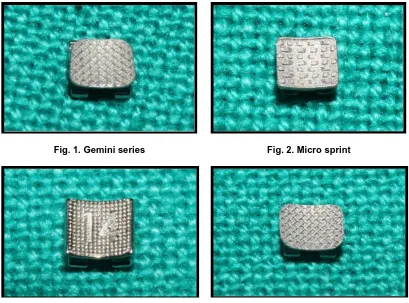
Unitek, Monrovia, Calif) (Fig. 1), Micro Sprint (G2) (Forestadent, Pforzheim, Germany) (Fig. 2), Equilibrium 2 (G3) (Dentaurum, Inspringen, Germany) (Fig. 3) and Mini-Master Series (G4) (American Orthodontics) (Fig. 4). One hundred brackets for each type were used. The mean base surface area of the brackets was calculated by measuring length and width and computing the area.
Four hundred human premolars were extracted, washed, and the pulp removed. The criteria of tooth selection were grossly perfect crown with absence of cracks caused by extraction forceps. The teeth were then embedded in self-curing acrylic resin (DPI), leaving the labial enamel exposed. The specimens were stored in normal saline solution for 1 week until testing.
A bracket placement site was prepared with pumice-powder paste-water with absence of fluoride content, rinsed it for 10 seconds, and dried. Before bonding, the enamel was etched with a 35% orthophosphoric acid gel solution for 15 seconds, sprayed for 20 seconds, and dried.
The adhesive primer (Transbond XT Primer, 3M Unitek) was uniformly applied on the enamel and sprayed with air to enhance the complete penetration of the resin matrix. After photopolymerization for 10 seconds, brackets with an adhesive (Transbond XT, 3 M Unitek) were positioned with the help of bracket positioning instrument and pressed it on the labial surfaces of the teeth. The excess of adhesive was removed, and the adhesive was cured by applying the light (Curing Light XL 3000, 3 M Unitek) for 10 seconds each (Fig. 5).
All specimens were tested on an Instron Universal Testing Machine (Star Testing
Systems, India. Model No. STS 248): A blade of an unit was placed at the bracket base-
enamel interface with a crosshead speed of 6 mm/min and a 50-kg load cell [20]. In this way all the brackets were shear tested to failure
(Fig. 6). The force producing failure was
recorded first in newtons and then converted it into megapascals by dividing the measured
force values by the mean surface area of the brackets.
Fig. 1. Gemini series Fig. 2. Micro sprint
Fig. 5. An extracted premolar with bracket bonded on exposed labial surface
Fig. 6. Instron universal testing machine with a sample
The ARI score was used to evaluate and compare the amount of adhesive left on the enamel surface after debonding and to
check the sites of adhesive fracture [21]. Brackets were observed with a
stereomicroscope at 10x magnification while the remaining part of adhesive was scored with respect to the amount of resin material left on the enamel:
ARI 0, less than 10% of the adhesive left on the enamel;
ARI 1, 10-50% of the adhesive left on the enamel;
ARI 2, 50-90% of the adhesive left on the enamel;
ARI 3, more than 90% of the adhesive remained on the enamel, where one can see a clear impression of the bracket base on the adhesive-enamel surface.
2.1 Statistical Analysis
The descriptive statistics was used to include mean and standard deviation (SD) for each and every group, in newton and in megapascal. A one-way analysis of variance (ANOVA) was used to compare the shear bond strength between four groups. The Tukey’s HSD Post hoc test was carried out to analyse the effect of bracket base design on mean shear bond strength of each group with other groups.
3. RESULTS
The overall mean shear bond strengths are shown in Table 1. The one-way ANOVA test was performed to compare the shear bond strength among four groups and it showed that there were statistically significant differences among the four groups (P = .001).
and G3 showed a significantly greater shear bonding force when data were expressed in megapascals.
The Tukey’s HSD post hoc test showed significant differences between brackets evaluated. The G2 and G3 presented significantly higher shear bond strength as compared to other samples (P = < 0.001). There were no significant differences between G1 and G4 (P = > 0.001) (Table 3).
The site of the fracture for each sample was evaluated with the Adhesive Remnant
Index (ARI) (Table 4). Three possible types of fractures were observed: (1) cohesive
fracture, within the body of the cement; (2) adhesive fracture, at the adhesive-bracket base or enamel-adhesive interface, and (3) mixed fracture.
4. DISCUSSION
In this study, all metal brackets had different types of base patterns and base surface areas, except G1 and G4, where the difference was only in base surface area.
Table 1. Descriptive statistics of shear bond strength (N) and nominal area (mm2)
Brackets Area
(mm2)
Mean (N)
SD SE 95% CI for mean
Minimum Maximum
Lower bound
Upper bound
Victory series G1 8.97 89.58 36.43 3.64 82.35 96.81 6.52 141.37 Micro sprint G2 7.90 150.49 35.11 3.51 143.53 157.46 110.11 218.79 Equilibrium 2 G3 10.4 153.03 83.87 8.39 136.38 169.67 80.41 368.14 Mini master series
G4
10.8 114.66 78.37 7.84 99.11 130.21 7.89 308.76
Table 2. Descriptive statistics of shear bond strength (MPa) and nominal area (mm2)
Brackets Area
(mm2)
Mean (N)
SD SE 95% CI for mean
Minimum Maximum
Lower bound
Upper bound
Victory series G1 8.97 9.99 4.06 0.41 9.19 10.80 0.72 15.78 Micro sprint G2 7.90 19.06 4.44 0.44 18.18 19.94 13.93 27.71 Equilibrium 2 G3 10.4 14.72 8.06 0.81 13.12 16.32 7.72 35.42 Mini master series
G4
10.8 10.65 6.10 0.61 9.44 11.86 4.02 25.61
Table 3. Statistical comparison (Tukey’s HSD post hoc test)
Bracket base comparison Newtons (N) Megapascals (MPa)
Victory series G1 G2 Micro sprint -60.9130000 -9.0660000 G3 Equilibrium 2 -63.4440000 -4.7280000 G4 Mini master
series
-25.0760000 -.6600000
Micro sprint G2 G1 Victory series 60.9130000 9.0660000 G3 Equilibrium 2 -2.5310000 4.3380000 G4 Mini master
series
35.8370000 8.4060000
Equilibrium 2 G3 G1 Victory series 63.4440000 4.7280000 G2 Micro sprint 2.5310000 -4.3380000 G4 Mini master
series
38.3680000 4.0680000
Table 4. Adhesive remnant index (ARI) scores in percentage
Value Criterion Interpretation G1 G2 G3 G4
ARI 0 No adhesive left on tooth (<10%)
Adhesive fracture at cement-enamel interface
0 70 0 40
ARI 1 Less than half of the adhesive left on the tooth
Mixed fracture 50 30 40 40
ARI 2 More than half of the adhesive left on the tooth
Cohesive fracture 40 0 30 10
ARI 3 All adhesive left on tooth (>90%)
Adhesive fracture at bracket-cement interface
10 0 30 10
G1 presents with a mesh foil having the most retentive size to allow more space for the penetration of the adhesive and the curing light during bonding procedure [11,22]. However, in some studies, the authors consider that the size of the bracket-base does not influence the retention significantly or that it also depends on the filler particles of the adhesive used [16,23] (Fig. 1).
G2 presents with the smallest base surface area among all those brackets selected in this study (7.9 mm2). In such brackets, a waffle base, which consists of metallic indentations coming out from the base of the bracket, enhances the retention. The photograph shows that waffle base is having unique design in which each indentation has been projected occluso-gingivally, creating adequate undercuts and the presence of free volume of space among the indentations allows the escape of air and excess resin (Fig. 2). The results for G2 obtained in this study are similar with previous study where such type of bracket base used [20].
G3 presents with laser-structured base with many hole-shaped cavities on the base of the brackets that are obtained by a laser beam scanned over the base surface, which provides the mechanical interlock and also enhances the retention. The photograph shows the presence of projecting metallic margins derived from the laser beam. It also presents with the quadrangular, anatomic and more concave base shape (Fig. 3).
G4 presents with similar mesh foil to G1, providing large space for the penetration of the adhesive and the curing light. The only difference is with the base surface area, which is larger than G1 (Fig. 4). In a previous study, similar brackets were used to evaluate shear bond strength and it was 10.68 MPa, which is similar to this study (10.65 MPa) [24].
Previous study [25] stated that the difference in designs of metallic meshes of bracket-bases gives contributions to more adherence of the adhesive material to the base of the bracket, achieving greater shear bond strength.
The shear bond strength also depends on the adhesive materials. Transbond XTTM is one of the most recommended products in current orthodontics. It has been a part of various comparative adhesion studies. In this study, all data were obtained with Transbond XTTM, which strongly associated with previous studies [26-29].
Reynolds and von Fraunhofer [30] stated that all the retentive designs used in the brackets tested should have an acceptable bond force levels (6– 8 MPa). However, there are various factors related to an oral environment or moisture contamination that may affect the shear bond strength. The moisture contamination of bracket-bases with water, saliva and blood has been shown to adversely affect the shear bond strength due to deposits of an organic adhesive layer immediately after exposure that is resistant to washing and subsequently it reduces the shear bond strength of brackets [17,31-33]. Ahmad Sheibaninia et al. [34] evaluated the effect of an acidic food simulating environment on shear bond strength of self-ligating brackets and stated that the margins of bracket-bases allows an acidic food to penetrate, which gradually decreases the shear bond strength. So care should be taken in predicting the results to those conditions. Arunima Goswami [35] et al. stated that moisture contamination did not affect the shear bond strength. It has been suggested that an adverse effect of moisture contamination on orthodontic bonding can be associated with water adsorption, which produce formaldehyde
So In vivo conditions have not been considered.
Generally, the shear bond strength is expressed in newton and the bonding force is expressed in megapascals while comparing the retention capacity of brackets selected. The values in megapascal can be obtained by dividing the values in newtons by the base surface areas, which directly reflect the effectiveness of the retention mechanism. When values are expressed in newton, G3 shows the highest shear bond strength. This value is not significantly greater than G2, but it is significantly greater than the values shown by the other specimens. When the values are expressed in megapascal, G2 shows the highest shear bonding force with respect to the others. Comparing the results, it is evident that the latter factor can influence the shear bond strength, which indicates that while increasing the surface area of the bracket, the load carrying capacity also increases, as observed in previous studies [11,16].
Cucu et al. performed an In vitro study to evaluate the effect of bracket-base size on shear bond strength and found no significant difference between bracket-base size and shear bond strength which confirms present findings where G2 having the smallest base surface area has the highest shear bond strength [36].
Finally, a large variability is seen in the fracture sites according to ARI index analysis in this study. G2 and G4 showed 70% and 40%, respectively, bond failures located at the enamel-adhesive interface (ARI 0) and 30-50% were having mixed fractures (ARI 1). There was no ARI 2 or ARI 3 score present for G2. This result is similar to previous study.[20] This seems to be a confirmation of the high retention of these bracket bases but there are high chances of enamel damage. Previous studies evaluated the shear bond strength of self-ligating brackets 24 h after immersion in water found higher frequencies of ARI score 3 [37,38]. Other study [31,39] stated that under moisture contamination water and saliva, there is a higher frequency of ARI score 0. In general, ARI results are very subjective and they should be evaluated carefully [40].
5. CONCLUSION
• In this study, shear bond strength of all four types of brackets tested is clinically acceptable.
• The waffle base of micro sprint brackets showed the highest shear bond strength followed by laser-structured base of equilibrium 2 brackets compared with other samples.
• By increasing the base surface area of the bracket, the load carrying capacity can be increased, but causes a decrease in adaptation.
• The ARI index score values showed a large variability. Waffle base of Micro sprint brackets showed 70% of adhesive fracture at the enamel-adhesive interface (ARI 0), which is the highest retention of these bracket-bases when compared with other brackets but on other side there are higher chances of enamel damage.
• So shear bond strength of the bracket does not solely depend on the base surface area, but it also depends on the base surface characteristics and the properties of the adhesive materials.
CONSENT
It is not applicable.
ETHICAL APPROVAL
Institutional Ethics Committee (IEC) for Research Manubhai Patel Dental College, Hospital and Oral Research Institute, Vadodara. REF. NO.: IEC/MPDC_069/Ortho-16/15
COMPETING INTERESTS
Authors have declared that no competing interests exist.
REFERENCES
1. Buonocore MG. A simple method of increasing the adhesion of acrylic filling materials to enamel surfaces. J Dent Res. 1955;34:849–853.
2. Newmann GV. Epoxy adhesives for orthodontic attachments: progress report. Am J Orthod Dentofacial Orthop. 1965; 51:901–912.
3. Bishara SE, Gordan VV, VonWald L, Olson ME. Effect of an acidic primer on shear bond strength of orthodontic brackets. Am J Orthod Dentofacial Orthop 1998;114: 243-7.
adhesives: Composite resin, hybrid GIC, and glass-filled GIC. Am J Orthod Dentofacial Orthop. 2001;119:36-42. 5. Grandhi RK, Combe EC, Speidel TM.
Shear bond strength of stainless steel orthodontic brackets with a moisture-insensitive primer. Am J Orthod Dentofacial Orthop. 2001;119:251-5. 6. Knox J, Hubsch P, Jones ML, Middleton J.
The influence of bracket base design on the strength of the bracket-cement interface. Br J Orthod. 2000;27:249-54. 7. Smith DC, Maijer R. Improvement in the
bracket base design. Am J Orthod Dentofacial Orthop. 1983;83:277-88. 8. Rossouw PE, Titley KC, Yamin C. The
relationship between bond strength and base surface area using conventional and micro-etched foil-mesh bases. Am J Orthod Dentofacial Orthop. 1988;113:276-81.
9. Lopez JI. Retentive shear strengths of various bonding attachment bases. Am J Orthod Dentofacial Orthop. 1980;77:669-78.
10. Maijer R, Smith DC. Variables influencing the bond strength of metal orthodontic bracket bases. Am J Orthod Dentofacial Orthop. 1981;79:20- 34.
11. Maijer R, Smith DC. Variables influencing the bond strength of metal orthodontic bracket bases. Am J Orthod Dentofacial Orthop. 1981;79:20–34.
12. Hanson GH, Gibbon WM, Shimizu H. Bonding bases coated with porous metal powder: a comparison with foil mesh. Am J
Orthod Dentofacial Orthop 1983;83:1-4.
13. Wang WN, Yeh CL, Fang BD, Sun KT, Arvystas MG. Effect of phosphoric acid concentration on bond strength. Angle Orthod. 1994;64:377-82.
14. Wang WN, Lu ZC. Bond strength with various etching times on young permanent teeth. Am J Orthod Dentofacial Orthop. 1991;100:72-9.
15. Wang WN, Li CH, Chou TH, Wang DDH, Lin LH, Lin CT. Bond strength of various bracket base designs. Am J Orthod Dentofacial Orthop. 2004;125:65–70. 16. Bishara SE, Oonsombat C, Ajlouni R,
Denehy G. The effect of saliva contamination on shear bond strength of orthodontic brackets when using a self-etch primer. Angle Orthod. 2002;72:554– 557.
17. Lopez JI. Retentive shear strengths of various bonding attachment bases. Am J
Orthod Dentofacial Orthop. 1980;77:669– 678.
18. Fernandez L, Canut JA. In vitro
comparison of the retention capacity of new aesthetic brackets. Eur J Orthod. 1999;21:71–77.
19. MacColl GA, Rossouw PE, Titley KC, Yamin C. The relationship between bond strength and orthodontic bracket base surface area with conventional and microetched foilmesh bases. Am. J. Orthod. Dentofacial Orthop. 1998;113: 276–281.
20. P. Cozza, Martucci L, de Toffol L, Pencoc SI. Shear bond strength of metal brackets on enamel. Angle Orthodontist. 2006; 76(5):851–856.
21. Artun J, Bergland S. Clinical trials with crystal growth conditioning as an alternative to acid-etch enamel pretreatment. Am J Orthod. 1984;85:333– 340.
22. Sharma-Sayal SK, Rossouw PE, Kulkami GV, Titley KC. The influence of orthodontic bracket base design on shear bond strength. Am J Orthod Dentofacial Orthop. 2003;124:74–82.
23. James JW, Miller BH, English JD, Tadlock LP, Buschang PH. Effects of high-speed curing devices on shear bond strength and microleakage of orthodontic brackets. Am J Orthod Dentofacial Orthop. 2003;123: 555–561.
24. Kazem Dalaie, Armin Mirfasihi, et al. Effect of bracket base design on shear bond strength to feldspathic porcelain. Eur J Dent. 2016;10(3):351–355.
25. Téssia Richelly Nóbrega Borja de Melo, Michel Nicolau Youssef, et al. Shear Bond Strength of Metallic Brackets: An In vitro Study. Brazilian Research in Pediatric Dentistry and Integrated Clinic. 2015, 15(1):319-325
26. Susanne Reimann, Judith Mezey, et al. The influence of adhesives and the base structure of metal brackets on shear bond strength. J Orofac Orthop. 2012;73(3): 184-93.
27. Amm EW, Hardan LS, BouSerhal JP, et al. Shear bond strength of orthodontic brackets bonded with self-etching primer to intact and preconditioned human enamel. J Orofac Orthop. 2008;69:383–392
bonding systems. Dent Mater J. 2008; 27:392–399
29. Vicente A, Bravo LA. Influence of an etchant and a desensitizer containing benzalkonium chloride on shear bond strength of brackets. J Adhes Dent. 2008; 10:205–209.
30. Reynolds IR, von Fraunhofer JA. Direct bonding of orthodontic attachments to teeth: The relation of adhesive bond strength to gauge mesh size. Br J Orthod. 1976;3:91–95.
31. Bianca Mota Santos, Matheus Melo Pithon, et al. Shear bond strength of brackets bonded with hydrophilic and hydrophobic bond systems under contamination. Angle Orthod. 2010;80: 963–967.
32. Ascensio´n Vicente, Ana Mena, et al. Water and saliva contamination effect on shear bond strength of brackets bonded with a moisture-tolerant light cure system. Angle Orthod. 2009;79:127–132.
33. Maria Francesca Sfondrini, Danilo Fraticelli, et al. Shear bond strength of orthodontic brackets and disinclusion buttons: Effect of water and saliva contamination. BioMed Research International 2013;2013:6. Article ID 180137.
34. Ahmad Sheibaninia, Sepehr Sepasi, et al. The effect of an acidic food-simulating environment on the shear bond strength of self-ligating brackets with different base
designs. International Journal of Dentistry. 2014;689-536.
35. Arunima Goswami, Borah Mitali, et al. Shear bond strength comparison of moisture insensitive primer and self-etching primer. J Orthod Sci. 2014;3(3): 89–93.
36. Cucu M, Driessen CH, Ferreira PD. The influence of orthodontic bracket base diameter and mesh size on bond strength. SADJ. 2002;57:16‑20.
37. Chalgren R, Combe EC, Wahl AJ, Effects of etchants and primers on shear bond strength of a self-ligating esthetic orthodontic bracket. Am J Orthod Dentofacial Orthop. 2007;132(5)577.e1– 577.e5.
38. Northrup RG, Berzins DW, Bradley TG, Schuckit W, Shear bond strength comparison between two orthodontic adhesives and self-ligating and conventional brackets. Angle Orthodontist. 2007;77(4)701–706,.
39. Sayinsu K, Isik F, Sezen S, Aydemir B, Effect of blood and saliva contamination on bond strength of brackets bonded with a protective liquid polish and a light-cured adhesive. American Journal of Orthodontics and Dentofacial Orthopedics. 2007;131(3):391–394.
40. Lalani N, Foley TF, Voth R, Banting D, Marnandras A. Polymerization with the argon laser: Curing time and shear bond strength. Angle Orthod. 2000;70:128–133. _________________________________________________________________________________ © 2016 Dholakiya et al.; This is an Open Access article distributed under the terms of the Creative Commons Attribution License (http://creativecommons.org/licenses/by/4.0), which permits unrestricted use, distribution, and reproduction in any medium, provided the original work is properly cited.
Peer-review history: