Copyright 0 1996 by the Genetics Society of America
Production of Androgenetic Zebrafish (Danio
rerio)
Graham
E.
Corley-Smith, Chinten James
Lim
and
Bruce
P.
Brandhorst
Institute of Molecular Biology and Biochemistry, Simon Fraser University, Burnaby, British Columbia V5A lS6, Canada Manuscript received October 11, 1995
Accepted for publication January 8, 1996
ABSTRACT
To help investigate the evolutionary origin of the imprinting (parent-of-origin mono-allelic expression) of paternal genes observed in mammals, we constructed haploid and diploid androgenetic zebrafish (Danio r h o ) . Haploid androgenotes were produced by fertilizing eggs that had been X-ray irradiated to eliminate the maternal genome. Subsequent inhibition of the first mitotic division of haploid andro- genotes by heat shock produced diploid androgenotes. The lack of inheritance of maternal-specific DNA markers (RAF'D and SSR) by putative diploid and haploid androgenotes confirmed the androgene- tic origin of their genomes. Marker analysis was performed on 18 putative androgenotes (five diploids and 13 haploids) from six families. None of 157 maternal-specific RAF'D markers analyzed, some of which were apparently homozygous, were passed on to any of these putative androgenotes. A mean of
7.7 maternal-specific markers were assessed per family. The survival of androgenetic zebrafish suggests that if paternal imprinting occurs in zebrafish, it does not result in essential genes being inactivated when their expression is required for development. Production of haploid androgenotes can be used to determine the meiotic recombination rate in male zebrafish. Androgenesis may also provide useful information about the mechanism of sex determination in zebrafish.
Z
EBRAFISH (Danio r h o , formerly known as Brachy- danio rerio; MEYER et al. 1993) are an important model for studying vertebrate development and are amenable to genetic analysis (STREISINGER et al. 1981;KIMMEL 1989; BARINAGA 1990, 1994; NUSSLEIN-VOL
HARD 1994; CONCORDET and INGHAM 1994; DRIEVER et
al. 1994; KAHN 1994). Their use as a model organism for genetic analysis is facilitated by a linkage map of DNA markers (POSTLETHWAIT et al. 1994) and large scale screens for mutations are underway (DRIEVER et al. 1994; KAHN 1994; MULLINS et al. 1994).
To investigate imprinting in vertebrates and to help develop zebrafish as a genetic system, we constructed haploid and diploid androgenotes. Construction of in- dividuals with uniparental inheritance can facilitate ge- netic analysis. Haploid and diploid gynogenotes have been produced by fertilizing zebrafish eggs with sperm irradiated to eliminate the paternal genome (STREI-
SINGER et al. 1981; HORSTGEN-SCHWARK 1993). Haploid
gynogenotes were used to produce a zebrafish linkage map based on rates of meiotic crossing over in oocytes (POSTLETHWAIT et al. 1994). Haploid gynogenotes com- plete embryogenesis and arrest as larvae. Thus, haploid embryos can be used for F1 mutant screening (KIMMEL
1989) : mutations in the stem cells of the maternal germ line are introduced by fertilization with mutagenized sperm or by mutagenesis of early embryos. Such screens require maintenance of considerably fewer progeny to recover an interesting recessive mutation from the ma-
Corresponding author Graham E. CorleySmith, Institute of Molecu- lar Biology and Biochemistry, Simon Fraser University, Burnaby, B.C.
V5A 1S6, Canada. E-mail: corley@sfu.ca
Genetics 142: 1265-1276 (April, 1996)
ternal stock than conventional diploid screens, which require production of an F3 generation (KAHN 1994; MULLINS et al. 1994). Diploid gynogenotes can be pro- duced by inhibiting extrusion of the second polar body or by inhibiting the first mitotic division of the gynogen- ote (STREISINGER et al. 1981; HORSTGEN-SCHWARK 1993). In the latter instance, the progeny are homozygous and clonal lines of gynogenetic zebrafish have been pro- duced (STREISINGER et al. 1981; KIMMEL 1989).
Androgenetic haploid progeny would result from fer- tilization of eggs that have been treated to eliminate the maternal genome. A method for production of an- drogenetic zebrafish has not been reported, but would facilitate determination of rates of meiotic recombina- tion in males, mapping of male specific DNA markers and linkage groups (if any), and analyses of sex determi- nation and genomic imprinting. Use of androgenetic haploids may have some usefulness for F1 mutant screens if interesting mutations can be reliably recov- ered from cryopreserved milt long after the screen was performed.
1266 G. E. Corley-Smith, C . J. Lim and B. P. Brandhorst
or mitochondrial DNA. Genomic imprinting of essen- tial genes that are irreversibly suppressed at a required developmental stage and derived from the paternal ge- nome as in mammals (MCGRATH and SOLTER 1984; SUR- ANI et al. 1984; SURANI 1986; BARRA and RENARD 1988; SAPIENZA 1990; RENARD et al. 1991; GOLD and PEDERSON 1994; CHAILLET et al. 1995) would make the survival of androgenotes impossible. Recessive lethal mutations could limit the successful production of androgenotes, but this is not expected to be an insurmountable prob- lem since gynogenetic homozygous zebrafish have been produced (STREISINGER et al. 1981; HORSTGEN-SCHWARK 1993), and inbred lines are available. We present ge- netic evidence that haploid and diploid androgenetic zebrafish can be constructed. To confirm androgenesis, lack of inheritance of maternal markers is a crucial part of the analysis. Markers relying on gene expression (e.g., phenotypic traits, isozymes, and allozymes) can be af- fected by many factors including imprinting, tissue spe- cific expression, and developmental specific expression. Failure to detect a maternal marker that results from gene expression in a putative androgenote can be at- tributed to lack of maternal DNA in the putative andro- genote or to lack of expression. Thus, we directly as- sayed the DNA of putative androgenotes for maternal specific markers using PCR methods.
METHODS AND MATERIALS
Production of androgenetic fish Androgenetic haploids are produced by irradiating eggs to destroy the maternal ge- nome followed by fertilization. By inhibition of the first mi- totic division, diploid androgenotes can be produced.
A Torrex 150D X-ray inspection system (Faxitron X-Ray Gorp., Buffalo Grove, IL) was used to irradiate eggs. X-ray dosimetry was performed with a MDH1515 dosimeter using a MDH model 10x5-180 ion chamber (paddle chamber). This was calibrated with a known I3'Cs source (NBS source No. 47455). The appropriate dose to eliminate the maternal DNA without unduly decreasing subsequent survival rates was deter- mined based on the Hertwig effect (HERTWIG 1911). Eggs were collected from fish anesthetized in 17 ppm (wv) Tricaine (3-aminobenzoic acid ethyl ester, Sigma A-5040; pH adjusted to -7 with sodium bicarbonate) by gently squeezing the abdo- men, Eggs were collected into a silanized capillary tube and placed into
-
100 p1 of coho salmon (Oncorhynchus kisutch)ovarian fluid (the fluid surrounding mature eggs) in a petri dish. The milt was collected just prior to being used for fertil- ization and was held i.n sperm extender: 80 mM KCI, 45 mM NaCI, 0.4 mM CaCl,, 0.2 mM MgCl,, 45 mM sodium acetate, and 10 mM HEPES, pH 7.7 (GIBBS et al. 1994).
In the first experiment, eggs were collected from one fe- male, and divided into eight groups. Each group of -I00 (88-1 12) eggs was held in coho salmon ovarian fluid and was exposed to a different total accumulated irradiation dose. All eggs were simultaneously inseminated, and survival was scored at 1 day postfertilization (p.f.). In the second trial, five groups of eggs (69-102 eggs) from a single female were irradiated with different total accumulated doses of X-rays. Living em- bryos were scored at 1 day p.f. and at 4 days p.f. according to appearance. Based on the results of these two dose response trials, a dose of 10,000 R was used in the following experi- ments to produce androgenotes.
The first mitotic division was inhibited by heat shock treat- ment. After fertilization, eggs were held at 28.5 ? 0.5" for 13 min, then heat shocked for 2 min at 41.4 ? 0.05", and re- turned to 28.5" (modified from STREISINCER et al. 1981). Tem- peratures were measured with a calibrated thermometer (Fisher Scientific, Cat. No. 15041A) having an uncertainty certified not to exceed 0.03'.
Families analyzed Fish from two laboratory lines of fish were used: *AB line (star AB line), which has been screened to reduce recessive lethals (C. WALKER, personal communica- tion) and the SFU line, which originated from zebrafish bought from pet stores on Vancouver Island, B.C., Canada.
Production of haploid or diploid androgenotes was at- tempted in 14 families (two families used for Hertwig experi- ments plus families A-H; Tables 4 and 5, plus seven other families). In six of these families (Table 5), inheritance of parental DNA markers was assessed in a sample of the putative androgenetic progeny, with family A being subjected to the most thorough genetic analysis.
Family A Eggs were collected from one female of the SFU line, and the milt was from one male of the *AB line. The eggs were held in coho salmon ovarian fluid at room tempera- ture for 50 min, the time required for irradiation of eggs. Of the 280 eggs collected, 76 eggs were not X-ray irradiated ( N l ) and 204 eggs were irradiated (I) with 10,000 R of X-rays. All eggs were then simultaneously inseminated. Of the irradiated eggs, 49 were not heat shocked (I/NHS; treatment to produce putative haploid androgenotes: PHA) and 155 were heat shocked (I/HS, treatment to produce putative diploid andro- genotes: PDA). The NI/NHS (not irradiated and not heat shocked) treatment was used to produce normal biparental diploid progeny: BDP.
DNA extraction: The following tissue samples were col- lected to prepare DNA extracts: both parents (caudal fin clips); 12 biparental diploid progeny (whole fish collected at 5 days postfertilization (p.f.); two putative haploid andro- genotes (whole fish collected at 5 days p.f.); one putative diploid androgenote (whole fish at 9 days p.f.); and a second putative diploid androgenote (caudal fin clip at 1.5 months p.f.). Whole fish were collected before feeding to decrease DNA contamination. All tissue samples were thoroughly rinsed with pure water before DNA extraction. DNA was pre- pared by phenol/chloroform extraction as described by ASH- BURNER (1989). The protocol was slightly modified by ex- tracting twice with phenol, twice with 1:l phenol/chloroform and once with chloroform; DNA precipitation was done by adding one-tenth volume 2.5 M sodium acetate (pH 5.5) and 2 volumes cold absolute ethanol. Quantification of DNA was done by spectrofluorometry using Pico-Green (Molecular Probes, Inc., Eugene, OR) on a SLM 4800 C Subnanosecond Spectrofluorometer (SLM Aminco, SLM Instruments Inc., Ur- bana, IL). DNA quantification standards were prepared from calf thymus DNA (Sigma, D 1501).
Androgenetic Zebrafish 1267
(NAPS) Unit, University of British Columbia, Vancouver, B.C., Canada.
PCR conditions: For RAPD amplifications, 4 ng of each template DNA was added to an aliquot of PCR cocktail con- taining: 1 X Idaho 3 mM Mg buffer (Idaho Technology Inc., Idaho Falls, ID), 100 p~ of each dNTP (Pharmacia) and 0.06 U/pl of Taq DNA polymerase (Promega in storage buffer B) and 1.6 p~ of one of the three fluorescendy labeled 10-mer primers. Thermocyling was performed in heat-sealed glass capillary tubes containing a total volume of 10 p1 in an Idaho 1605 Air-Thermo-Cycler. Two cycles of 91" for 60 sec, 42" for
7 sec, and 72" for 70 sec was followed by 38 cycles of 91" for
1 sec, 42" for 7 sec, and 72" for 70 sec, which was followed by a Smin hold at 72". SSR (simple sequence repeats) conditions were identical except that annealing was at 69" for 10 sec.
Analysis of RAPD-PCR products: Aliquots of each PCR re- action were separated by electrophoresis on agarose and poly- acrylamide gels. One aliquot of each PCR reaction was loaded onto 1.8% agarose gels in 0.5X TBE (0.045 M Tris base, 0.045 M boric acid, 0.001 M EDTA) containing 0.5 pg/ml of ethid- ium bromide. Electrophoresis was performed at 3 V/cm. Ethidium bromide staining was visualized with a 300-nm trans- illuminator, and the images were captured on a W Gel Documentation System (W, San Gabriel, C A ) . These im- ages were analyzed using the NCSA GelReader program, Mac- intosh version 2.0.5 (National Center for Supercomputing Applications, University of Illinois at Urbana-Champaign). The second aliquot of each RAPD-PCR reaction was loaded onto a 4% native polyacrylamide gel on an AB1 373 DNA Sequencer. ABI's fluorescent ROX-GS2500 inlane size stan- dard was also loaded onto all lanes. Data were collected with the AB1 GeneScan Collection Software (version 1.1) and ana- lyzed with the AB1 GeneScan Analysis Software (version 1.2.2- 1). RAPD markers are dominant markers and were scored as either present or absent. By scoring for presence or absence of each marker in 12 normal biparental diploid progeny, it was determined if the marker was heterozygous or apparently (see below) homozygous in the parent. To check that the markers were segregating in a normal Mendelian fashion, for each of the heterozygous markers the number of BDP containing that marker were scored. The observed present:ab- sent ratio in the F, was then checked against the theoretical ratio of 1:l using chi-square analysis, employing the Yates correction for continuity. To check that markers were a s sorting independently, each heterozygous marker was then tested for cis or trans linkage with each other marker. Markers were named according to the convention adopted for the zebrafish RAPD linkage map (POSTLETHWAIT et al. 1994): markers are prefixed with the RAPD primer used and suffixed with the size designation obtained from the Genescan soft- ware. For example, 21ObcJ453 is a marker amplified with the sequence of primer 210 from the UBC primer series, the F designates a FAM labeled primer, and 453 designates the size of the amplified fragment.
MHC and SSR pedigree analysis: To further investigate inheritance to putative androgenetic progeny, other PCR- based methods were used to screen for polymorphisms be- tween parental DNAs. The MHC (Major Histocompatibility Complex class I1 genes) primers Tu360 and Tu385 (ONO et
al. 1992) were used as they often show polymorphisms be- tween the AB line of zebrafish and other zebrafish strains (POSTLETHWAIT et al. 1994). We also used a set of 16 forward and reverse simple sequence repeat (SSR) primers (GOFF et
al. 1992). The fragments amplified by the MHC primer Tu360 and Tu385, and the fragments amplified by the SSR primers have been placed on the zebrafish linkage map (POSTLE-
THWAIT et al. 1994).
100
80
n
8
=
60 ((I>
5
40v)
.-
20
0 100 1000 10000 100000
Irradiation Dose
(R)
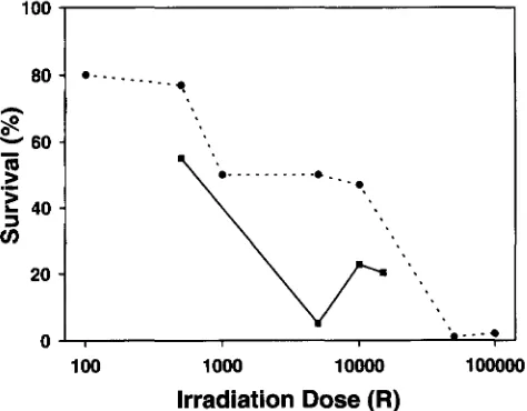
FIGURE 1.-Irradiation dose response curve. The percent- age of eggs irradiated at each dose surviving at 24 hr p.f. is shown. 0 , first experiment in which embryos were considered surviving if the chorions were transparent and some cleavage was observed; W, a second experiment in which only those embryos having transparent chorions, some cleavage and a distinct body axis were scored as survivors.
RESULTS
Irradiation dosage: In the first Hertwig dose re- sponse survival trial (Figure 1) , a shoulder was observed a t 10,000
R.
Survival in the first trial included embryos that developed as masses of cells without any discernible body axis a t 24 h r p.f. In the second trial, only those surviving embryos that developed a body axis are repre- sented in Figure 1. For 5000 R dosage group at 24 hr,5% had developed a body axis a n d 52% had developed as a mass of cells with n o body axis. These were both scored as alive in experiment 1. When viewed a t 4 days p.f., in the unirradiated group, all embryos alive a t 24
h r were still alive a n d all appeared to be normal diploids (see below and Figure
2 ) ,
the group irradiated with500 R had four surviving embryos that appeared to be normal diploids, and the rest of the group had moder- ate to severe abnormalities, none of which displayed the haploid syndrome (see below). All embryos from the 5000 R group were dead and had arrested as grossly abnormal embryos. In the 10,000 R group, all embryos displayed the haploid syndrome (see below), 13 ap- peared to be normal haploids and 12 appeared to be anatomically abnormal haploids. In the 15,000
R
group,1 normal haploid and 13 abnormal haploids were seen.
No embryos with a diploid appearance were seen in the 5000, 10,000, n o r 15,000
R
groups. Based on these results, 10,000R
was used in later experiments to pro-duce androgenotes.
Production o f androgenotes: No embryos with a dip- loid phenotype were observed among 49 eggs for the
I/NHS (irradiated and not heat shocked) group a n d two were observed among the 155 eggs for the I/HS
1268 G. E. Corley-Smith, C . J . Lim and R. P. Rrandhorst
TABLE 1
Progeny of family A surviving at 24 hr
Initial Surviving at 24 hr
(No. of Haploids Diploids
group size
Treatment eggs) (No. of embryos) (No. of embryos)
NI/NHS 76 0 55
I/NHS 49 5 0
I/HS 155 0 2
I, irradiated; NI, not irradiated; HS, heat shocked; NHS, not heat shocked. Data for family A.
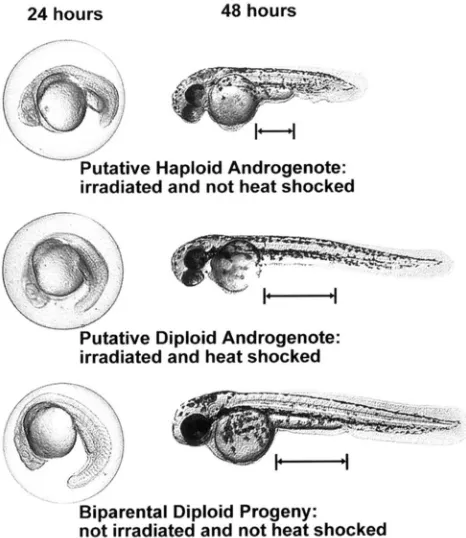
drome similar to the haploid syndrome of gynogenetic haploids (STREISINGER d nl. 1981; HORSTGEN-SCHWARK 1993) was seen at 24 hr as a shortened body phenotype (Figure 2), which was obvious at 48 hr in I/NHS em- bryos. Melanocytes are characteristically smaller in h a p loid embryos. This became noticeable at 48 hr (Figure 2) and was pronounced by 96 hr (not shown). The development of putative androgenetic diploid embryos was initially slightly retarded (Figure 2). However, by the end of the first month, the PDA fish in this experi- ment, and several in other experiments, were approxi- mately the same size as the diploid control fish. In this experiment, the percentage of haploid and diploid an- drogenotes produced relative to our control group was 14 and 2%, respectively.
Evaluation of the pedigree analysis technique: Analy- sis of DNA polymorphisms was used to determine the inheritance of maternal and paternal DNA to putative androgenetic offspring. Using RAPD primer 208BCF, three maternal-specific markers, but no paternal-spe- cific markers, were detected by agarose gel electropho- resis (Figure 3). None of the maternal markers was inherited by any of the four putative androgenetic prog- eny. The AB1 373 Automated DNA Sequencer allows for the separation of PCR products with greater resolu- tion and sensitivity than agarose electrophoresis, and the use of inlane fluorescent size markers allows for more precise sizing of fragments, facilitating identifica- tion of markers. Thus, most of our genetic analyses were based on fluorescent RAPD products separated on the AB1 sequencer.
Figure 4 shows output from the Genescan program. A comparison of two separate RAPD-PCR reactions r e p licated for each of two DNA templates (parents of puta- tive androgenotes) using the same fluorescent primer are shown. Although some peak heights vary slightly, all major peaks can be seen in both PCR reactions that contained an aliquot of the same DNA template, dem- onstrating that fluorescent RAPD-PCR markers are am- plified reproducibly and that they can be reproducibly detected. While the resolution is much better than on agarose gels, some peaks overlap. The zoom feature in the GeneScan software allows resolution of more peaks than can be seen in Figure 4. For our analysis, we used
24 hours 48 hours
,”
-u
Putative Haploid Androgenote: irradiated and not heat shocked
W
Putative Diploid Androgenote: irradiated and heat shocked
Biparental Diploid Progeny:
not irradiated and not heat shocked
FIGURE 2.-Putative haploid and diploid androgenetic em- bryos and biparental diploid embryos. Developing embryos of each type were photographed at 24 hr (left). The same embryos were photographed again at 48 hr (right) after re- moving the chorion. The distance between the posterior yolk sac margin and the anal pore (as shown by horizontal bars) is greater for the diploid phenotype than for the haploid phenotype. The appearance of two eyes in the putative andro- genotes and not in the biparental diploid progeny is the result of differences in the angle of photography and is not a pheno- typic difference.
only those markers that were clearly distinguishable. In all the
AI31
GeneScan electropherograms we have viewed to date, we have never detected a RAPD-PCR product in a progeny that was not detected in one of the parents, which is consistent with our RAPD markers acting as Mendelian markers. A clearly polymorphic peak specific to the father is seen in the top two panels at 799 bp in Figure 4. A small amount of amplification product was observed when no template was included in the PCR reaction (Figure 5 ) . Amplification products appearing in the absence of template DNA, which dis- appeared when template DNA was included in the PCR reaction, have been previously noted for RAPD reac- tions ( e g . , WILLIAMS et al. 1990). PCR markers used in our analyses are clearly distinct in mobility from those amplified in the absence of DNA template.~\ntlrogc.nctic Zchrafish 1269
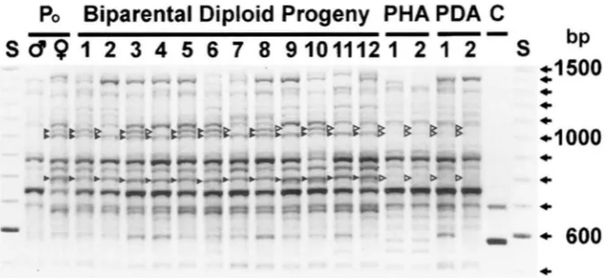
FI(;~:KE 3.-Inheritance of three maternal polymorphic markers for U P D primrr ‘LOXB<:F. The inheritance of three maternal RAPD markers is shown for 12 hiparental diploid progeny and four putative androgenctic progeny. The PCR products were separated hv electrophoresis on a 1.8% agarose gel and stained w i t h ethidium hromitle. PHA. putative haploid androgenote ( 1
and 2); PDA, putative diploid androgenote ( 1 and 2); C, control (no tcmplatc); S. sizing stantlard (Gihco BRL, 100-hp ladder); b, presence of band; D, ahsence o f band. Three hands (RAPD markers) in the matcrn;ll ( I : , 9 ) lane are marked with b. These Iyands are ahsent from the paternal (/;)
6)
lane and were tlcsignatcd a s maternal. Onc o f the maternal markers is seen in only some of the hiparental diploid progeny antl is thus considrrccl hctrrozygo1rs i n the fcmale parent. Two of these maternal hands are seen in all 12 hiparental diploid progeny and thus arc presumed homozygous i n the femalc parent. None o f these three maternal markers was detected i n any o f the f o w putative anc1rogenotc.s tcstetl.Figure
5 )
and are presumed to be homozygous in the parent (see Table 2). Henceforth, any marker referred to as maternal or paternal, will refer to a marker of a particular size that was observed in only one of the parents. The apparently homozygous maternal marker (Figure 5) was not inherited by any of the four putative androgenstes (only one of which is shown in Figure li), while the apparently homozygous paternal marker was inherited by all four putative androgenotes tested.Inheritance of RAPD-PCR markers by putative andro-
genotes: Using three fluorescent primers, 16 maternal
( 1 1 heterozygous antl five apparentlv homozygous) and
seven paternal (four heterozygous and three apparently homozygous) markers were itlentificd (Table 2). Mark- ers were considered heterozygous if only some of the progeny received the marker and apparently homozy- gous i f a l l 12 test RDP received the marker (Table 2, footnote n).
To test whether these RAPD markers were segregating in a Mendelian fashion, the 11 heterozygous maternal
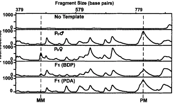
FI(:L;KE 4.-Detection of DNA polymorphisms h y RAP11 ;unalysis using fluorescent primcrs. Replicate P(:R reactions (Rep 1
antl Rep 2) are shown for the paternal and maternal DNA temp1;ltes. E:;wh templ;w was PCR ;~mplilietl with the fluorescent (&
FAM) RAPD primer 210BCF. Fluorescent RAP11 products were sep;mtcd ;wd tleterted during electrophoresis on an AB1 373 DNA sequencer. Fragment sizing was performed hv AB1 Genescan software, using fluorescent inlanc size standards. Each panel is an electropherogram output by the AB1 Genescan program. Genescan elcctropherogra~ns were captured as print files and imported into Photoshop version 3.0 t o acld Iahels and thicken lines t o allow for I)llotoretlrlction. Fluorescencc is shown in arbitrary units. Arrows and associated numhcrs indicate sizes o f parentally polymorphic peaks used i n the single Family analysis to assess the androgenctic nature o f putative androgenetic progeny. Three p i 1 t e r n ; d specific and six maternal specific markers
are shown. Although maternal marker 2/1)0$300 appears i n the figurr t o he pres<-nt i n paternal elrctropllcrograms, the peak
1270 G. E. Corley-Smith, C. J. Lim and B. P. Brandhorst
Fragment Size (base pairs)
379 579 779
1000 I No
Template
I
I
f
'"""1
h
A
I-1 uuu -r
I
Ff (PDA)MM I PM I
FIGURE 5.-Inheritance of fluorescent RAPD markers by putative androgenotes for same primer (210BCF) as used in Figure 4. This example shows a maternal marker (MM) 2 1 0 b 6 4 5 3 and a paternal marker (PM) 21Obcf: 799 and their inheritance to a biparental diploid progeny (BDP) and a putative diploid androgenote (PDA). Both markers were found in all 12 biparental diploid progeny (not shown in this figure) and are presumed to be homozygous in the parents (see footnote to Table 2). The reduced peak height from the parent where the marker is homozygous (PM and MM) to the BDP is consistent with there being a single allele, as expected for a heterozygote, in the BDP. However, our results are based only on presence or absence of a marker and do not rely on quantitative PCR. The bottom panel shows that the homozygous maternal marker is not inherited by the putative diploid androgenote and that the homozygous paternal marker is inherited by the putative diploid androgenote.
markers were scored in the 12 BDP, and the four hetero- zygous paternal markers were scored in the 16 progeny (12 BDP
+
two PHA+
two PDA). For each marker, the present:absent count in the F, progeny was tested using chi-square analysis for goodness of fit (ZAR 1974) to the theoretical ratio of 1:l. The null hypothesis of no differ- ence to a 1:l ratio was not rejected, with a = 0.05, for 14 of the 15 heterozygous markers. Thus, 14 of the 15 heterozygous markers appear to behave as Mendelian factors in our analysis. Marker 269bcf: 793 was clearly present in both replicates for the maternal template and was clearly present in the one BDP progeny it was o b served in. It was considered a statistical outlier and was not excluded from the data. Its exclusion would have very little effect on our androgenetic analysis.It is important for our analysis to show that each
marker represents a different locus, rather than some of them being length variants of the same locus. The 11 maternal heterozygous markers were tested for inde- pendent assortment against all other maternal markers. A similar analysis was performed for the four heterozy- gous paternal markers. For this analysis, it was assumed there was a recessive (unamplified) allele for each of the dominant RAPD markers. As the markers are domi- nant and parent specific, the cross can be viewed as a test cross. By arbitrarily assigning the two markers being compared as A and B, we use the notation for the cross as AaBb X aabb. If unlinked, the four categories in the cross will have a ratio approximating 1:l:l:l. If the two dominant markers are closely linked on the same chro- mosome (&linked), the AaBb and aabb categories would strongly dominate. If the dominant form of A is
TABLE 2
Number of parentally polymorphic RAPD markers in family A
Maternal markers Paternal markers
Primer Homozygous" Heterozygous Homozygous Heterozygous
208BCF 2 1 OBCF 269BCF
Total
3
5
3
11
Markers were designated homozygous if they occurred in all 12 biparental diploid progeny tested. The probability ( P ) of making an error by calling a marker homozygous, based on it being found in 12 BDP, was calculated: P = 1/2n with n = number of progeny tested. P = ( = 0.00024414. Thus, the chance that a marker designated as homozygous is in fact heterozygous, is 0.00024414 and the chance that it is homozygous
Androgenetic Zebrafish 1271
TABLE 3
Inheritance of heterozygous paternal markers by four
putative androgenetic progeny tested in family A
PHA PDA
Marker 1 2 1 2
21 ObcJ:23 7
+
+
- -2IObcJ313 269bcfi 615 269bcJ: 637
-
+
+
+
+
+
+
+
- -
-
+
210BCF, the fluorescent RAF'D primer used and the digits after the decimal indicate the size of the amplified fragment; PHA, putative haploid androgenotes (Nos. 1 and 2); PDA, putative diploid androgenotes (Nos. 1 and 2):
+,
marker pres-ent; -, marker absent.
on the same chromosome as the recessive form of B (translinked), the Aabb and aaBb categories would strongly predominate. In the 55 comparisons done be- tween maternal heterozygous markers, only one pair of markers appeared cislinked and this was not complete. The degree of linkage was not calculated due to the small sample size. None of the paternal marker compar- isons indicated linkage. In summary, our heterozygous RAF'D markers appear to be segregating as Mendelian markers and (with perhaps one exception) appear to be assorting independently.
To verify the androgenetic nature of PHA and PDA progeny of family A, the inheritance of maternal and paternal markers by these progeny was analyzed. All three of the homozygous paternal markers were inher- ited by all four of the putative androgenetic progeny tested, whereas none of the 16 maternal markers, five of which are probably homozygous, were detected in any of the four putative androgenetic progeny. Hetero- zygous paternal markers were inherited by putative an- drogenotes 10 times out of a possible 16 (Table 3 ) .
Inheritance of MHC and SSR-PCR markers by puta- tive androgenotes: The MHC primer pair did not pro- duce an informative marker as it was monomorphic between the parents. Likewise, 15 of the 16 SSR primers tested did not detect parental polymorphisms. SSR primer set 29 (GOFF et al. 1992), fluorescently labeled, detected two maternal-specific and two paternal-specific markers. The paternal markers are ssr29J153 and ssr29J 189, and the maternal markers are ssr29J 164 and ssr29J: 179. The two maternal markers appear to be dif- ferent loci as both markers occur in some BDP. Likewise the paternal bands are assumed to be different loci as both markers occurred in some BDP. Maternal marker ssr29'164 was found in four out of five BDP tested and is assumed to be heterozygous. Maternal marker ssr29J 179was found in five out of five BDP tested, and is assumed to be homozygous. Neither of these maternal markers were found in any of the four putative andro- genotes. Both paternal markers were found in all four putative androgenotes.
Confirmation of fertility of a diploid androgenote: We have produced several putative androgenotes that have survived to adulthood. The androgenetic nature of a male fish (progeny of family B ) that has sired hundreds of offspring was analyzed using fluorescent RAPD markers (primer 208BCF was used). Two pater- nal markers, which were designated as homozygous based on their occurrence in all seven BDP tested, were both found in the breeding putative male diploid an- drogenote. Two homozygous and one heterozygous ma- ternal markers were not transmitted to this breeding male diploid androgenote.
Efficiency of production of androgenotes: Five crosses were made to test the percentage of normal haploid appearing embryos resulting from irradiating eggs with 10,000 R of X-rays; the range varied from 8 to 28% (Table 4). If normalized relative to the control groups, the percentages range from 10 to 37%. The first four crosses were performed in one morning and the fifth the following morning.
I/NHS (irradiated and not heat shocked) embryos, when viewed at 24 and 48 hr p.f., displayed a range of morphological phenotypes, ranging from haploid ap- pearing with no noticeable morphological abnormali- ties (scored as A in Table 4, with example of one shown in Figure 2) to balls of cells that had arrested develop- ment before 24 hr. Examples of phenotypes observed more than once in embryo scored as category B (Table 4) included: developed head with diminished body and no tail, developed body and head with no tail, and body and tail with little or no head. The occurrence of certain morphological abnormalities was more common in some families than others.
When the milt for use in producing androgenotes was obtained from a fish of the SFU line, which has not been screened for recessive lethals as has the *AB line, the efficiency of production of putative haploid andro- genotes (category A in Table 4) was similar to that when milt was obtained from a *AB fish.
We have scored
>
1200 embryos from 12 families to date that resulted from eggs irradiated with 10,000 R of X-rays and not heat shocked. We have never observed an embryo with a diploid appearance resulting from this I/NHS treatment. Data on seven of these 12 fami- lies are not presented in Table 4, as the morphological characterization of abnormal androgenotes was less thorough.To date we have produced 44 putative diploid andro- genotes; thirteen of them survived past 20 days. Produc- tion of large numbers of diploid androgenotes has not been attempted, as our rearing facility is not large.
Genetic analysis in multiple families: Using RAPD primers 208BCF and ZlOBCF, we surveyed a sample of putative haploid androgenotes (category A in Table 4)
1272 G. E. Corley-Smith, C. J. Lim and B. P. Brandhorst
TABLE 4
Production rates of androgenetic haploids
PO Control Irradiated
Family Male Female n AA (%) n A
(%I
€3 (%) C/D (%)D SFU SFU 67 76 101 28 27 14
E *AB SFU 85 66 62 15 18 23
F *AB SFU 39 69 131 21 14 11
G *AB SFU 27 78 89 8 20 12
H *AB SFU 57 72 87 24 7 22
Eggs in the control group were held at room temperature and fertilized at same time irradiated eggs were fertilized. Milt and eggs for the five families were collected from five separate males and females. The five groups of eggs were irradiated separately at 10,000 R. Embryos were scored 2 days after fertilization based on appearance. Percentages are not normalized relative to control groups. n, sample size; AA, diploid phenotype with no morphological abnormalities apparent; A, haploid phenotype as described in Figure 2; B, haploid phenotype with noticeable morphological imperfections such as bent tail or missing part of tail; C/D, grossly abnormal embryos (classification based on WALKER and STREISINCER 1 9 9 4 ~ ) . In the control groups, only dead eggs and embryos that were normal diploid in appearance were observed. In the irradiated group, no embryos having a diploid appearance were observed. Data from families A-C, inclusive, are not included in this table as the progeny were not double checked by a second observer for agreement of classification into above categories.
lyzed. No maternal markers were found in any of the DISCUSSION
18 embryos analyzed that had been irradiated with The genetic analysis presented here, and discussed
10,000 R of X-rays. below, demonstrates the successful production of d i p
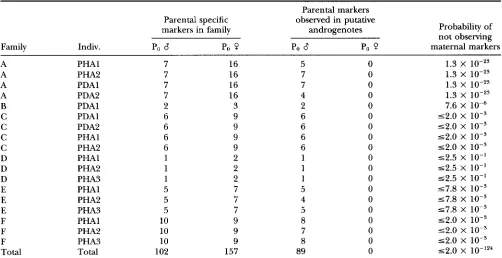
TABLE 5
S u m m a r y of genetic marker analysis for six families of androgenotes
Parental markers
Parental specific observed in putative
markers in family androgenotes Probability of
not observing
Family Indiv. Po Po Q Po 8 Po Q maternal markers
A PHAl 7 16 5 0 1.3 X
A PHA2 7 16 7 0 1.3 X 10-z3
A PDAl 7 16 7 0 1.3 X
lo-''
A PDA2 7 16 4 0 1.3 X 1 0 - 2 ~
B PDAl 2 3 2 0 7.6 X
C PDAl 6 9 6 0 ~ 2 . 0 X 1 0 - ~
C PDA2 6 9 6 0 52.0 x 10⁻³
C PHAl 6 9 6 0 52.0 X 1 0 - ~
C PHA2 6 9 6 0 52.0 x 10⁻³
D PHAl 1 2 1 0 52.5 X 10"
D PHA2 1 2 1 0 52.5 X 10"
D PHA3 1 2 1 0 52.5 X 10"
E PHAl 5 7 5 0 57.8 X lo-'
E PHA2 5 7 4 0 57.8 X 1 0 - ~
E PHA3 5 7 5 0 57.8 X 1 0 - ~
F PHAl 10 9 8 0 52.0 X 1 0 - ~
F PHA2 10 9 7 0 52.0 X 1 0 - ~
F PHA3 10 9 8 0 52.0 X 1 0 - ~
Total Total 102 157 89 0 5 2 . 0 X
Androgenetic Zebrafish 1273
loid androgenetic zebrafish surviving to adulthood. The results are significant for the use of androgenotes in genetic research and the evolutionary origin of genetic imprinting.
Confirmation of androgenetic inheritance in family
A:
The androgenetic nature of progeny in the one family experiment was confirmed by lack of inheritance of
12
(11 RAPD+
one SSR) heterozygous maternal and six (five RAPD+
one SSR) apparently homozygous, mater- nal DNA markers to all four androgenetic progeny tested. Although it is possible that some DNA leakage from the mother occurred, none was detected and the results strongly indicate that genomic DNA inheritance to the progeny was mostly or entirely from the male parent.A RAPD marker was presumed homozygous in the parent if it occurred in all 12 biparental diploid progeny tested (Table 2, footnote a ) . The chance of a biparental diploid progeny inheriting a marker designated as ho- mozygous, based on it being found in all 12 previously tested biparental diploid progeny, is': (1.0 X chance of marker being homozygous)
+
(0.5 X chance of marker being heterozygous) = (1 X 0.99975586)+
(0.5 X 0.00024414) = 0.99987793. Thus, the chance of not finding one of these presumed homozygous markers in a biparental diploid progeny is 1 - 0.99987793 = 0.00012207, or -1 in 10,000. The chance that none of the 11 heterozygous, and none of the five apparently homozygous maternal markers, being inherited by a single biparental diploid progeny can also be estimated: (0.5)''x
(0.00012207)5 = 1.3235 Xlo-".
This calcula- tion assumes all markers are segregating as Mendelian markers and are independently assorting. Although some of the maternal heterozygous markers may be weakly linked, our analysis of random assortment of markers showed no complete linkage between any twomarkers. The chance that four biparental progeny would receive none of these 16 maternal RAPD markers is: (1.3235
x
10-23)4 = 3.068x
lo-".
The androgenetic nature of the putative androgene- tic progeny is further supported by the lack of maternal SSR-PCR markers and presence of paternal SSR-PCR markers in these progeny. The chance that neither the maternal heterozygous nor the apparently homozygous SSR markers (see RESULTS) would be found in four biparental diploid progeny is 3.7 X
lo-'.
Combining the RAPD and SSR maternal marker data, the chance that none of the markers would be found in four bipa- rental diploid progeny is 1.1 X This strongly suggests androgenetic inheritance.All of the apparently homozygous, paternal RAPD markers and a proportion of the heterozygous paternal markers were inherited by all four putative andro-
'
The large number of significant figures are included for calcula- tion purposes only and do not indicate an exact probability as some markers, as discussed in text, may not he assorting independently to progeny.genotes analyzed. The proportion of paternal RAPD markers inherited by the progeny is consistent ( P = 0.45) with the Mendelian expectation that heterozygous markers will be inherited by half the progeny by chi- square testing for goodness of fit (ZAR 1974). Thus, it appears that androgenotes are inheriting paternal markers in a Mendelian fashion and not inheriting ma- ternal markers.
The androgenetic nature of these fish is further sup- ported by the phenotype of the irradiated embryos. Only severely abnormal embryos or embryos exhibiting the haploid syndrome were observed when irradiated eggs were inseminated. Following insemination of eggs irradiated with 10,000 R, in >1200 embryos observed, we have never observed the diploid phenotype (Figure 2), unless the zygotes were subsequently treated to in- hibit the first mitotic division. This evidence suggests that the irradiation dose is sufficient to eliminate most or all the maternal DNA and also that the heat shock procedure is effective in restoring embryos to the nor- mal diploid phenotype. That the irradiation dose used (10,000 R) is sufficient to prevent inheritance of mater- nal DNA is further supported by the coincidence of this dose with the secondary peak on a plot of survival, as a function of dosage (Figure l ) , known as the Hertwig effect (HERTWIG 1911). The initial decline in survival is thought to be due to partial destruction of the maternal genome leading to aneuploidy, while further irradia- tion leads to complete destruction of the irradiated ge- nome (ARAI et al. 1979; DON and AVTALION 1988). Al- though the Hertwig effect was originally observed for irradiated sperm (HERTWIG 1911), we have noted simi- lar survival curves for irradiated zebrafish, chinook salmon (Oncmhynchus tshawytscha)
,
and rainbow trout(0. mykiss) eggs.
In combination, the RAPD marker evidence, the SSR marker evidence, the absence of the normal diploid phenotype in the irradiated and not heat shocked group of progeny, and the observation of the Hertwig effect, provide strong evidence to support androgenetic inheritance.
Confirmation of fertility of a diploid androgenote:
Genetic analysis of a putative androgenetic breeding male zebrafish, indicated it has an androgenetic ge- nome. The chance of a BDP not inheriting two homozy- gous maternal (found in all seven BDP), nor one het- erozygous maternal marker is 7.6 X indicating that this breeding fish has an androgenetic genome.
Morphological appearance of haploid androgenotes:
1274 G. E. Corley-Smith, C. J . Lim a n d B. P. Brandhorst
suggests that some of the abnormalities result from background mutations in the
*AB
line.Efficiency of production of androgenotes: The ob- served efficiency of production of haploid andro- genotes (category A, Table 4) in five families (D-H) ranged from 8 to 28%. If categories A and B (Table 4) are combined, the production efficiency of haploid androgenotes ranged from 28 to 55%. Although we initially used milt from
*AB
males because this line was screened to reduce recessive lethals, we have achieved good results with milt from the SFU line of fish which are believed to be relatively heterozygous as they origi- nated from several pet stores and presently are not ho- mogenous in appearance. This suggests that milt useful for producing androgenotes, does not need to be o b tained from a line of fish screened for recessive lethals.The efficiency of production of diploid androgenotes in family A was 1.3%. To date, we have achieved success rates up to 2.1% for production of diploid andro- genotes. In our facility, if fish live past the first 20 days, they usually survive through adulthood. This applies both to diploid biparental and diploid androgenetic progeny. Thus, survival was measured at day 25. At
2%
efficiency, six diploid androgenotes can be expected from a batch of 300 eggs.Some abnormalities observed i n haploid andro- genotes are likely to have resulted from irradiation dam- age to cytoplasmic components of the oocyte. Since cytoplasmic components are known to be damaged by soft (low energy) X-rays, the efficiency of production of androgenotes might be increased by: filtering out soft (low energy) X-rays, using an X-ray machine with a higher KeV output, or using a gamma irradiation source (e.g., " t o or
""cs).
Androgenesis in other teleosts: Attempts to produce androgenetic fishes have been reported by several groups (reviewed by IHSSEN et al. 1990). Putative hap- loid' androgenetic embryos did not survive to the active feeding larval stage (ROMASHOV and BELYAEVA 1964; AM et al. 1979; PARSONS and THORGAARI) 1984). The production and survival of diploid androgenetic salmo- nids has been reported (PARSONS and THORGAARD 1985; SCHEERER et al. 1986,1991; MAY et al. 1988). These fishes were reported to be androgenetic based on their being homozygous at several loci, as determined from enzyme expression assays. However, the use of DNA polymorphisms allows for direct assessment of parental alleles, irrespective of their state of expression. Thus, it provides more compelling genetic evidence for lack of maternal inheritance to androgenetic progeny.
Androgenesis as a genetic tool: The production of androgenetic zebrafish has significance for investiga-
Haploid is used here to designate the set of chromosomes found
in one normal gamete. I t has been speculated that pacific salmon
may have four sets ol' chromosomes (KI.OSE rt al. 1968; BAILEY et al.
1969). Thus, in our usage, haploid is not necessarily equivalent to
onc set o f chromosomes.
tion of several biological phenomena and provides a useful genetic tool. The process of collecting eggs and milt and irradiating and fertilizing them can be accom- plished by one person in <1 hr. If heat shocking is performed, an additional 20 min is required. Tens of thousands of eggs can be irradiated simultaneously in the X-ray machine we use.
Male-spec@c meiotic recombination rates: Knowledge of the meiotic recombination rate in each sex during ga- metogenesis is important for genetic studies. In humans and mice, the male meiotic crossover rate is approxi- mately half that found in females and no crossing over occurs during meiosis in Drosophila males. POSTLETH-
WAIT et al. (1994) has determined the female specific
cross over rate for numerous RAPD markers on all 25 zebrafish chromosomes by analyzing markers inherited by gynogenetic haploid zebrafish. The male specific cross over rates could be determined using a similar procedure, except that inheritance would be assessed in haploid androgenetic rather than haploid gynogenetic progeny.
As
the present RAPD map is based on female meioses, male specific markers (if any exist) would not have been observed. Thus any new linkage group that might show up during mapping with androgenetic hap- loids, might be male specific, and might include sex determining genes.Only 94 gynogenetic haploid embryos were used to produce the zebrafish linkage map (POSTLETHWAIT et
al. 1994). To produce 100 androgenotes for a linkage map based on male cross over rates would require irra- diating 1000 eggs, assuming a 10% production rate of androgenetic haploids. Assuming 100 eggs/female, eggs would need to be collected from 10 females. Be- cause >800 eggs can on occasion be collected from one female, a 28% efficiency of production would pro- duce 224 haploid androgenotes from a single cross.
Androgenetic Zebrafish 1275
sex determination in zebrafish, which must be consid- ered in interpreting sex ratios of progeny from andro- genotes.
Cryopreservation of allelic combinations: Androgen- esis may be useful for storing and retrieving desirable combinations of certain alleles, clonal lines or wild (e.g., salmon) stocks. Zebrafish milt can be cryopreserved, (HARVEY et al. 1982; WALKER and STREISINGER 1994a,b) but we are not aware of any reports of successful fertil- ization of any previously frozen teleost eggs. Frozen sperm is generally not as effective in fertilizing eggs as normal sperm (HARVEY et al. 1982 report that frozen sperm on average was 51% as effective as fresh sperm in fertilizing eggs). However, even very low rates of pro- duction of diploid androgenotes may be acceptable for some applications as the milt from one fish can be used to attempt fertilization of thousands of eggs. Although we have never attempted to produce diploid andro- genotes from frozen sperm, we cannot foresee any rea- son why it should not be possible. We are hopeful that in the future the efficiency of both fertilization using frozen sperm and efficiency of production of diploid androgenotes will improve.
Mutation screening: Androgenetic F1 haploid screens in theory might have certain advantages over gynoge- netic haploid screens in mutagenesis protocols. Muta- tions can be induced, as for gynogenetic screens, by irradiation of sperm, eggs, or early embryos. Part of the milt obtained from the male containing the mutagen- ized germ-line could be used to produce androgenetic haploids for the F, screen, and the rest frozen and used only if mutations of interest were detected. Thus, in principle, the mutagenized parent need not be retained as the mutation can be recovered from cryopreserved sperm following mutation screening.
If a haploid androgenesis screen is attempted, a back- ground set of haploid abnormalities is expected, similar to those which are found during mutation screening using gynogenetic haploids (C. WALKER, personal com- munication). Induced mutations can be identified by the new appearance of specific haploid abnormalities that are particular to a certain family.
Genomic imprinting: Completion of mouse em- bryogenesis requires both the maternal and paternal genomes because of imprinting (parent-of-origin mono-allelic expression) of essential genes in male and female gametes (MCGRATH and SOLTER 1984; SURANI et al. 1984; SURANI 1986; BARRA and RENARD 1988; SAPIE- NZA 1990; & , N A R D et al. 1991; GOLD and PEDERSON
1994; OHLSSON et al. 1994; CHAILLET et al. 1995). This does not appear to be the case for zebrafish. Diploid homozygous gynogenotes not only complete em- bryogenesis, but survive to adulthood (STREISINGER et al. 1981). Our results show that this is also true of diploid homozygous androgenotes. These results suggest that imprinting, in either of the parental gametic genomes,
does not result in essential genes being irreversibly inac- tivated during a time when required for development.
While parent-of-origin (gametic) inactivation of es- sential genes has been ruled out in zebrafish, parent- of-origin effects on a transgene have been detected
(MARTIN and MCGOWAN 1995b). A decrease in methyla-
tion with maternal passage and an increase in methyla- tion with paternal passage of a transgene in zebrafish was consistently observed. Thus, it appears that epige- netic phenomena associated with genomic imprinting occur in zebrafish and that parent-of-origin imprinting may occur in zebrafish but not for genes essential for development.
Zebrafish androgenetic haploid embryos are morpho- logically slightly abnormal (Figure
2)
and arrest around day four. Zebrafish gynogenetic haploid embryos exhibit a typical “haploid syndrome”: they have short, stocky bodies, their eyes are incompletely formed at the ventral furrow and the brain is poorly sculptured. Cell size is often smaller in gynogenetic haploids than in diploids, as observed for melanocytes. Eventually they become edematous and die after 4-5 days (C. WALKER, personal communication). Haploid androgenotes are indistin- guishable in appearance from haploid gynogenotes, sug- gesting that the abnormalities are not due to parent-of- origin, but may be dependent on gene dosage.We believe that this is the first report of the produc- tion of a viable and fertile androgenetic diploid verte- brate in which the extent of elimination of the maternal genome has been assessed by the use of DNA markers. There have been other reports of production of fertile androgenetic and gynogenetic adult teleost fishes (re- viewed by IHSSEN et al. 1990) and amphibians (e.g., GIL-
LESPIE and ARMSTRONG 1981). Collectively, these re-
ports indicate that the failure of androgenesis and gynogenesis reported for some mammals (MCGRATH and SOLTER 1984; SURANI et al. 1984; SURANI 1986; GOLD and PEDERSON 1994) is not characteristic of verte- brates in general. Thus, genomic imprinting of genes essential for development may be a specialized phe- nomenon that arose during mammalian evolution.
We thank Dr. JOHN POSTLETHWAIT for helpful suggestions on the
manuscript; CHARI.INE WAI.K!LR for supplying the *AB line of zebrafish
and for her numerous suggestions on raising zebrafish; SUSAN GILLES
PIE for help in rearing zebrafish; and Dr. C. CRISTOFRE MARTIN, Dr.
V EK. ~LLOYD and Ms. KATHLEEN A. FITZPATRICK, for helpful discus-
sions. This research was supported in part by a grant from the Natural
Sciences and Engineering Research Council of Canada.
LITERATURE CITED
A K A I , K., H. ONOZATO and F . YAMAZAKI, 1979 Artificial androgenesis
induced with gamma irradiation in Masu Salmon, Oncorhynchus
masou. Bull. Fac. Fish. Hokkaido Univ. 30: 181-186.
ASHBURNER, M., 1989 Isolation of DNA from adult flies, pp. 106-
107 in Drosophila-A Laboratmy Manual. Cold Spring Harbor
Laboratory Press, Cold Spring Harbor, NY.
BAILEY, G. S., G. T. COCKS and A. C. WIISON, 1969 Gene duplication
in fishes: malate dehydrogenases of salmon and trout. Biochem.
1276 G. E. Corley-Smith, C. J. Lim and B. P. Brandhorst
BARINAGA, M., 1990 Zebrafish: swimming into the development
mainstream. Science 250: 94-35.
BARINM:A, M., 1994 Looking to development's future. Science 2 6 6
561-564.
BARRA, J., and J. P. R E N . ~ , 1988 Diploid mouse embryos con- structed at the late 2-cell stage from haploid parthenotes and
androgenotes can develop to term. Development 102: 773-779.
<:lIAII.I.I<T, J. R., D. S . BADL'R and P. L ~ D E K , 1995 Regulation of genomic imprinting by gametic and embryonic processes. Genes
Dev. 9: 11 77- 1187.
(:ON(:ORI)ET J. P., and P. INGIIAM, 1994 Catch of the decade. Nature
369: 19-20.
D O N , J., and R. R. ACTAI.ION, 1988 Production of F, and F, diploid
gynogenetic tilapias and analysis of the "Hertwig curve" ob-
tained using ultraviolet irradiated sperm. Theor. Appl. Genet.
76: 253-259.
DRIEVER, W., D. STEM PI.^, A. SCHIL'R and L. SOI.NI(A-KRE:ZF.I., 1994
Zebrafish: genetic tools for studying vertebrate development.
Trends Genet. 10: 152-159.
GIHHS, P. D. L., A. PEEK and G. TEIORMARI>, 1994 An in aivo screen for the luciferase transgeue in zebrafish. Mol. Mar. Biol. Biotech.
3: 307-316.
G I [ J . F S I W , 1.. l.., and J. B. ARMSTRONG, 1981 Suppression of first
cleavage in the mexican axolotl (Ambystoma mexiranum) by heat
shock or hydrostatic pressure. J. Exp. Zool. 218: 441-445.
1992 Identification of polymorphic simple sequence repeats in
the genome of the zebrafish. Genomics 14: 200-202.
GoI.I), J. D., and R. A. PEDERSON, 1994 Mechanisms of genomic
imprinting in mammals. Curr. Top. Dev. Biol. 29: 227-280.
HAKVL'Y, B., R. N. &:I.I.EY and M. J. ASHMW)I)-SMI~'H, 19F2 <:yo-
preservation of zebra fish spermatozoa using methanol. Cau. .J.
Zool. 60: 1867-1870.
HIXTWI(;, O., 191 I Die Radiumkrankheit tierischer Keimzellen.
Arch. Mikr. Anat. 77: 1-97.
H~~RSTGL'N-SCHWARK, G., 1993 Production of homozygous diploid
zebra fish (Brarhydanio rm.0). Aquaculture 112: 25-37.
Ploidy manipulation aud gynogenesis in fishes: cytogenetic and
fisheries applications. Trans. Am. Fish. Soc. 119: 698-717.
KUIN, P., 1994 Zebrafish hit the big time. Science 264: 904-905.
KIMW.I., C. R., 1989 Genetics and early development of zebrafish.
Trends Genet. 5: 283-288.
K I . ~ s F ,
,I.,
U. WOI.F, H. H~EEROTH and H. RITTER, 1968 Duplica- tion o f the LDH gene loci by polyploidization in the fish orderclupeiformes. Humangenetik 5: 390-196.
MARTIN, C . C., and R. MCG~WAK, 199Sa Genotype-specific modifi-
crs of transgene methylation and expression in the zebrafish,
IkLnio rm'o. Genet. Res. <:amb. 65: 21-28.
MARTIN, C. C . , and R. M&ow.m, l995b Parent-of-origin specific
effects on the methylation o f a transgeue i u the zebrafish, Danio
rm'o. Dev. Genet. 17: 233-239.
MAY, B., K..J. HENI.EY,C. C. K K U L ( : E K ~ I I ~ S . P. Gr.oss, 1988 Androgen-
esis as a mechanism for chromosome set manipulation i n Brook
Trout (Suluulzntc5 /onlinclli.s). Aquaculture 75: 57-70.
M&IL\IFI, J., and D. SOI.TEK, 1984 Completion of mouse em- hlyogenesis requires both thc maternal and paternal genomes.
Cell 37: 179-183.
MEYKR, A,, C:. B. BIEKMANN and G. ORTI, 1993 The phylogenetic
position of the zebrafish (Duniu rniu), a model system in develop-
mental biology: an invitation to the comparative method. Proc.
R. Soc. Lond. B Bid. Sci. 252: 231-236.
\ ' 0 1 . k i 2 u u ) , 1994 Large-scale mutagenesis in the zebrafish: in search of genes contl-olling development i n a Vertebrate. Curr.
Biol. 4: 189-202.
GOFF, D..]., K. GAI.WK, H. K Y I ' Z , M . WESrF,RFlEl.l), E. S. LANDER et al.,
IIISSICN. P. E., L. R. MCKAY, I . , MCMILIAN and R. B. PHII.I.lI'S, 1990
MIII.I.INS, M. C., M . HAMMEKS(:HhlIL)T, P. HAFFTER and c:. NOSSI.EIN-
NUSSI.EIN-VOI.W, C., 1994 Of flies and fishes. Science 266: 572-
574.
OHISSON, R., D. B A R I . ~ ~ and A. SURANI, 1994 Impressions of i n -
prints. Trends Genet. 10: 415-417.
0 x 0 , H., D. KI.EIN, V. VINCEK, F. FICWEROA, C. O'HUIGIN. rl rcl., 1992
Major histocompatibility complex class I1 genes of Lebrafish.
Proc. Natl. Acad. Sci. USA 89: 11,886-11,890.
PARSONS, J. E., and G. H. TIIORGMRD, 1984 Induced androgenesis
in rainbow trout. .J. Exp. Zool. 231: 407-412.
PARSONS, J. E., and G. H. THOKGAARU, 1985 Production of andro-
genetic diploid rainbow trout. J. Hered. 76: 177-181.
POSTI.ETHWAIT, J. H., S. L. JOHNSON, C. N. MIDSON, W. S. TAL.HOI',
M. GATES rt al., 1994 A genetic linkage map for the zebrafish.
Scieuce 264: 699-703.
RENARD, J. P., C. B.ZH1NE-r and J. BARKA, 1991 Participation of the
paternal genome is not required before the eightcell stage for
fdl-term development of mouse embryos. Dev. Biol. 1 4 3 199-
202.
RoMi\sr{ov, D. D., and V. N. BELYAEVA, 1964 Cytology of radiation
gynogenesis and androgenesis in the loach ( M i s p r n u s fossilis L..).
Dokl. Akad. Nauk SSSR Biol. Sci. Sect. (Eng. Transl.) 157: 503-
506.
SAPIENZA, C., 1990 Sex-linked dosage-sensitive modifiers as im-
printing genes. Development 1990 (Suppl.): 107-1 13.
SCHEERER, P. D., G. H., THORGAARD, F. W. AI.I.ENI)OW aud K. L . KNLIL)SE:N, 1986 Androgenetic rainbow trout produced from in- bred and outbred sperm sources show similar survival. Aquacul-
ture 57: 289-298.
SCHL'EWK, P. D., G. H. THOK(:AARD and F. W. AI.I.L'NI)OKF, 1991 Ge-
netic analysis of androgenetic rainbow trout. J. Exp. Zool. 260:
382-300.
1981 Production of clones of homozygous diploid zebra fish
(Hrarhydanio r m ' o ) . Nature 291: 293-296.
SITRANI M. A. H., 1986 Evidences and consequences of differences between maternal and paternal genomes during embryogenesis
in the mouse, pp. 401 -435 in Expm'mental Approccchn tu Mommcl-
licm Emhyonic Dmebpmmt, edited by J. ROSSANT and R. A. PEI).
IIRSEN. C:atnbridge University Press, New York.
S l l R A N I M . A. H., S. C. BARTON and M. L. NORRIS, 1984 Development of reconstituted mouse eggs suggests imprinting of the genome
during gametogenesis. Nature 308: 548-550.
W,ZI.KEK, C., and G. STREISIN(;ER, 1994a Freezing sperm, pp. 7.22-
7.23 i n The 2 l m $ s h Book-A ( h i d e Jbr the Lrcboratuy Usr of Zebm
j . r h (Hrarlyianio rmo), Ed. 2.1, edited by M. WESTERFIEIJ). Insti- tute of Neuroscience, University of Oregon, Eugene, OR.
W A I K E K , C., and G. STREISINGER, l994b Thawing and using frozen
sperm for in vitro fertilization, p. 7.24 in T/ZP Z&rrclfish Book--A
h i d e f u r thr Laboratory Usr of 2braJish (Brarhvrlnnio rm'o), Ed. 2. I ,
edited by M. WESTF.RFIF.I.I>. Institute of Neuroscience, University
of' Oregon, Eugcnc, OR.
W,ZI.KER, C., and G. STREISINGER, 1994c Embryo productiou by in
vitro fertilization, p. 72.1 1 i n Thr fiirrajsh Book-A h i d r /or Ihc
Laboratoly llsr (IJ %Iwajish (Brachydanio rm'u), Ed. 2. I , edited by M.
WTes-rl;.wmx.u. Institute of Neuroscience, Uuiversity of Oregon, Eugene, OR.
WII.I.IAMS, .J. C;. K., A. R. K t r m k , K. J. LIVAK, J. A. RAFAI.SKI aud
S . V. TINGEY, 1990 DNA polymorphisms amplified by arbitrary
primers are useful as genetic markers. Nucleic Acids Res. 18:
6531-6535.
WII.I.IAMS, J. G. K., M. K. HANAF~Y, J. A. WAI.SKI and S. V. TINGEY,
1993 Genetic analysis using random amplified polymorphic
DNA markers. Methods Enzymol. 218: 704-740.
ZAR, J. H., 1974 BiostutisticalAnalysis. Prentice-Hall, Inc., Englewood Cliff& N.J.
%rREISINGER, G., C. U'AI.KEK, N. DOWER, D. K N ~ Z I J H E K and F. SIN(;t:K,