Article
Atomic Surface Segregation and Structural
Characterization of PdPt Bimetallic Nanoparticles
Carlos A. Rodríguez-Proenza1, Juan P. Palomares-Báez2,*, Marco A. Chávez-Rojo2, Amado F. García-Ruiz3, Cristy L. Azanza-Ricardo4, Alan Santoveña-Uribe1, Gabriel Luna-Bárcenas5, José L. Rodríguez-López6,* and Rodrigo Esparza4,*
1 Posgrado en Ciencia e Ingeniería de Materiales, Centro de Física Aplicada y Tecnología Avanzada, Universidad Nacional Autónoma de México, Campus Juriquilla, Querétaro, Qro, México;
[email protected]; [email protected]
2 Facultad de Ciencias Químicas, Universidad Autónoma de Chihuahua, Circuito Universitario s/n, Campus II, Chihuahua, Chih., 31125, México; [email protected]; [email protected]
3 UPIICSA-COFAA, Instituto Politécnico Nacional, Te 950, Col. Granjas-México, Iztacalco, México, D. F.,
08400, México. On sabbatical year at CFATA-UNAM; [email protected]
4 Centro de Física Aplicada y Tecnología Avanzada, Universidad Nacional Autónoma de México, Boulevard Juriquilla 3001, Santiago de Querétaro, Qro., 76230, México; [email protected];
5 Centro de Investigación y Estudios Avanzados (CINVESTAV) del Instituto Politécnico Nacional (IPN), Unidad Querétaro, Fracc. Real de Juriquilla, Santiago de Querétaro, Qro., 76230, México;
6 Departamento de Materiales Avanzados, Instituto Potosino de Investigación Científica y Tecnológica, A.C.,
San Luis Potosí, SLP, 78216, México; [email protected]
* Correspondence: [email protected]; Tel.: +52-695-110-0315; [email protected]; Tel: +52-442-321-4865; [email protected]; Tel: +52-444-130-9861
Abstract: Bimetallic nanoparticles are of interest since they lead to many interesting electrical, chemical, catalytic, and optical properties. They are particularly important in the field of catalysis since they show superior catalytic properties than their monometallic counterparts. The structures of bimetallic nanoparticles depend mainly on the synthesis conditions and the miscibility of the two components. In this work, PdPt alloyed-bimetallic nanoparticles (NPs) were synthesized through the polyol method, and characterized using spherical aberration (Cs) corrected scanning transmission electron microscopy (STEM). High-angle annular dark-field (HAADF)-STEM images of bimetallic nanoparticles were obtained. The contrast of images shows that nanoparticles have an alloy structure with an average size of 8.2 nm. Together with the characterization of nanoparticles, a systematic molecular dynamics simulations study, focused on the structural stability and atomic surface segregation trends in 923-atom PdPt alloyed-bimetallic NPs was carried out.
Keywords: Bimetallic nanoparticles; Electron microscopy; Molecular dynamics simulation; Gupta potential; PdPt alloys; Nanostructures
1. Introduction
Bimetallic nanoparticles are of wide interest since they show physical and chemical properties that are distinct from those of their monometallic counterparts, and they present a combination of
the properties associated with the two metals and create synergistic effects, which signifies the whole being greater than the sum of its parts [1]. Bimetallic nanoparticles have emerged as a new class of catalysts mainly in energy issue, with potential applications in advanced energy conversion and storage (ECS) devices, including fuel cells, photo-electrochemical water splitting cells, solar cells, Li-ion batteries, supercapacitors, among others [2].
It is known that platinum (Pt) is one of the best single metal catalysts and is used mainly in the chemical industry, the petrochemical industry, automobile exhaust purification, and fuel cells because of its excellent reactivity and stability [3]. Improving the activity and utilization efficiency of Pt catalysts is therefore the key issue in development of relevant catalysis fields. Normally, there are two types of target products to reduce Pt loading: Pt monometallic nanoparticles (NPs) with open surface structure [4] and based bi- (or multi-) metallic NPs [5]. Meanwhile, synthesis of Pt-based bimetallic NPs, as an alternative strategy, has received considerable attention as well because of their enhanced catalytic activities as compared to conventional Pt monometallic NPs [6-9]. For example, Pt-Au bimetallic NPs exhibited higher electrocatalytic activity and durability for oxygen reduction reactions than Pt monometallic catalysts [6,7]. Moreover, the superior activities of both Pt-Ru and Pt-Rh bimetallic NPs were also ascertained in preferential oxidation of CO in hydrogen [8,9].
Among Pt-based nanocatalysts, Pt-Pd is one of the most attractive bimetallic systems due to its highly promising application as a catalyst in formic acid oxidation and fuel cells [10-12]. Furthermore, Pt-Pd nanocatalysts have been considered as preferred substitutes of Pt-Ru catalysts for low-temperature direct formic acid fuel cell (DFAFC) technology owing to the much cheaper price of Pd [12]. PdPt bimetallic NPs can be produced generally by either successive co-reduction of Pt and Pd species or a seed-mediated growth method [13].
As it is known, the structure of bimetallic nanoparticles mainly depends on the synthesis methods and the miscibility of the two components [14]. The experimentally synthesized Pt-Pd bimetallic NPs can be classified into three configurations: core/Pd-shell, Pd-core/shell, and Pt-Pd alloyed nanostructures [10,15,16]. It is well-known that the catalytic activity of these NPs strongly depends not only on the particle size but also the Pt/Pd ratio [17]. Therefore, the effects of core sizes, shell thicknesses, and alloy compositions are urgent topics that should be addressed for further tailored design of highly active Pt-Pd nanocatalysts. Furthermore, considering that catalysts are generally used at different temperatures, the structural and thermal stabilities of PdPt bimetallic NPs are also important issues that need to be clarified [18]. Although considerable experimental and theoretical studies have been dedicated to design, synthesis, characterization and performance evaluation of Pt-Pd bimetallic NPs [19], there is still lacking a full atomic-level understanding of structural and thermal stabilities of PdPt bimetallic NPs with different configurations and compositions.
2. Materials and Methods
2.1. Synthesis of bimetallic nanoparticles
PdPt nanoparticles were prepared by oil-bath heating in ethylene glycol (EG) using a one-step reduction method. Briefly, 3 ml of EG were preheated until the temperature was stabilized at 160 °C in a 3-neck round bottom flask. Over the stirred EG, 1.5 ml of 50 mM H2PtCl6·6H2O in EG, 1.5 ml of 50 mM K2PdCl4 in EG and 6 ml of 100 mM PVP in EG were added slowly in aliquots of 200 µl (H2PtCl6·6H2O and K2PdCl4) and 400 µl (PVP), respectively, each 2.5 minutes until the volume of the precursor and surfactant was completed, then the mixture was kept on stirring at 160 °C for 2 hours. The cleaning process of nanoparticles was performed both with hexane and deionized water in ultrasound baths and this process was repeated for several times.
2.2. Characterization
All the samples were characterized using the synthesized colloidal solution. Copper grids with carbon film were prepared with a drop of the colloidal solution for electron microscopy images. The samples were analyzed using aberration-corrected scanning transmission electron microscopy (STEM) with a Jeol ARM200F (200 keV) FEG-TEM/STEM microscope equipped with a CEOS Cs-corrector on the illumination system. High-angle annular dark field (HAADF)-STEM images were obtained. The probe current used in STEM mode was 23.2 pA using a condenser lens aperture size of 40 microns. The HAADF images were registered using a camera length of 80 mm and a collection angle of 50 - 180 mrad. Energy-dispersive X-Ray spectroscopy (EDS) was performed by means of Oxford system. Parallel beam X-Ray diffraction (XRD) analysis of the bimetallic nanoparticles was carried out on a Rigaku Ultima IV X-Ray diffractometer equipped with a Cu Kα source (λ = 0.154 nm) at 40 keV and 30 mA. The 2θ angular region between 30 and 90° is recorded at a scan rate of 0.05°/step with a speed of 2°/min.
2.3. Image simulations
HAADF-STEM image simulations have been performed using the QSTEM software package [20], which uses the multislice algorithm based on the physical optics theory of Cowley and Moodie [21]. The QSTEM code is based on the methods described by E.J. Kirkland [22]. The parameters considered for the simulation correspond to the optical conditions of the electron microscope.
2.4. Molecular dynamics simulation
addition, temperature-dependent bond order parameters and potential energy for each nanostructure were calculated and plotted. Furthermore, the cone algorithm [28] was used to compute the number of Pd and Pt atoms residing on the nanoparticle's surface. Both for heating and cooling processes, the same research protocol was followed; that is, all the parameters used in the molecular dynamics simulations were kept on the same. For each system, simulations were replicated ten times and all the graphs shown throughout the work resulted from the averages made with these replicas.
3. Results and Discussion
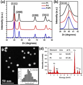
Figure 1. (a) XRD patterns of Pt, Pd and PdPt nanoparticles, (b) magnified zone of the (111) reflection, (c) low magnification HAADF-STEM image and (d) EDS spectrum of the bimetallic nanoparticles.
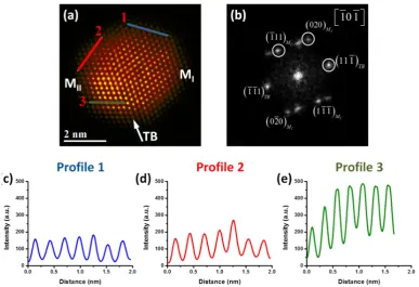
PdPt bimetallic alloy nanoparticles using HAADF-STEM images has been reported previously by our group [35]. This study showed a method semi-quantitative to analyze the atomic distribution of the elements which is dependent of their atomic number. Intensity profiles were performed to analyze the intensity brightness of the different atomic columns, it is important to mention that the graphics have the same intensity scale for a better comparison. Intensity profile 1 (Figure 2c) was measured along the MI crystal of the nanoparticle, the intensity columns is uniform, indicating that the atomic columns are from the same type of atom, which according to the HAADF-STEM image could be associated with the Pd atoms. Intensity profile 2 (Figure 2d) was measured along the MII crystal of the nanoparticle, which shows more diverse intensity columns than profile 1. This behavior is because of there are Pd rich columns and Pt rich columns which appear much brighter in the image and with more intensity scale in the graphic. Intensity profile 3 (Figure 2e) was measured in the same crystal of the nanoparticle (MII) but with different direction. In this profile, the difference in intensity among the first and last columns is higher, indicating clearly that some columns are Pt rich than others ones and that the Pt atoms is not uniform in the surface.
Figure 2. (a) HAADF-STEM image of a PdPt nanoparticle, (b) FFT showing the twin boundary plane, and intensity profiles along: (c) blue line, (d) red line and (e) green line, respectively.
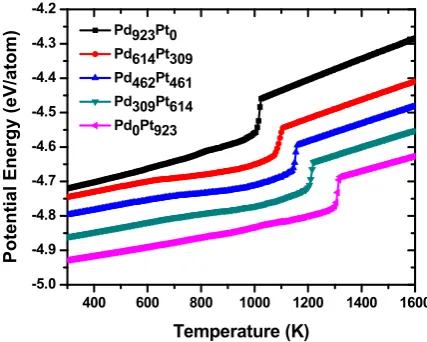
Figure 3. Temperature-dependent average potential energy of nanoparticles. In the graph, the values of the potential energy for each nanostructure were rescaled so that the distinctive features of each curve could be better appreciated.
For the case of bimetallic NPs, melting points for both nanoalloys are the following: 1087 K for PdPt (2:1), 1140 K for PdPt (1:1) and 1200 K for PdPt (1:2) and each one is included between the values corresponding to the monometallic systems. However, the significant differences observed in melting points could be explained not only considering the number of atoms of each species in nanoalloys but by the way in which the Pd and Pt atoms are distributed in the clusters.
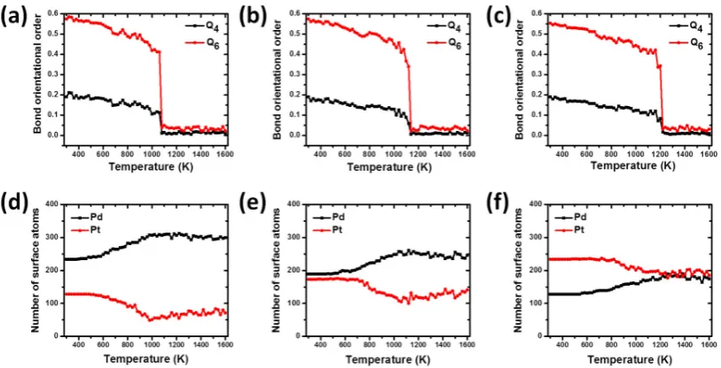
Figure 4 shows the temperature dependences of bond order parameter Q4 and Q6, and the number of surface Pd and Pt atoms as a function of temperature for the Pd-Pt-alloyed bimetallic nanoparticles. As known from previous works, the bond order parameter is a method very useful to investigate the structural evolution of nanoparticles [28,39]. Before starting the relaxation and simulation of nanoalloys, the values for the bond order parameters Q4 and Q6 were calculated for the designed models, obtaining a good agreement with the values reported for ideal structures [28]. As can be seen from the figure, the bond order parameter curves for the bimetallic nanoparticles show a slight decrease from 300 K until 1060 K for PdPt (2:1) (Figure 4a), 1080 K for PdPt (1:1) (Figure 4b), and 1160 K for the PdPt (1:2) (Figure 4c) nanoparticles. These results show that in this temperature range, the structures do not show significant structural changes and the nanoparticles are experiencing contractions and expansions around their centers of mass. At this stage, slight oscillations are observed in each of the curves and generally they can be attributed to the fact that all atoms are experiencing chaotic and disordered movements because to excess thermal energy absorbed by systems. However, after the mentioned temperatures, a rapid and sudden drop in these values occurs until the phase transition temperature from solid to liquid is reached. During this solid-liquid transition particles undergo strong changes in their morphology and they are in an amorphous state. The last temperature values are in good agreement to those observed in the caloric curves, where they were identified as the melting points for both nanoparticles.
One of the methodologies used to investigate the atomic segregation on the surface of Pd-Pt alloyed nanoparticles is the “cone” algorithm [28] which is based on identifying the number of atoms on the particle surface from their geometric positions. The dependence of the number of Pd and Pt atoms on the surface particles as a function of temperature can be observed in Figure 4. As can be seen, Figure 4d shows that for the nanoalloy PdPt (2:1) there is a greater amount of Pd atoms on the surface in relation to those of Pt at the beginning of heating process. These Pd atoms remain all the time on the particle surface, while those in the interior keep on migrating to the surface
400 600 800 1000 1200 1400 1600
during the premelting stage, which is longer than in the case of the alloy PdPt (1:1) and PdPt (1:2). It is known that melting phenomenon starts in the surface and evolves towards the interior of the particle until that this is completely melted. At this point, the structure begins to undergo considerable structural transformations and the phenomenon of atomic diffusion begins to occur showing a clear tendency of the Pd atoms to segregate on the particle surface and in addition, of Pt atoms to diffuse towards its inner core.
A similar behavior is observed for nanoalloy PdPt (1:1) (Figure 4e) in the variation of the number of the atomic species on the particle surface: the Pd atoms prefer to remain on the particle surface throughout the heating process. In this case, the amount of surface Pd atoms at the end of the heating process is lower than that observed in the alloy PdPt (2:1) (246 and 300, respectively); and this could be explained by the fact that the last nanostructure has the highest number of Pd atoms and this metallic species prefers to diffuse towards the surface of nanoparticles.
Figure 4. (a), (b) and (c) Temperature-dependent bond order parameter, and (d), (e) and (f) number of surface atoms, for the for bimetallic nanoparticles PdPt (2:1), PdPt (1:1), and PdPt (1:2), respectively.
Figure 5. Final configurations after 1000 ps MD run of the nanoparticles at various temperatures for: (a) PdPt (2:1), (b) PdPt (1:1), and (c) PdPt (1:2) nanoparticles, respectively (Pd=blue and Pt=grey).
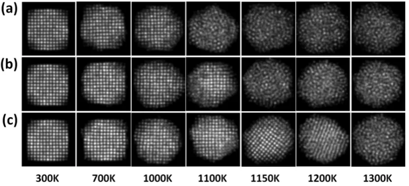
Figures 6a, b and c show the simulated HAADF-STEM images of PdPt (2:1), PdPt (1:1), and PdPt (1:2) nanoparticles at various temperatures, respectively, which were acquired using the optical parameters of the microscope. The aim of simulated HAADF-STEM images is to understand the effect of the contrast associate to the Pd and Pt elements (Z-contrast) and to analyze with detail the structure in the image. The images were recorded using the same intensity scale, this with the purpose of comparison the intensity contrast among them. As can be observed from the simulated images, in all the cases the intensity contrast decrease with respect to the temperature, it is due to increase of Pd atoms on the surface, which produces low contrast compare with the Pt atoms. This behavior is more notable in the PdPt (2:1) structure because exist more amount of Pd atoms on the surface compared with the PdPt (1:1) and PdPt (1:2) structures.
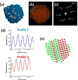
After the completion of cooling processes, significant surface reconstructions can be made for the final nanostructures at 300 K given that from the melted nanostructures a great diversity of structures could occur after the liquid to solid phase transition. Something important that must be pointed out is that after carrying out the cooling processes, of the total of simulated replicas, seven of them fell into FCC twinned-type bimetallic NPs while the rest ended in decahedral nanostructures. Figure 7 shows the results obtained at 300 K after the completion of cooling process (crystallization) for the PdPt (1:1) nanoparticle. In this work, only the results corresponding to the PdPt composition (1: 1) are shown to have a greater similarity with the experimental results and consequently, a better comparison criterion. Figure 7a shows the model of the PdPt (1:1) alloyed bimetallic nanoparticle. In the image, most of Pd atoms are located on the particle surface and it was constructed from the calculation of the average of atomic positions of each metallic species at 300 K (ten samples were considered, as mentioned above). Figure 7b shows the simulated HAADF-STEM image of model; to visualize better the contrast as case of the experimental image, the Apply_CLUT (color look-up table) script was used. Simulated image displays some crystalline defect (twin boundary), which causes perturbation in the nanoparticle. FFT of the simulated image (Figure 7c) shows a dual-spot diffraction characteristic of twinning and the nanoparticle is viewed along the [-10-1] zone axis. Figure 7d shows the intensity profiles measurement over two directions; the intensity profiles show diverse intensities; this behavior is characteristic of Pd rich columns and Pt rich columns. Finally, Figure 7e shows the atoms colored after a common neighbor analysis (CNA) using OVITO [43]; green and red coloring represent atoms with an FCC and HCP (twinning) stacking, respectively. As can be observed there is good agreement between the experimental and the simulation results.
HAADF-STEM image, (d) intensity profiles along two directions, and (e) Atoms colored according to their crystal stacking structure calculated using a common neighbor analysis (green=FCC and red=HCP).
4. Conclusions
PdPt bimetallic nanoparticles with alloy structure and homogeneous particle size of 8.2 nm were synthesized by polyol method. The bimetallic nanoparticles have been characterized by XRD and HAADF-STEM, which is dependent of atomic number of the elements; both techniques showed that the nanoparticles have an alloy structure. Profiles intensity of HAADF-STEM images show that exists atomic columns with higher content of Pd than Pt. Temperature-dependent average potential energy, bond order parameters, and number of surface atoms for PdPt nanoparticles, have been simulated by molecular dynamics calculations based on the Gupta potential. The simulated results show that from a thermodynamic point of view, the nanoalloys PdPt (2:1), PdPt (1:1) and PdPt (1:2) exhibit thermal stabilities higher than Pd but lower than that of Pt monometallic nanoparticles. Bond order parameters show that the nanoalloys experiment morphological and structural changes as the temperature increases. On the other hand, for analyzing the surface segregation phenomena, the number of atoms on particle surface was evaluated. The criteria predict clearly a trend of Pd atoms to segregate on the nanoparticle surface when the temperature increases. This behavior is attributed to the fact that Pd has a lower surface energy than that of Pt atoms. The importance of to study the atomic surface segregation of the PdPt bimetallic nanoparticles is because this system is attractive as electrocatalyst for fuel cells, since exhibits enhanced catalytic activity and good stability for oxygen and ethanol reduction reactions.
Author Contributions: Conceptualization and Funding Acquisition, J.L.R.-L.; R.E.; Methodology, Validation and Formal Analysis, C.A.R.-P.; J.P.P.-B.; M.A.C.-R.; A.F.G.-R.; C.L.A.-R; A.S.-U.; Supervision, J.L.R.-L.; R.E.; Investigation, C.A.R.-P.; A.F.G.-R.; A.S.-U.; Writing-Original Draft Preparation, C.A.R.-P., Writing-Review & Editing, J.P.P.-B.; G.L.-B.; R.E.
Acknowledgments: The authors thank to A. Angeles-Pascual, J.A. Pérez-Guzman, and A.L. Rodríguez-Morales for the technical support. One of the authors, AFGR, wants to thank to UPIICSA-IPN for supporting his sabbatical stay at CFATA-UNAM. The authors would like to acknowledge to the Laboratorio Nacional de Caracterización de Materiales (LaNCaM) at CFATA-UNAM, to the Laboratorio Avanzado de Nanoscopía Electrónica (LANE) at CINVESTAV-Zacatenco, and Laboratorio Nacional de Visualización Científica Avanzada (LAVIS) at UNAM-Juriquilla. This work was supported by UNAM-PAPIIT with grant IN113317.
Conflicts of Interest: The authors declare no conflict of interest.
References
1. Liao, H.; Fisher, A.; Xu, Z.J. Surface segregation in bimetallic nanoparticles: A critical issue in electrocatalyst engineering. Small 2015, 11, 3221-3246.
2. Gao, M.-R.; Xu, Y.-F.; Jiang, J.; Yu, S.-H. Nanostructured metal chalcogenides: Synthesis, modification, and applications in energy conversion and storage devices. Chem. Soc. Rev. 2013, 42, 2986-3017.
3. Bell, A.T. The impact of nanoscience on heterogeneous catalysis. Science 2003, 299, 1688-1691.
4. Huang, R.; Wen, Y.-H.; Zhu, Z.-Z.; Sun, S.-G. Pt–pd bimetallic catalysts: Structural and thermal stabilities of core–shell and alloyed nanoparticles. J. Phys. Chem. C 2012, 116, 8664-8671.
5. Sharma, G.; Kumar, D.; Kumar, A.; Ala'a, H.; Pathania, D.; Naushad, M.; Mola, G.T. Revolution from monometallic to trimetallic nanoparticle composites, various synthesis methods and their applications: A review. Mater. Sci. Eng. C 2017, 71, 1216-1230.
6. Kim, Y.; Hong, J.W.; Lee, Y.W.; Kim, M.; Kim, D.; Yun, W.S.; Han, S.W. Synthesis of aupt heteronanostructures with enhanced electrocatalytic activity toward oxygen reduction. Angew. Chem.
7. Ataee-Esfahani, H.; Wang, L.; Nemoto, Y.; Yamauchi, Y. Synthesis of bimetallic Au@Pt nanoparticles with au core and nanostructured pt shell toward highly active electrocatalysts. Chem. Mater. 2010, 22, 6310-6318.
8. Alayoglu, S.; Nilekar, A.U.; Mavrikakis, M.; Eichhorn, B. Ru–Pt core–shell nanoparticles for preferential oxidation of carbon monoxide in hydrogen. Nat. Mater. 2008, 7, 333-338.
9. Alayoglu, S.; Eichhorn, B. Rh−Pt bimetallic catalysts: Synthesis, characterization, and catalysis of core− shell, alloy, and monometallic nanoparticles. J. Am. Chem. Soc. 2008, 130, 17479-17486.
10. Tao, F.; Grass, M.E.; Zhang, Y.; Butcher, D.R.; Renzas, J.R.; Liu, Z.; Chung, J.Y.; Mun, B.S.; Salmeron, M.; Somorjai, G.A. Reaction-driven restructuring of Rh-Pd and Pt-Pd core-shell nanoparticles. Science 2008,
322, 932-934.
11. Lim, B.; Xia, Y. Metal nanocrystals with highly branched morphologies. Angew. Chem. Int. Edit. 2011, 50, 76-85.
12. Koenigsmann, C.; Santulli, A.C.; Gong, K.; Vukmirovic, M.B.; Zhou, W.-P.; Sutter, E.; Wong, S.S.; Adzic, R.R. Enhanced electrocatalytic performance of processed, ultrathin, supported Pd–Pt core–shell nanowire catalysts for the oxygen reduction reaction. J. Am. Chem. Soc. 2011, 133, 9783-9795.
13. Wang, L.; Nemoto, Y.; Yamauchi, Y. Direct synthesis of spatially-controlled Pt-on-Pd bimetallic nanodendrites with superior electrocatalytic activity. J. Am. Chem. Soc. 2011, 133, 9674-9677.
14. Toshima, N.; Yonezawa, T. Bimetallic nanoparticles—novel materials for chemical and physical applications. New J. Chem. 1998, 22, 1179-1201.
15. Sanchez, S.I.; Small, M.W.; Zuo, J.-M.; Nuzzo, R.G. Structural characterization of Pt−Pd and Pd−Pt core− shell nanoclusters at atomic resolution. J. Am. Chem. Soc. 2009, 131, 8683-8689.
16. Wang, L.; Yamauchi, Y. Controlled aqueous solution synthesis of platinum–palladium alloy nanodendrites with various compositions using amphiphilic triblock copolymers. Chem. Asian J. 2010, 5, 2493-2498.
17. Ding, Y.; Fan, F.; Tian, Z.; Wang, Z.L. Atomic structure of Au−Pd bimetallic alloyed nanoparticles. J. Am.
Chem. Soc. 2010, 132, 12480-12486.
18. Long, N.V.; Asaka, T.; Matsubara, T.; Nogami, M. Shape-controlled synthesis of Pt–Pd core–shell nanoparticles exhibiting polyhedral morphologies by modified polyol method. Acta Mater. 2011, 59, 2901-2907.
19. Sankaranarayanan, S.K.; Bhethanabotla, V.R.; Joseph, B. Molecular dynamics simulation study of the melting of Pd-Pt nanoclusters. Phys. Rev. B 2005, 71, 195415.
20. Koch, C.T. Determination of core structure periodicity and point defect density along dislocations. 2002.
21. Cowley, J.M.; Moodie, A.F. The scattering of electrons by atoms and crystals. I. A new theoretical approach. Acta Crystallogr. 1957, 10, 609-619.
22. Kirkland, E.J. Advanced computing in electron microscopy. Springer Science & Business Media: 2010. 23. Plimpton, S. Fast parallel algorithms for short-range molecular dynamics. J. Comput. Phys. 1995, 117, 1-19. 24. Gupta, R.P. Lattice relaxation at a metal surface. Phys. Rev. B 1981, 23, 6265.
25. Cleri, F.; Rosato, V. Tight-binding potentials for transition metals and alloys. Phys. Rev. B 1993, 48, 22-33. 26. Evans, D.J.; Holian, B.L. The nose–hoover thermostat. J. Chem. Phys. 1985, 83, 4069-4074.
27. Paterlini, M.G.; Ferguson, D.M. Constant temperature simulations using the langevin equation with velocity verlet integration. Chem. Phys. 1998, 236, 243-252.
28. Wang, Y.; Teitel, S.; Dellago, C. Melting of icosahedral gold nanoclusters from molecular dynamics simulations. J. Chem. Phys. 2005, 122, 214722.
29. Dorofeev, G.; Streletskii, A.; Povstugar, I.; Protasov, A.; Elsukov, E. Determination of nanoparticle sizes by x-ray diffraction. Colloid J. 2012, 74, 675-685.
30. Cullity, B. Elements of xrd. USA Edison-Wesley P Inc 1978.
31. Esparza, R.; García-Ruiz, A.F.; Salazar, J.V.; Pérez, R.; José-Yacamán, M. Structural characterization of Pt– Pd core–shell nanoparticles by Cs-corrected STEM. J. Nanopart. Res. 2013, 15, 1342.
33. Pennycook, S.; Rafferty, B.; Nellist, P. Z-contrast imaging in an aberration-corrected scanning transmission electron microscope. Micros. Microanal. 2000, 6, 343-352.
34. Mitchell, D.; Schaffer, B. Scripting-customised microscopy tools for Digital Micrograph™. Ultramicroscopy
2005, 103, 319-332.
35. Esparza, R.; Santoveña, A.; Ruíz-Baltazar, A.; Angeles-Pascual, A.; Bahena, D.; Maya-Cornejo, J.; Ledesma-García, J.; Pérez, R. Study of PtPd bimetallic nanoparticles for fuel cell applications. Mater. Res.
2017, 20, 1193-1200.
36. Sankaranarayanan, S.K.; Bhethanabotla, V.R.; Joseph, B. Molecular dynamics simulation of temperature and strain rate effects on the elastic properties of bimetallic Pd-Pt nanowires. Phys. Rev. B 2007, 76, 134117.
37. Huang, R.; Shao, G.-F.; Zeng, X.-M.; Wen, Y.-H. Diverse melting modes and structural collapse of hollow bimetallic core-shell nanoparticles: A perspective from molecular dynamics simulations. Sci. Rep. 2014, 4, 7051.
38. Kittel, C.; McEuen, P.; McEuen, P. Introduction to solid state physics. Wiley New York: 1996; Vol. 8.
39. Steinhardt, P.J.; Nelson, D.R.; Ronchetti, M. Bond-orientational order in liquids and glasses. Phys. Rev. B
1983, 28, 784.
40. Guisbiers, G.; Mendoza-Cruz, R.; Bazán-Díaz, L.; Velázquez-Salazar, J.J.; Mendoza-Perez, R.; Robledo-Torres, J.A.; Rodriguez-Lopez, J.-L.; Montejano-Carrizales, J.M.; Whetten, R.L.; José-Yacamán, M. Electrum, the gold–silver alloy, from the bulk scale to the nanoscale: Synthesis, properties, and segregation rules. ACS Nano 2015, 10, 188-198.
41. Mobedpour, B.; Rajabdoust, S.; Roumina, R. Melting of graphene supported Pd-Pt core-shell nanoparticles: A molecular dynamics study. Comput. Mater. Sci. 2018, 151, 132-143.
42. Fernández Navarro, C.J.; Mejía Rosales, S. Molecular dynamics of free and graphite-supported Pt-Pd nanoparticles. Adv. Nanopart. 2013, 2, 323-328.