Electronic Thesis and Dissertation Repository
8-15-2012 12:00 AM
Human Equilibrative Nucleoside Transporter Subtype 1:
Human Equilibrative Nucleoside Transporter Subtype 1:
Structure-Function Analysis Using Cysteine Mutagenesis and Thiol
Function Analysis Using Cysteine Mutagenesis and Thiol
Modifying Techniques
Modifying Techniques
Jamie ParkThe University of Western Ontario Supervisor
Dr. James R. Hammond
The University of Western Ontario
Graduate Program in Pharmacology and Toxicology
A thesis submitted in partial fulfillment of the requirements for the degree in Doctor of Philosophy
© Jamie Park 2012
Follow this and additional works at: https://ir.lib.uwo.ca/etd
Part of the Biochemistry Commons, Laboratory and Basic Science Research Commons, Molecular Biology Commons, Other Biochemistry, Biophysics, and Structural Biology Commons, and the
Pharmacology Commons
Recommended Citation Recommended Citation
Park, Jamie, "Human Equilibrative Nucleoside Transporter Subtype 1: Structure-Function Analysis Using Cysteine Mutagenesis and Thiol Modifying Techniques" (2012). Electronic Thesis and Dissertation Repository. 758.
https://ir.lib.uwo.ca/etd/758
This Dissertation/Thesis is brought to you for free and open access by Scholarship@Western. It has been accepted for inclusion in Electronic Thesis and Dissertation Repository by an authorized administrator of
using Cysteine Mutagenesis and Thiol Modifying Techniques
(Spine Title: Cysteine directed structure-function analysis of hENT1)
(Thesis Format: Monograph)
by
Jamie Park
Graduate Program of Pharmacology and Toxicology
A thesis submitted in partial fulfillment
of the requirements for the degree of
Doctor of Philosophy
The School of Graduate and Postdoctoral Studies
The University of Western Ontario
London, Ontario, Canada
ii
THE UNIVERSITY OF WESTERN ONTARIO
SCHOOL OF GRADUATE AND POSTDOCTORAL STUDIES
Certificate of Examination
Supervisor
______________________________ Dr. James R. Hammond
Supervisory Committee
______________________________ Dr. Rommel Tirona
______________________________ Dr. Jane Rylett
______________________________ Dr. Brian Shilton
Examiners
______________________________ Dr. Susan Cole
______________________________ Dr. Lina Dagnino
______________________________ Dr. Jeff Dixon
______________________________ Dr. David Heinrichs
The thesis by
Jamie S. Park
entitled:
Human Equilibrative Nucleoside Transporter Subtype 1: Structure - Function Analysis
Using Cysteine Mutagenesis and Thiol Modifying Techniques
is accepted in partial fulfillment of the requirements for the degree of
Doctor of Philosophy
iii Abstract
Human equilibrative nucleoside transporter 1 (hENT1) is the main mediator of
bi-directional nucleoside flux into and out of cells and is found ubiquitously in all tissues.
Inhibitor and substrate interactions with ENT1 are known to be affected by
cysteine-modifying reagents. Our aim was to investigate the importance of cysteine residues in
hENT1 function and identify which residues were sensitive to thiol modification for
further application of cysteine scanning mutagenesis on extracellular loop 5. Transporter
function was assessed by the binding of [3H] nitrobenzylmercaptopurine riboside
(NBMPR) and the cellular uptake of [3H]2-chloroadenosine. Treatment of hENT1 with the
neutral sulfhydryl-modifier methyl methanethiosulfonate (MMTS) enhanced [3H]NBMPR
binding but decreased [3H]2-chloroadenosine uptake. The membrane impermeable
positively charged reagent [2-(trimethylammonium)ethyl] methane-thiosulfonate
(MTSET), but not the negatively charged reagent
sodium-(2-sulfonatoethyl)-methanethiosulfonate (MTSES), inhibited [3H]NBMPR binding and enhanced [3
H]2-chloroadenosine uptake. Furthermore, all three sulfhydryl modifiers decreased
[3H]NBMPR binding when allowed cytoplasmic access. Site-directed mutagenesis on
Cys222 eliminated the effect of MMTS on NBMPR binding. Mutation of Cys378 abolished
the effect of MTSET on NMBPR binding and indicated that Cys378 is an
extracellularly-located residue. Mutation of Cys414 led to an enhancement of the ability of MTSET to
inhibit NBMPR binding and this effect was eliminated by co-mutation of Cys378.
Mutation of Cys416 abolished the effect of charged sulfhydryl reagents to inhibit NBMPR
binding in isolated membranes. Additionally, Cys416 to serine also eliminated transport
function supporting a conformational linkage between the fifth intracellular loop and
the NBMPR binding domain, and implicates this region in the translocation function of
hENT1. To further confirm the importance of this region, extracellular loop 5 (EL5) was
examined by cysteine scanning mutagenesis as residues in EL5 were individually
mutated to cysteines. Mutation of N379, F390, E391, H392, and D393 to cysteine
abolished uptake of [3H]2-chloroadenosine indicating their role in the transport
iv
in all but the V389C mutant. Co-incubation of NBMPR with MTSET was able to protect
N379C from thiol modification while co-incubation of adenosine with MTSET protected
R384C, Y385C, and L386C from MTSET effects. Our results indicate that adenosine may
bind in close vicinity or in direct contact to these residues to prevent MTSET to attain
access.
Key Words: Equilibrative nucleoside transport, methanethiosulfonate, cysteine
v
Acknowledgements
Thank you to Dr. Hammond for allowing me to pursue my graduate studies in his
laboratory, for all his help, and pints of Guinness at the Grad Club. Thank you to
Catherine for those warm welcomes and words of encouragements in the end. I would
also like to acknowledge my advisory committee members for their input and
suggestions throughout this project. To Dr. Chidiac and Dr. DiGuglielmo, thank you for
being so generous in both lab supplies and in counselling. I am extremely grateful for all
the members of the Hammond Lab for their endless assistance and help they provided.
Sumeeta, Milica, and Arielle the girls of my twilight years, thank you for keeping the lab
busy and alive with your scientific queries and upbeat natures. Diana, I am enormously
grateful for all your wit and humour, kind words of wisdom, and of course your amazing
technical skills. Scott and Frances, thank you for all the happy memories blaring bad
music in the lab, walk dancing to Einsteins, cracking off beat jokes, and basically being
amazing lab mates. To Mistre, Jeff, Ciric, Eddie, Adrian, Boun, and Sarah, I am indebted
to you for keeping me sane and laughing. Your friendships, positivity, and advice have
meant the world to me. To my parents, Jess and Thomas; thank you for all your love and
support, for constantly asking when I will graduate, and finally encouraging me to do so.
Finally, to Remi, I am truly thankful for your love and patience throughout this time in
vi Co-authorship
A portion of the results from this thesis presented in Chapter 4 have been previously
published in the peer-reviewed journal of Molecular Pharmacology.
Park J.S., Hughes S.J., Cunningham F.K.M., Hammond J.R. (2011). Identification of
Cysteines Involved in the Effects of Methanethiosulfonate Reagents on Human
Equilibrative Nucleoside Transporter 1. Molecular Pharmacology. 80(4):735-746.
Park J.S and Hammond J.R. (2012) Cysteine residues in the TM9-TM11 region of the
human equilibrative nucleoside transporter subtype 1 play an important role in inhibitor
binding and translocation function. Molecular Pharmacology. Epub ahead of print July
26, 2012 as doi:10.1124/mol.112.079616.
The remaining results from this thesis presented in Chapter 4 are in a manuscript in
progress.
J. Park contributed to experimental design, manuscript preparations, and performed
experiments for Figures 4.1-4.9, 4.11-4.56.
J.R. Hammond contributed to experimental design, manuscript preparation, and
performed experiments for Figure 4.10.
F. Cunningham (MSc.) contributed to experiments performed in Figures 4.14-4.16.
S. Hughes (4th year honors student, MSc.) contributed to experiments performed in
Figures 4.17-4.19.
L. Frieburger (4th year honors student) contributed to experiments performed in Figures
4.39 and 4.40.
All experiments performed by F. Cunningham, S. Hughes, and L. Frieburger were
vii
Hammond in the Department of Physiology and Pharmacology at the University of
viii
Table of Contents
Certificate of Examination ... ii
Abstract ... iii
Acknowledgements ... v
Co-authorship ... vi
Table of Contents ... viii
List of Tables ... xii
List of Figures ... xiii
List of Appendices ... xvii
List of Abbreviations ... xviii
Chapter 1: Introduction ... 1
1.1 Nucleosides ... 1
1.2 Nucleoside analogues ... 4
1.3 Nucleoside transporters... 5
1.4 ENT1 subtype ... 9
1.4.1 Characterization of ENT1 ... 13
1.4.2 Homologues of ENT1 ... 15
1.5 Clinical relevance of hENT1 ... 19
1.5.1 Inhibitors: NBMPR and the vasodilators ... 19
1.5.2 Substrates: Cytotoxic nucleoside analogues ... 19
1.6 Molecular characteristics of hENT1 ... 21
1.6.1 Membrane topology and protein structure determinants ... 21
1.6.2 Pharmacophore modeling ... 24
1.6.3 Mechanism of translocation function ... 26
1.6.4 Predicted 3-D topology of ENT1 ... 28
1.6.5 Thiol modifications ... 32
1.7 Substituted cysteine accessibility method ... 35
Chapter 2: Rationale ... 37
2.1 Hypothesis #1: ... 37
ix
2.3 Hypothesis #3: ... 39
2.4 Specific Aims: ... 40
Chapter 3: Materials and Methods ... 43
3.1 Materials: ... 43
3.2 Plasmid generation: ... 44
3.3 Single amino acid mutagenesis ... 44
3.4 Stable cell line generation: ... 45
3.5 Transient transfections: ... 47
3.6 Crude Cell Membrane Preparations: ... 48
3.7 Treatment with MTS reagents: ... 48
3.8 [3H]NBMPR binding assay: ... 49
3.9 5’-S-[2-(1-[(fluorescein-5-yl) thioureido] hexanoamido) ethyl]-6-N (2-nitrobenzyl) -5’-thio adenosine (FTH-SAENTA) Inhibition Assay: ... 49
3.10 [3H]2-chloroadenosine uptake assay: ... 49
3.11 Inhibition studies: ... 50
3.12 Cell Surface Biotinylation: ... 51
3.13 Western Blot Analysis: ... 51
3.14 Data analysis and statistics: ... 52
Chapter 4: Results ... 53
4.1 Validation that hENT1-p3XFlag functions in PK15-NTD cells: ... 53
4.2 Optimization of MTS reagents ... 58
4.3 Effects of MTS reagents on hENT1 function and ligand binding: ... 58
4.3.1 Effects of MMTS on NBMPR Binding: the NBMPR binding site is sensitive to neutral thiol modification ... 60
4.3.2 Effects of MMTS on 2-chloroadenosine uptake: substrate transport by hENT1 is sensitive to neutral thiol modification ... 65
4.3.3 Protection of hENT1 from the effects of MMTS ... 67
x
4.3.5 Effects of MTSET on NBMPR binding and 2-chloroadenosine uptake: the NBMPR binding pocket and substrate translocation site are sensitive to a thiol
modifier with a positive charge ... 71
4.4 Summary of MTS effects ... 71
4.5 Mutation of 10 endogenous cysteines ... 73
4.5.1 Mutation of C87 to serine ... 75
4.5.2 Mutation of C193 to serine ... 75
4.5.3 Mutation of C213 to serine ... 81
4.5.4 Mutation of C222 to serine ... 87
4.5.5 Mutation of C297 to serine: ... 93
4.5.6 Mutation of C333 to serine: ... 98
4.5.7 Mutation of C378 to serine: ... 98
4.5.8 Mutation of C414 to serine: ... 103
4.5.9 Mutation of C416 and C439 to serines and alanines: ... 109
4.7 SCAM analysis of Extracellular loop 5 ... 124
4.7.1 Assessment of [3H]NBMPR binding for EL5 mutants ... 125
4.7.2 Assessment of [3H]2-chloroadenosine uptake for EL5 mutants ... 125
4.7.3 Effects of MTSET on extracellular loop five mutants ... 129
4.8 Summary of EL5 mutants ... 133
Chapter 5: Discussion ... 135
5.1 Wild-type hENT1 is sensitive to neutral and positively charged thiol modification 137 5.2 Assessment of single cysteine to serine mutants ... 140
5.2.1 Mutation of C416 to alanine alters the transport mechanism ... 143
5.2.2 Cysteine 222 is responsible for MMTS effects ... 146
5.2.3 Cysteine 378 is responsible for MTSET effects ... 149
5.2.4 Cysteine 416 is responsible for MTSES effects in membrane preparations ... 153
5.3 Role of residues in EL5 ... 154
5.3.1 N379C, F390C, E391C, H392C, and D393C are critical in transporter function ... 155
xi
5.4 General conclusions ... 157
Chapter 6: References ... 159
Appendices ... 171
xii List of Tables
Table Title Page
1.1 Characteristics of the Equilibrative Nucleoside Transporter (ENT) Family
members
10
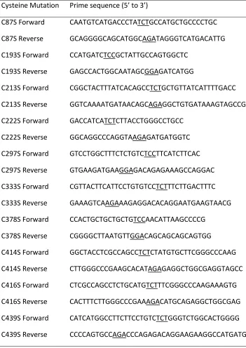
3.1 List of the PCR primers used for single site-directed mutagenesis 46
4.1 Summary of the effects of MTS reagents on [3H]NBMPR binding and
[3H]2-chloroadenosine uptake by hENT1
74
4.2 Effect of cysteine mutations on the binding of [3H]NBMPR and the
uptake of [3H]2-chloroadenosine by hENT1
77
4.3 Effects of MTS reagents on [3H]NBMPR binding by cell membranes
transfected with hENT1 and with C416A or C439A cysteine mutants
xiii List of Figures
Figure Title Page
1.1 Chemical structures of physiological nucleosides 2
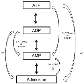
1.2 Schematic pathways of adenosine breakdown and formation 3
1.3 Chemical structures of anti-cancer cytotoxic nucleoside analogues 6
1.4 Chemical structures of antiviral cytotoxic nucleoside analogues 7
1.5 Chemical structures of ENT inhibitors 16
1.6 The Equilibrative Nucleoside Transporter family rootless phylogenetic
tree
18
1.7 Predicted 2-D topology of hENT1 created in TMPPres2D 22
1.8 Topology model of hENT1 with amino acid residues that have been
identified as structurally or functionally important determinants
25
1.9 Generated pharmacophore model aligned against NBMPR obtained
from PHASE
27
1.10 Schematic depicting the alternating access model for ENT1 29
1.11 Structural models of LdNT1.1 based on ab initio analysis 31
1.12 Chemical structures of methanethiosulfonate reagents 34
2.1 Predicted 2-D topology of hENT1 with location of 10 endogenous
cysteine residues
41
4.1 Characterization of PK15-hENT1 55
4.2 Inhibitor profile for PK15-hENT1 56
4.3 Effect of DTT on PK15-hENT1 activity 57
4.4 Varying incubation times with MTS reagents to PK15-hENT1 cells 59
4.5 Treatment of hENT1 with MMTS 61
4.6 Effects of isolating membranes to the treatment of hENT1 with MMTS 62
4.7 MMTS effects are dependent on pH 63
4.8 Effects of the treatment of hENT1 with MMTS to inhibitor binding 64
xiv
Figure Title Page
4.10 Effects of the treatment of hENT1 with MMTS on inhibition of substrate
uptake
68
4.11 NBMPR and adenosine are unable to protect against MMTS effects 69
4.12 Treatment of hENT1 with MTSES 70
4.13 Treatment of hENT1 with MTSET 72
4.14 Characterization of PK15-C87S 76
4.15 Treatment of C87S with MMTS 78
4.16 Treatment of C87S with MTSET and MTSES 79
4.17 Characterization of PK15-C193S 80
4.18 Treatment of C193S with MMTS 82
4.19 Treatment of C193S with MTSET and MTSES 83
4.20 Characterization of PK15-C213S 84
4.21 Treatment of C213S with MMTS 85
4.22 Treatment of C213S with MTSET and MTSES 86
4.23 Characterization of PK15-C222S 88
4.24 Treatment of C222S with MMTS: NBMPR binding to C222S is insensitive
to MMTS
89
4.25 Treatment of C222S with MTSET and MTSES 90
4.26 Cell membrane treatment of C222S with MMTS 91
4.27 Cell membrane treatment of C222S with MTSET 92
4.28 NBMPR binding to C222S is insensitive to pH 94
4.29 Characterization of PK15-C297S 95
4.30 Treatment of C297S with MMTS 96
4.31 Treatment of C297S with MTSET and MTSES 97
4.32 Characterization of PK15-C333S 99
4.33 Treatment of C333S with MMTS 100
xv
Figure Title Page
4.35 Characterization of PK15-C378S 102
4.36 Treatment of C378S with MMTS 104
4.37 Treatment of C378S with MTSET and MTSES: NBMPR binding to C378S
is insensitive to MTSET
105
4.38 Characterization of PK15-C414S 106
4.39 Treatment of C414S with MMTS and MTSES 107
4.40 Treatment of C414S with MTSET: enhanced inhibition of NBMPR
binding
108
4.41 Characterization of PK15-C378-C414S 110
4.42 Treatment of C378-414S with MMTS and MTSES 111
4.43 Treatment of C378-414S with MTSET: NBMPR binding to C378-414S is
insensitive to MTSET
112
4.44 Treatment of PK15-hENT1 with 2-Br 114
4.45 Characteristics of PK15-C416A and PK15-C439A: PK15-416A does not
transport 2-chloroadenosine
115
4.46 Analysis of the plasma membrane expression of hENT1 and
PK15-C416A via biotinylation and FTH-SAENTA competition assays
117
4.47 2-chloroadenosine can effectively block NBMPR binding to C416A 118
4.48 Cell membrane treatment of C439A with MTSET and MTSES 120
4.49 Treatment of C416A with MMTS, MTSET, MTSES: NBMPR binding to
C416A is insensitive to MTS reagents
121
4.50 NBMPR and adenosine are unable to protect against MTSES effects in
membranes
123
4.51 EL5 mutants bind NBMPR with high affinities 126
4.52 N379C, F390C, E391C, H392C, and D393C show no measurable uptake
of 2-chloroadenosine
127
4.53 Quantification of cell-surface hENT1 by competitive inhibition assay of
NBMPR and FTH-SAENTA of N379C, F390C, E391C, H392C, or D393C
xvi
Figure Title Page
4.54 2-chloroadenosine can effectively block NBMPR binding to N379C,
F390C, E391C, H392C, or D393C
130
4.55 NBMPR binding of all EL5 mutants except V389C are sensitive to MTSET 131
4.56 NBMPR is able to protect N379C against MTSET effects in cells 132
4.57 Adenosine is able to protect R384C, Y385C, and L386C against MTSET
effects in cells
134
5.1 Predicted topology of hENT1 with C378 highlighted in grey as the target
residue for positively charged thiol modification with MTSET
141
5.2 Simulated topologies of hENT1 based on the ab initio model of
LdNT1.1.
145
5.3 Predicted topology of hENT1 with C222 highlighted in grey as the target
residue for neutral thiol modification with MMTS
147
5.4 Simulated topology of hENT1 based on the ab initio model of LdNT1.1.
highlighting extracellular negatively charged amino acids
xvii
List of Appendices
Title Page
Permission to reprint Figures from J.Park et al. (2011) Molecular Pharmacology.
80(4):735-746
207
Permission to reprint Figures from J.Park and J.R. Hammond (2012) Molecular
Pharmacology. mol.112.079616. published ahead of print July 26, 2012
208
Permission to reprint Figure 1.6 209
Permission to reprint Figure 1.9 210
Permission to reprint Figure 1.11 212
xviii
List of Abbreviations
2-Br 2-Bromohexadecanoic acid
3TC (-)-β-L-2′,3′-dideoxy-3′-thiacytidine
A1, 2a, 2b, 3 Adenosine receptor subtype 1, 2a, 2b, 3
AP Alkaline phosphatases
Ara-C Arabinofuranosyl Cytidine
AZT 3′-azido-3′-deoxythymidine
Bmax Maximum number of binding sites
cAMP Cyclic adenosine monophosphate
CD73 Ecto-5’ nucleotidase
CeENT1-2 Caenorhabditis elegans equilibrative nucleoside transporter subtype1,
2
cGMP Cyclic guanosine monophosphate
CK2 Casein kinase II
CNS Central nervous system
CNT Concentrative nucleoside transporters
ddI 2′,3′-dideoxyinosine
DMSO Dimethyl sulfoxide
DNA Deoxyribonucleic acid
dNTP Deoxynucleoside triphosphate
DTT Dithiothreitol
EL5 Extracellular loop 5
E-NPP pyrophosphatase/phosphodiesterases
ENT equilibrative nucleoside transporters
ENT1-4 Equilibrative nucleoside transporter subtype 1, 2, 3, 4
ENT1-KO Equilibrative nucleoside transporter subtype 1 knock out mouse
E-NTPDase Triphosphate diphosphohydrolases
FLAG N-DYKDDDDK-C peptide tag
xix
ethyl]-6-N(2-nitrobenzyl) -5’-thio adenosine
G1 Gap 1 phase
G2 Gap 2 phase
G418 Geneticin
GFP Green fluorescent protein
GlpT Glycerol-3-phosphate transporter
GltT Glutamate transporter
Glut1 Glucose transporter
Gly Glycine
GTP Guanosine triphosphate
hENT1 Human equilibrative nucleoside transporter subtype 1 HIF-1 Hypoxia inducible factor 1
HMEC-1 Human microvascular endothelial cells
hOCT2 Human organic cationic transporter subtype 2
HUVEC Human umbilical vein endothelial cells
IC50 Half maximal inhibitory concentration
kDa Kilodalton
Ki Inhibition constant
Km Substrate affinity
LacY Lactose permease
LdNT1.1.-2 Leishmania donovani nucleoside transporter subtype 1.1 and 2
L-FTC (-)-β-L-2′,3′-dideoxy-3′-thia-5-fluoro-cytidine
MAP kinase Mitogen activated protein kinase MAZ Myc-associated zinc finger protein
MCF-7 cells Michigan Cancer Foundation – 7 breast cancer cell
MDCK Madin-Darby Canine Kidney Cells
MEM Modified Eagle`s Medium
mENT1 Mouse equilibrative nucleoside transporter subtype 1
xx
MFS Major Facilitator Superfamily
MMTS Methyl methanethiosulfonate
MPP+ 1-methyl-4-phenylpyridinium
MTS methanethiosulfonate
MTSES sodium (2-sulfonatoethyl)methanethiosulfonate
MTSET 2-(trimethylammonium)ethyl]methanethiosulfonate bromide
NBMPR 6-S-[(4-Nitrophenyl)methyl]-6-thioinosine
NBTGR Nitrobenzylthioguanosine
NEM N-ethyl maleimide
NHE1 Na+/H+ exchanger
NMG N-methyl-D-glucamine
NTP Nucleoside triphosphate
P1 Purinoceptor subtype 1
P1 and P2 Typanosoma brucei nucleoside transporter subtype 1 and 2
PBS Phosphate-buffered saline
pCMBS P-chloromercuribenzene sulfonate
pCREB cAMP response element binding protein
PfENT1-4 Plasmodium falciparum equilibrative nucleoside transporter subtype
PK15-NTD Pig kidney nucleoside transporter-deficient cells
PKC Protein kinase C
PMAT Plasma membrane monoamine transporter
QSAR Quantitative structure-activity relationships
RNA Ribonucleic acid
SCAM Substituted cysteine accessibility method
SDS-Page Sodium dodecyl sulfate-polyacrylamide gel electrophoresis
SLC28 Solute carrier gene for concentrative nucleoside transporter
SLC29 Solute carrier gene for equilibrative nucleoside transporter
TM Transmembrane
Chapter 1: Introduction
1.1 Nucleosides
Nucleosides are endogenous purine and pyrimidine heterocyclic nitrogenous bases
attached to a ribose or 2-deoxyribose sugar. The main naturally occurring nucleosides
include adenosine, guanosine and inosine (purines) and thymidine, uridine, and cytidine
(pyrimidines) (Figure 1.1) [1, 2]. The primary functions of nucleosides are to form the
base structural unit of nucleotides and nucleic acids. Once formed, nucleotides are then
involved in multiple events such as DNA/RNA formation (NTP, dNTP), energy supply
(ATP/GTP), and signaling pathways (cAMP, cGMP). The main source of nucleoside
formation is through a series of resourceful enzymatic cascades involving the breakdown
of nucleotides. These pathways are performed by 5’ nucleotidases found intracellularly
and by ectonucleotide hydrolyzing enzymes such as triphosphate diphosphohydrolases
(E-NTPDase), pyrophosphatase/phosphodiesterases (E-NPP), ecto-5’nucleotidase (CD73)
and alkaline phosphatases (AP) located extracellularly on plasma membranes [3-7]
(Figure 1.2). In this cyclical manner, there is a continuous supply of nucleosides and
nucleotides under basal conditions. The generation of these nucleoside pools can have
an important impact on their secondary function as signaling molecules, specifically as
purinergic agonists. For example, adenosine is a ubiquitous signaling molecule in
purinergic pathways by binding to its purinergic receptors (P1) also known as adenosine
receptors that are widely distributed throughout the body [8-10]. These P1 receptors are
a class of G protein-coupled receptors further divided into 4 subtypes (A1, A2a, A2b, and
A3) [11]. The four adenosine receptors subtypes differ in their molecular structure,
tissue distribution, and pharmacological profile and mediate diverse biological effects
[12-16].
For instance, the A1 receptor subtype is found largely in the central nervous
system (CNS) particularly in the cerebral cortex, hippocampus, cerebellum, and spinal
Figure 1.1. Chemical structures of physiological nucleosides
Figure 1.2. Schematic pathways of adenosine breakdown and formation
neurotransmitter release to cause a depression of neuronal activity. This is especially
important in times of hypoxia and ischemia because adenosine which is released in high
doses during times of stress can act as a neuroprotective agent to reduce neuronal
activity and oxygen consumption [17-19]. Adenosine receptors expressed in the
cardiovascular system can also elicit responses such as cardiac depression and
vasodilation when activated by adenosine. A1 receptor activation in the sinoatrial and
atrioventicular nodes can result in bradycardia and heart block to slow down the heart
rate; this event has been applied to treat supraventricular tachycardia. Alternatively,
adenosine binding to the A2a receptor subtype located on vascular smooth muscles of
coronary arteries can elicit a relaxation response by activation of adenylate cyclase [9,
20]. Thus it is clear that endogenous adenosine plays an important role in human
physiology and can impact a wide variety of processes including cardiovascular function,
neurotransmission, inflammatory reactions, and immune responses.
1.2 Nucleoside analogues
Given the importance of nucleosides as metabolic precursors to biologically important
molecules, their properties have been capitalized upon for the treatment of many
diseases by the design of nucleoside analogues. Cytotoxic nucleoside analogues are used
as antimetabolites that interfere with the synthesis of nucleic acids. These agents can
exert cytotoxicity either by being incorporated into and altering the DNA and RNA
macromolecules themselves, or by interfering with various enzymes involved in
synthesis of nucleic acids, or even by modifying the metabolism of physiological
nucleosides [21-26]. In this manner, nucleoside analogues can be used as antiviral,
chemotherapeutic, and immunosuppressive agents. Currently there are several
analogues that are clinically used for their anticancer properties (Figure 1.3). Specifically,
cladribine and fludarabine are two purine analogues used for their treatment of
low-grade malignant disorders of the blood [27, 28]. Pyrimidine analogues, such as
cytarabine and gemcitabine, are extensively used in the treatment of acute leukaemia;
the fluoropyrimidines 5-fluorouracil and capecitabine have shown to have activity
against colorectal and breast cancers [34-37].
Cytotoxic nucleosides are also used in anti-viral therapy against various highly
active viral diseases such as acquired immunodeficiency syndrome (AIDS) and diseases
caused by the herpes simplex virus (HSV). Anti-viral nucleoside analogues include 2′,
3′-dideoxyinosine (ddI, didanosine), 3′-azido-3′-deoxythymidine (AZT, zidovudine, Retrovir),
(-)-β-L-2′, 3′-dideoxy-3′-thiacytidine (3TC, lamivudine), and (-)-β-L-2′,
3′-dideoxy-3′-thia-5-fluoro-cytidine (L-FTC, emtricitabine) (Figure 1.4). Once again, they produce their
therapeutic effects by becoming phosphorylated intracellularly and inhibiting viral DNA
synthesis or by involving mitochondrial toxicity. However, all of these anti-viral agents
and the anti-cancer agents described above utilize membrane transporters to gain
access to target cells for further activation by intracellular kinases and cytosolic
metabolic reactions in forming their active phosphate derivatives.
1.3 Nucleoside transporters
Given the importance of nucleosides and their analogues in their roles in extracellular
signaling and intracellular nucleotide generation, the ability of cells to effectively
accumulate these molecules relies on their efficient movement across membranes.
Nucleosides and their analogues are hydrophilic due to the hydrogen bonding nature of
the hydroxyl groups found on the sugar moiety and consequently, the presence of
specialized transporters are necessary to effectively facilitate their import. Additionally,
cells that lack de novo nucleoside synthesis capabilities such as enterocytes, bone
marrow cells, and certain brain cells, rely heavily on these nucleoside transporters to
salvage nucleosides from the extracellular milieu [38-40]. Nucleoside transporters are
characterized into two separate gene families that differ in their structure and transport
mechanism, and are termed concentrative nucleoside transporters (CNT) and
Figure 1.3. Chemical structures of anti-cancer cytotoxic nucleoside analogues
Cladribine, cytarabine, fludarabine, capecitabine, and gemcitabine are nucleoside
Figure 1.4. Chemical structures of antiviral cytotoxic nucleoside analogues
The concentrative nucleoside transporters (gene SLC28) are sodium-dependent
symporters that move nucleosides unidirectionally into cells in an active energy-costly
process [44, 45]. CNTs are generally found in apical membranes of specialized epithelial
cells of the intestine and kidney and can play a major role in active absorption or
reabsorption processes. There are three sub-families of CNTs that have been cloned,
CNT1, CNT2 and CNT3, that differ in their substrate selectivity and sodium:nucleoside
stoichiometry [46, 47]. CNT1 selectively transports pyrimidine nucleosides and
adenosine while CNT2 transports purines and uridine. CNT3 transports both purine and
pyrimidine nucleosides and functions by translocating two sodium molecules per
nucleoside. These three concentrative nucleoside transporters are plasma membrane
transporters and share a general topology based on 13 putative transmembrane
domains, a long intracellular N-terminus, and an extracellular C-terminus [48].
The equilibrative nucleoside transporters (gene SLC29) are sodium-independent
facilitated diffusers that have been confirmed to transport nucleosides bi-directionally
down concentration gradients [49]. These transporters are found in most if not all cell
types, and can transport a wide variety of purines and pyrimidines and in some cases
nucleobases. There are four subtypes of ENTs (ENT1-4) that have been cloned to date
and differ in their substrate selectivities and inhibitor sensitivities [43, 50] (Table 1.1).
ENT1 and ENT2 were the first transporters to be characterized by their differential
inhibition by nitrobenzylthioinosine (NBMPR), ENT1 being sensitive to NBMPR at a nM
range [51]. ENT2, insensitive to NBMPR, transports nucleosides as well as nucleobases
such as hypoxanthine and is found predominantly in skeletal muscle, although its
expression has been detected in brain, heart, placenta, and kidney. ENT3 and ENT4 have
recently been characterized as members of the ENT family of transporters that are
active in acidic pH. ENT3 is found intracellularly and contains an endosomal/lysosomal
targeting motif and is shown to have elevated expression in human placenta [52, 53].
ENT4 found abundantly in heart and brain is also termed plasma membrane monoamine
while only transporting adenosine at acidic pH [54]. All four equilibrative nucleoside
transporters transport adenosine and therefore can influence the many physiological
processes described above such as cardiovascular tone and neurotransmission.
1.4 ENT1 subtype
The ENT1 subtype has been suggested to be the main mediator of adenosine flux and
cytotoxic accumulation of nucleoside analogues, as inhibition of ENT1 has been proven
to increase adenosine levels and adenosine signaling in cardiovascular tissues, CNS, and
kidney [55-57]. Since the ENT1 subtype is highly and widely expressed and mediates the entry of cytotoxic nucleoside analogues, it is not surprising that the loss of ENT1 expression has been correlated to drug resistance in in vitro models of malignant cancer cells. Additionally, studies with the ENT1-knock out mouse (ENT1-KO) have found higher levels of circulating adenosine and ribavirin (a nucleoside drug) compared to their
wild-type counterparts suggesting that ENT1 is a major contributor to extracellular adenosine
concentrations and uptake of nucleoside drugs [58, 59]. The ENT1 knock-out mouse was
first created by Choi et al. (2004) through deletion at exons 2-4 of the protein-coding
region of the ENT1 gene. The ENT1 knock-out mice in those studies that were less than 4
months of age, reproduced normally, were viable, showed apparent normal mortality
rates, and had normal brain anatomy [60]. However, these mice had a lower body
weight (8.7% less than wild-type littermates), and were found to show a slower rate of
intoxication and increased preference for ethanol consumption. This enhancement for
ethanol consumption was associated with increased levels of cAMP response element
binding protein (pCREB) in the striatum through an increase in glutamate signaling.
Furthermore, when examining the behaviour of the ENT1-KO mice, it was shown that
they showed less of an anxiety phenotype when compared to their wild-type
counterparts. They also showed a lowered natural aversion to the centre of an open
field indicating that they showed less anxiety; however, the locomotor activity was
Table 1.1. Characteristics of the Equilibrative Nucleoside Transporter (ENT) Family
(specific ENT1 inhibitor) in the amygdala, they also showed reduced anxiety indicating
that the behavioural effects in the ENT1-KO mice were due to a loss of ENT1 and not
through developmental changes. Additionally, phenotypic changes have been recently
identified in older ENT1-KO mice (12 months of age) in our lab. Current studies from our
lab (unpublished work from Bone and Warraich et al.) have found ENT1-null mice
acquired spinal stiffness, hind limb dysfunction and eventual paralysis by 12 months of
age. Upon further examination, it was found that the mice showed signs of ectopic
mineralization of paraspinal tissues in the cervical-thoracic region (as early as 2 months
of age) forming lesions that contained high amounts of calcium and phosphorus. These
unpublished studies by Bone and Warraich et al., are the first to identify ENT1 as playing
a role in regulating the calcification of soft tissues.
The cardiovascular system has also been studied in the ENT1-null mouse, as its
substrate adenosine is a significant contributor to vascular tone and heart function.
Initial examination of the ENT1-null mouse found it to be cardioprotected such that
myocardial infarcts were significantly smaller after subjected to ischemia (coronary
occlusion for 30 min and reperfusion for 2h) [55]. Cardiomyocytes isolated from these
mice showed no significant differences in gene expression profiles of the other ENT
subtypes or adenosine receptors indicating that there was no compensation of the loss
of ENT1 in these cells [58]. However, when examining isolated microvascular endothelial
cells from the ENT1-KO mice, there was a 2 fold increase in expression of the A2a
receptor and adenosine deaminase enzyme was observed [62]. Given that there is an
increase in circulating adenosine in KO mice, increased expression of these genes may
reflect compensatory mechanisms in the animal to handle the excess adenosine.
phorbol ester treatment by activation of protein kinase C (PKC) isoforms [67-70]. The specific targets on mouse ENT1 (mENT1) for PKC-mediated phosphorylation have been shown to involve serines 279 and 286 and threonine 274 located in the large intracellular loop betweetn transmembrane 6 and 7 [71]. Alternatively, hENT1 also
contains casein kinase II (CK2) consensus sites which are known to play a role in
regulating proliferation [72, 73]. Inhibition of CK2 phosphorylation was shown to
increase hENT1 activity and NBMPR binding in human osteosarcoma cells [74]. Our own
lab has demonstrated that the expression of a catalytically inactive CKII subunit which
inhibits endogenous CKII activity caused an enhancement in hENT1-specific NBMPR
binding and transport of the substrate 2-chloroadenosine in U2OS cells [75]. Taken
together, these data have demonstrated that ENT1 is a phosphoprotein that can be
directly phosphorylated at several sites which shows that it is involved in a complex
array of pathways in its regulation.
Besides post-translational regulation by phosphorylation, pre-transcriptional events also regulate hENT1 expression. The promoter sequence of hENT1 (involving one transcriptional initiation site 58 base pairs downstream of the TATA box) has been shown to contain consensus sites for ERE, MAZ, Sp1, AP-2, and CREB transcription factors [76]. Studies investigating hENT1 expression and activity have shown that human umbilical vein endothelial cells (HUVEC) isolated from gestational diabetic pregnancies showed a decrease in hENT1 expression [77]. A second study found hENT1 expression was reduced in HUVECs when exposed to hyperglycemic conditions by the engagement of nitric oxide, MAP kinase, and PKC. Incubation with N(G)-nitro-L-arginine methyl ester (L-NAME, nitric oxide synthase inhibitor), PD-98059 (MEK1/2 inhibitor), or calphostin C
(PKC inhibitor) prevented hENT1 downregulation in the hyperglycemic environment
[78]. Additionally, when measuring Sp1 protein levels, they found Sp1 expression
increased when hENT1 promoter activity decreased, suggesting that Sp1 may be a
negative transcriptional factor for hENT1 [79]. These studies link the importance of
adenosine-modulated placenta to fetus blood flux is damaged and loss of hENT1 causes
a loss of the endothelium ability to remove adenosine from the extracellular space.
Furthermore, ENTs are also predominantly expressed in endothelial cells of the cardiovascular system with minimal contribution from the CNTs [80]. In human microvascular endothelial cells (HMEC-1), it was found that, under hypoxic conditions, ENT1 expression was downregulated in a HIF-1 (hypoxia inducible factor 1)-dependent manner [81]. These findings indicate that an innate protective mechanism is present that serves to enhance adenosine signaling in times of cellular stress by decreasing uptake of nucleosides into cells by ENT1.
1.4.1 Characterization of ENT1
The NBMPR-sensitive transporter ENT1 was first purified from human erythrocytes
which allowed for the cloning of hENT1 from human placental cDNA [82]. Previous
studies examining cells that transported nucleosides relied on the use of radioligand
binding and uptake assays. These initial studies on ENT1 activity found that
nitrobenzylthioinosine bound to high-affinity sites on human and sheep erythrocyte
membranes and on rat, mouse, guinea pig, and dog cortical membranes in a saturable
manner (Kd ~ 0.1-1 nM) [83-88]. Binding data from these studies indicated that NBMPR
had a specific interaction with functional nucleoside-transport sites that could be
inhibited by nitrobenzylthioguanosine (NBTGR), dipyridamole (a vasodilator), and
uridine (substrate). Additionally, transport processes examined in human and sheep
erythrocytes showed [3H]NBMPR inhibition of [14C]uridine influx, consistent with a
simple competitive inhibition model (apparent Ki = 1 nM). Binding of inhibitor to these
sites was competitively blocked by uridine, a well characterized substrate for the
nucleoside transporter (apparent Ki = 1.25 and 0.9 mM, respectively). These apparent Ki
values were found to be close to the apparent Km for uridine equilibrium exchange in
human erythrocytes, indicating that NBMPR competes directly with nucleosides for the
permeation site of the nucleoside transporter, and that the inhibitor binds preferentially
Human ENT1 is 456 amino acids and is 78% identical in sequence to rat ENT1 and
79% identical to mouse ENT1 [82, 89, 90]. Splice variants of hENT1 are not reported,
however, there are multiple variants found in mouse shown to possess different
functional characteristics [75, 91]. One functional splice variant mENT1.2 altered at the
end of exon 7, lacks a potential casein kinase II phosphorylation site and has shown to
be widely expressed with mENT1.1. This mouse variant, mENT1.2, was also found to
have an altered affinity for the prototypical ENT1 inhibitor NBMPR. Another splice
variant of mouse ENT1 involving the exclusion of exon 11 during pre-RNA processing is
widely distributed in multiples tissues. This functional variant termed mENT1Δ11 bound
inhibitors and transported substrates with high affinities and was predicted to possess
nine TM domains and cytoplasmic COOH and NH2 termini. Additionally, rat ENT1 is
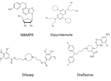
found to be inhibited by NBMPR but is resistant to inhibition by the vasodilator
compounds dilazep, draflazine and dipyridamole [82] (Figure 1.5).
Human, mouse and rat ENT1, transport a wide range of purine and pyrimidine
nucleosides with affinities ranging from 0.05 mM for adenosine to 0.6 mM for cytidine.
However ENT1 subtypes are unable to transport the pyrimidine base uracil. Human and
rat ENT1 also poorly transport the antiviral nucleosides ddC, ddI and AZT (compared to
the anti-neoplastic analogues). These anti-viral drugs are pyrimidine nucleoside
analogues that lack the C3- hydroxyl group, revealing the importance of the hydroxyl
group for permeant recognition by ENT1 [92]. In contrast, the anticancer analogues such
as gemcitabine and fludarabine are transported readily and efficiently by the ENT1
subtype [93].
ENT1 is expressed ubiquitously in all tissues but at differing levels. For example,
hENT1 is found in brain tissue with higher expression in the frontal and parietal lobes of
the cortex [94]. In the rat kidney cortex, rENT1 is found on the basolateral surface of the
tubular epithelial cells similarly seen in hENT1-GFP tagged proteins in MDCK cells in vitro
[45]. Additionally, rENT1 found in high abundance in the sinoatrial node of the heart is
suggested to play a role in modulating the chronotropic effects of adenosine [95].
on tissue and cell location. Furthermore, though ENT1 is primarily expressed as a plasma
membrane transporter, there are also studies that suggests it can also be detected in
nuclear membranes and endoplasmic reticulum [96]. Functional human ENT1 is also
found in the mitochondria where it has been suggested to play a part in the
mitochondrial toxicity effects of the antiviral agents [97]. This feature seems to be
specific for the human homologue, as rat and mouse ENT1 lack the
mitochondrial-targeting motif (PEXN). These subpopulations of intracellular ENT transporters are
thought to contribute to the nucleoside passage between the cytosol and lumen of
cellular compartments and could also correspond to a pool of intracellular transporters
available for membrane recruitment at crucial time points.
1.4.2 Homologues of ENT1
Following the initial cloning of hENT1, homologues of mammalian ENTs have been
detected in protozoa, fungi, plants, nematodes, and insects due to their sequence
similarity to mammalian ENTs [50, 98] (Figure 1.6). Within the nematode Caenorhabditis
elegans genome, there are five genes encoding equilibrative nucleoside transporters,
two of which (CeENT1 and Ce ENT2) are closely related with 94% sequence similarity.
The substrate specificities of the CeENTs closely resemble those of hENT1 and hENT2.
However, their sequence similarities to hENT1 and hENT2 are between 15-24% and they
both differ from the mammalian transporters in that they are not sensitive to NBMPR,
dilazep, or draflazine. Dipyridamole, on the other hand, does show moderate inhibition
of CeENT1 and Ce ENT2 at an IC50 300 nM, indicating that inhibitors with different
structures interact with the protein at different residues. The CeENTs also are capable of
transporting the cytotoxic dideoxynucleosides (ddI, ddC, AZT) with high efficiency [99,
Figure 1.5. Chemical structures of ENT inhibitors
The parasite Plasmodium falciparum, which is the causative microbe in malaria,
also possess ENT family members that have been designated as PfENT1-4 [40, 101, 102].
The PfENTs have broad substrate specificity and have 18% sequence identity to hENT1
and hENT2. Similar to the CeENTs, PfENTs are not proton dependent and have
conserved sequence motifs in the region of the transmembrane spanning segments
confirmeing that they are members of the ENT protein family. The majority of
transporter expression has been detected during the erythrocytic stages of the parasite
which are known to be responsible for the clinical pathogenesis of the disease. PfENTs
are efficient in transporting natural nucleosides with apparent affinities (Km) of 320 µM
similarly reported for mammalian ENTs. However, PfENTs are not sensitive to NBMPR or
the inhibitory vasodilators up to the mM concentrations.
Mammalian ENTs are also homologous to the active, proton-linked transporters
in kinetoplastid protozoa in Leshmania and Trypanosoma [103-105]. Transporters in the
Trypanosoma brucei, include two high affinity transporters (P1 and P2) that differ in
their substrate selectivity [106-108]. The P1 transporters mediate the movement of
adenosine and inosine with higher affinity (low Km) than the PfENTs. P2 transporters
passage adenosine and the nucleobase adenine and was initially identified through its
sequence similarity from the Leishmania donovani nucleoside transporter LdNT1.1.
In Leishmania donovani, there are also two nucleoside transport processes, one
selective for adenosine and pyrimidine nucleosides (LdNT1.1 and LdNT1.2) and the other
for inosine and guanosine (LdNT2) [109-111]. Comparing LdNT1.1 and LdNT1.2, they
have almost identical sequences and transport adenosine at high affinities of Km < 1 µM
however uridine is also transported at a much lower affinity. The LdNT2 transporter
selectively carries inosine with a high affinity (Km 0.3 µM) as well as guanosine (Km 1.7
µM). Despite the functional difference from mammalian transporters in the fact that
they are proton linked and therefore not equilibrative diffusers, they are confirmed to
Figure 1.6. The Equilibrative Nucleoside Transporter family rootless phylogenetic tree.
The blue box highlights hENT1 and the red boxes highlight homologs of hENT1 described in Section 1.4.1
1.5 Clinical relevance of hENT1
1.5.1 Inhibitors: NBMPR and the vasodilators
The influence of ENT1 on the extracellular levels of adenosine, a nucleoside with
physiological activity, indicates that it is a viable target for drug therapy in multiple
pathologies. Inhibition of hENT1 is of particular importance because it is the main
contributor of adenosine uptake and clearance from the extracellular space [49, 98]. By
blocking the removal of adenosine, there is enhancement of adenosine signaling
through adenosine receptors which can impact the neurological, cardiovascular, and
immunological systems [113]. NBMPR and the coronary vasodilators such as
dipyridamole, dilazep, and draflazine (Figure 1.5) inhibit ENT1 leading to enhanced
extracellular concentrations of adenosine [114-117]. Studies in murine cardiomyocytes
show that adenosine uptake is sodium independent, saturable, and inhibited by NBMPR,
dilazep, and dipyridamole [118]. In endothelial cells, the inhibition of hENT1 by
draflazine provided increased A1/A3 signaling shown to be beneficial in the
ischemic/reperfused myocardium [119, 120]. Additionally, administration of
dipyridamole during percutaneous transluminal coronary angioplasty in humans also
reduced the incidence of abrupt vessel closure by inhibition of ENTs [121]. The ENT1-KO
mice also been have shown to have a cardioprotected phenotype especially during times
of ischemia/reperfusion by having enhanced circulating adenosine levels compared to
wildtype [55].
In addition to blocking adenosine reuptake, hENT1 inhibitors have shown to be
useful in anti-cancer therapy as well. For example, the cytotoxic effect of cladribine
uptake by nucleoside transporters is complemented with co-treatment of NBMPR to
prevent drug efflux [122]. Therefore selective inhibition of ENTs may be useful in
combined drug therapy in the treatment of many cancers to improve drug efficacy.
1.5.2 Substrates: Cytotoxic nucleoside analogues
A frequent avenue in drug therapy for cancer and viral diseases utilizes cytotoxic
the target cells. Specifically, human ENT1 has been shown to enhance the transport of
chemotherapeutic agents such as cladribine, cytarabine, fludarabine, gemcitabine, and
capecitabine (Figure 1.3) [93, 123, 124]. These nucleoside analogues function in a variety
of ways: by incorporation into nucleic acids, through interfering with the nucleic acids
synthesis, and by modifying the metabolism of endogenous nucleosides. By depleting
the endogenous pools of nucleosides, the cytotoxic nucleosides increase their chances
for incorporation into newly forming DNA and RNA. Expression of hENT1 in highly
proliferating cells such as the malignant cancerous cells contributes to the selectivity for
nucleoside analogues since they require higher transport of nucleosides for their
replication [125]. For example, in acute lymphoblastic leukaemia cells, hENT1 expression
was correlated to increased sensitivity to cladribine [126]. In accord with this evidence,
the downregulation of hENT1 was also suggested to contribute to clinical resistance of
cytarabine and gemcitabine given that ENT1 is the major route of entry for these drugs
[127]. Specifically, leukemic cells resistant to Ara-C treatment showed a downregulation in hENT1 gene expression [128]. Moreover, hENT1 is now known as a positive predictive marker of patients receiving gemcitabine treatments for pancreatic cancer and
metastatic lung disease [127, 129].
Recent studies highlight the importance in measuring hENT1 levels as a
predictive tool for better drug therapy protocols that are specific to individual patients
as levels of hENT1 in patients with different breast cancers, Hodgkin’s disease, and
pancreas adenocarcinoma have shown a significant range of distribution. Additionally,
the expression of hENT1 has been shown to be positively correlated with a three-fold
increase in the survival of patients receiving gemcitabine treatment [130]. Imaging
analogues of NBMPR for specific binding to hENT1 have proven useful in determining
the abundance of transporter expression at the plasma membrane to guide drug
treatment protocols [131]. Given that cancer cells have a higher demand for
extracellular nucleosides to maintain their increased proliferation rates, nucleoside
predictive marker in pancreatic cancer and non-small cell lung cancer, it is of significant
importance to improve the selectivity and specificity of drugs for cancer cells to help
decrease normal cell toxicity and death.
1.6 Molecular characteristics of hENT1
1.6.1 Membrane topology and protein structure determinants
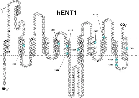
The original hydropathy plot of hENT1 indicated a 2-D topology of an intracellular
N-terminus, 11 transmembrane domains (TM), an extracellular C-N-terminus, and a large
intracellular loop linking TM6 and 7 (Figure 1.7) [90]. This generated figure was then
confirmed and supported using biochemical studies using antibodies as topological
probes in combination with glycosylation scanning mutagenesis [132]. Additionally,
hENT1 is shown to have a glycosylation site in the extracellular loop 1 at residue Asn48,
however, this modification does not seem to have an essential role in either activity or
expression at the plasma membrane [133]. Given that rat ENT1 had a different inhibitor
profile sensitivity to vasodilator compounds [134], studies based on human and rat
chimeras identified regions containing TM 3-6 to have a significant role in hENT1
functionality in both inhibitor binding and substrate interactions [135]. This region is
thought to form the major site of interaction with NBMPR and substrates as multiple
studies have shown their ability to competitively inhibit each other [86, 136]. Mutational
analyses within this region have also validated the importance of this domain. For
example, mutations of Gly154 and Ser160 in TM4 affected permeant translocation and
NBMPR binding, indicating that they possessed dual roles recognizing inhibitors and
substrates [137, 138].
Mutation of Gly154 to serine caused a loss of NBMPR binding and decreased
affinities of hENT1 for adenosine and cytidine. The important roles of glycine residues
have also been implicated in hENT1 structure as mutations at the conserved Gly179 and
Gly184 residues altered hENT1 activity and reduced plasma membrane expression
respectively [139]. Although TM3 is crucial for ENT1 function, characterization of other
Figure 1.7. Predicted 2-D topology of hENT1 created in TMPPres2D [140]
structural/functional roles for other regions of hENT1. In particular, mutations of Met89
and Leu92 in TM2 produced changes in transporter affinities for adenosine, guanosine,
NBMPR and dipyridamole [138, 141]. Additionally, Leu442 in TM11 was found to be
involved in dipyridamole sensitivity when Met33 in TM1 was first mutated to isoleucine
[100]. This study indicated a functional interaction between TM1 and TM11, regions
outside the predicted crucial domain (TM3-6) and implicated Met33 in dipyridamole and
NBMPR binding interactions. Additionally, the highly conserved residue Trp29 was found
to have a selective role in pyrimidine transport activity (uridine and cytidine) [142].
Mutation of Trp29 also decreased the ability of the inhibitors to interact with hENT1. A
helical wheel projection of this transmembrane helix (TM1) suggested that Trp29 and
Met33 were in close proximity and therefore validates the importance of this region in
hENT1 activity.
Further evidence for the involvement of the terminal domains was revealed by
mutations at Phe334 and Asp338 in TM8 altered the ability of hENT1 to be inhibited by
the coronary vasodilators [143]. Specifically, Phe334 (TM8) mutated to tyrosine
increased the rate of transport of 2-chloroadenosine suggesting an altered conformation
state of hENT1 to accept the substrate. Mutational analysis of the LdNT2 transporter has
also implicated a role for TM8 in ENT functions, wherehydrophilic residues Asp341 and
Arg345 (corresponding to Phe334 and Asp338 in hENT1) are essential for expression and
function of the transporter [110]. As a result of these studies (summarized in Figure 1.8),
it is suggested that though there is one overlapping recognition site for inhibitors and
substrates, there are multiple regions that contribute to ENT1 function. In effect, each
individual mutation that was examined did not alter all interactions with different
inhibitors or substrates which indicates that each ligand has its own individual points of
contact with hENT1. As hENT1 is shown to have therapeutic potential through its
inhibition as well as its cytotoxic nucleoside translocation, development of specific drugs
to recognize and preferentially select for hENT1 may prove helpful. Understanding the
how they interact with the protein can help in the design of such drugs. However, there
are no 3-D models of hENT1 given its unyielding nature for biophysical techniques such
as x-ray crystallography, therefore the advancement of such rational drug design has
been slow moving.
1.6.2 Pharmacophore modeling
One way to gain an understanding of the binding determinants of nucleosides and
inhibitors to their transporters is to use a computational approach to model in vitro
affinity data. In silico studies can be used as a tool to direct the exploration of new ligand
to nucleoside transporters. Given that the structure of ENTs have not yet been
elucidated, studies predicting ligand interactions have employed the use of ligand-based
quantitative structure-activity relationships (QSAR). Current biological tested models of
inhibitor and substrate interactions have found that on NBMPR, the nitrobenzyl moiety
is critical for high-affinity binding to the transporter. Specifically, electron-withdrawing
substituents at the 6-position benzyl substituent have been indicated to contribute to
high affinity binding of the transporter [144, 145]. Addition of a nitro group at that
position enhanced affinity by 50-fold compared to NBMPR itself. A specific feature of the
nitro group was its electron-withdrawing capabilities as well as its negative charge,
suggesting that negative charges interact with positively charged moieties in the binding
site of the transporter.
Given the size of the nitrobenzyl moiety and the enhanced interaction with the
nitro group, these data suggest that the area where NBMPR binds is in a large pocket
that is able to accommodate its chemical structure containing an area of positively
charged residues. A separate study analyzing the ability of C2-purine position-substituted
analogs of NBMPR to inhibit ENT1 has identified that substitutions at the C2 lead to a
general decrease in the ability of the analogues to inhibit hENT1 activity compared to
NBMPR [146]. These data suggest that C2 interacts with the transporter in a very specific
Figure 1.8. Topology model of hENT1 with amino acid residues that have been identified as structurally or functionally important determinants.
ability of the purine portion of NBMPR to bind with hENT1. Additionally, examination on
the structure-activity relationship of ENTs to substrates found the requirement of sugar
moiety for transporter interaction specifically with the presence of the C3 hydroxyl [92].
Selective structural determinant for ENT1 also include C2 and C5 in comparison to ENT2
requiring only the C5 interaction [147, 148]. Further investigations using bioinformatics
found incorporation of electrostatic and steric features at the C3 position and a negative
charge at the 2,7 position of the purine (3,5-position pyrimidine) to contribute to
enhanced affinity to hENT1, validating again a positively charged region within the ligand
binding pocket [149, 150]. These 3-D QSAR models are based on correlations between
ligand affinities and variations on their structural features and validated the importance
of the substrates C3 position and the pentose ring structure for hydrogen bond
formation (Figure 1.9). As mentioned previously, these models should assist the design
of high-affinity nucleoside transporter inhibitors and substrates; however, caution
should be taken when interpreting these data before assessment in biological models.
1.6.3 Mechanism of translocation function
It is believed that nucleoside transporters share a common evolutionary origin with the
MFS (Major Facilitator Superfamily) of transporters of which the majority function as
monomers, transporting substrates in an “alternating access mechanism” [85, 151, 152].
In this manner, ENT1 is also predicted to have an extracellular and intracellular substrate
site that when bound, produces a conformational change in the transporter to reorient
itself and release the substrate on the opposite side of the membrane (Figure 1.10). In
this case, there would be two primary conformations for ENT1 that alternated regardless
of substrate binding, one inward facing and one outward facing. However, only one site
would be accessible at any time. NBMPR binds specifically to the extracellular site of the
protein and would potentially lock it into this conformation in this model [46, 153].
Additionally, depending on the permeant bound, hENT1 may be able to alter the rate at
Figure 1.9. Generated pharmacophore model aligned against NBMPR obtained from PHASE.
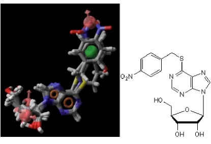
Chemical structure of NBMPR shown on the right beside the Pharmacophore model. Red points indicating hydrogen-bond acceptors (oxygen), orange points for aromatic groups, green indicated for hydrophobic regions, blue points indicate nitrogen, yellow points showing sulfur, gray is showing carbon and white highlights hydrogen.
erythrocytes showed a rapid conformation change with pyrimidine nucleosides and a
slower change with 2-chloroadenosine [51]. However, there are other complex models
of hENT1 that have been proposed where the transporter may exist as an oligomer with
allosteric sites. Several studies have found the presence of higher molecular weight
bands on immunoblots probing for ENT1 in multiple tissues in rats. From our own lab,
photoaffinity labelling of mouse ENT1 with [3H]NBMPR found that mENT1 was present
at a higher molecular weight band at approximately 100 kDa, or twice the size of the
ENT1 monomer. Additionally, unpublished data from Cunningham F. et al., found hENT1
at a higher molecular mass complex (147-180 kDa) compared to its monomer size (55
kDa) under native conditions using the blue native gel electrophoresis technique. It has
been previously suggested that there are two permeant recognition sites for hENT1, one
that is a high affinity site for NBMPR and substrates and a second lower affinity site
which may allosterically modulate the higher affinity site. Data from [3H]NBMPR
inhibition studies using dipyridamole and the lidoflazine analogues as competitive
inhibitors found them to have pseudo-Hill coefficients that were not equal to unity
indicating the presence of co-operativity or multiple sites. Additionally, studies
examining the rates of dissociation of [3H]NBMPR binding found nucleosides to enhance
dissociation rates versus inhibitors such as dipyridamole and dilazep decreased
dissociation rates. This suggests the presence of a second site that can influence the first
high affinity site which could indicate either multiple sites or co-operativity. However,
there is still not enough evidence to distinguish between the two possibilities and
mechanisms.
1.6.4 Predicted 3-D topology of ENT1
Clearly, understanding the structure, function, and mechanism of hENT1 would be of
considerable value in drug discovery. Current homology and comparative modeling of
ENT1 have generated several putative configurations of the transporter since the
Figure 1.10. Schematic depicting the alternating access model for ENT1.


![Figure 1.7. Predicted 2-D topology of hENT1 created in TMPPres2D [140]](https://thumb-us.123doks.com/thumbv2/123dok_us/7785326.1287708/43.612.111.542.102.407/figure-predicted-d-topology-hent-created-tmppres-d.webp)