Retinal images vessel width using spline interpolation
G.Shivakumari
1; N.Veeraiah
2& D. Vijay Kumar
31 M.Tech, Dept of ECE, Vijaya Engineering College, Telangana,
India. Email:[email protected]
2Associate Professor,Dept of ECE,Vijaya Engineering college, Telangana, India,
3Associate Professor, HOD, Dept of ECE ,Vijaya Engineering college, Telangana, India, Email: [email protected]
Email: [email protected]
Abstract
Vasculature segmentation and vessel caliber measurements in retinal images can improve early diagnosis of several diseases, such as diabetes, retinopathy of prematurity and hypertension. The aim of this thesis is to present a novel algorithm for improving the vessel contours obtained from binary vessel maps. This is useful for quantitative evaluations like width and tortuosity estimation. Two algorithms are described in this document. Firstly, a simple vessel segmentation strategy ltering the image using a Gaussian kernel and producing a binary vessel mask from the response image by the application of a thresholding step. Secondly, a procedure _tting the two contours of each vessel in the binary map with a cubic spline curve, under a parallelism constraint between the two splines. The second algorithm is the main focus of this work. The performance of the algorithm has been evaluated on the publicly available REVIEW database, which contains a set of images with vasculature showing different characteristics. Images also include several manual measurements made by three independent observers.
Keywords: Retinal images; vessel width; spline interpolation 1. Introduction
The key components of Vessel segmentation
algorithms are automated radiological
diagnostic systems. The essential step in Blood vessel delineation on medical images to solving several practical applications such as diagnosis of the vessels (e.g. stenosis or malformations) and registration of patient images obtained at different times. Segmentation methods vary depending on the application domain, imaging modality method being automatic or semi-automatic, and other specific factors. There is no single segmentation method that can extract vasculature from every medical image modality. While some methods employ pure intensity-based pattern recognition techniques such as thresholding followed by connected component analysis some other methods apply explicit vessel models to extract the vessel contours. Depending on the image quality and
the general image artifacts such as noise, some segmentation methods may require image preprocessing prior to the segmentation algorithm . On the other hand, some methods apply post-processing to overcome the problems arising from over segmentation. In current vessel segmentation tracking based method, covering both early and recent literature related to vessel segmentation . We introduce active contour segmentation method for extracting the vessels from the CT cardiac images.
step is taken along vessel direction and the procedure is repeated until stop conditions are satisfied The results demonstrate the good performance of method in the whole tracking process and detecting more complete vessel network in the ocular fundus photograph. Hanwei Shen[2], present a semiautomatic image segmentation tool which combines conventional manual segmentation utilities with a novel automatic image segmentation algorithm. We use a bimodal thresholding algorithm to determine the boundary segments in the local region .When the user picks an initial point, a small local window is placed around this point. A local histogram is then computed according to the values of pixels located inside the window. The pixel value in a histogram that separates the pixels of an image into two major groups determines the most significant value. Image segmentation, the process of defining boundary domains in 2D images, the boundaries of interesting regions must be defined before surface reconstruction mesh generation, and other modeling operations begins. This approach works only in 2D images.
2. Related Work 2.1 Previous method:
The method produces segmentations by
classifying each image pixel as vessel or
nonvessel, based on the pixel’s feature vector. Feature vectors are composed of the pixel’s intensity and continuous two-dimensional Morlet wavelet transform responses taken at multiple scales. The Morlet wavelet is capable of tuning to specific frequencies, thus allowing noise filtering and vessel enhancement in a single step. We use a Bayesian classifier with
minimum squared error classifier. The probability distributions are estimated based on a training set of labeled pixels obtained from manual segmentations.
2.2 Proposed Method:
This paper describes a novel algorithm for smoothing raw vessel contours in binary retinal vessels masks. For each processed vessel, after identifying its spline-smoothed centreline, the algorithm finds two cubic spline curves fitting the jagged contours. The coefficients of these splines are computed by solving an over constrained system including both standard spline formulae and a parallel-tangent constraint. This constraint is very well suited for retinal vessels, because it ensures _Research partially supported by Lever hume trust grant RPG-419 that the vessel profile is as similar as possible to a 2-D curvilinear pipe with parallel borders. The algorithm performs effectively on both linear and curved vessels. Our algorithm does not aim to improve vessel detection but to improve the accuracy of width estimation from binary maps obtained from vessel detection algorithms.
3. Implementation
3.1 Vessel segmentation algorithm:
segmentation, this methodology turns out to be e_ective and easy to implement because it does not require any previous training step. An approach similar to the one described below was _rst proposed by Chaudhuri et al. [6]. In image processing, convolving an image with a mask requires moving the kernel through all the pixels of the image. Let I be an image of size M _N and g the convolution mask of size p _ p, where p is an odd integer; then for each pixel (i; j) in I the _lter response at that pixel is
The goal is to devise a convolution mask that is suitable for vessel detection in retinal images. The key point is to notice that the intensity pro_les of the cross-section perpendicular to a vessel at any point can often be modelled as a Gaussian curve. Hence, a set of Gaussian-shaped _lters turns out to be appropriate for vessel segmentation (but see [1,2,5] for a discussion of the limitations of this approach). The results presented below were obtained using the _lter shown in Figure 1 and described by the function
Since vessels in retinal images have di_erent amplitudes and orientations, the kernel has to be rotated by a _nite set of angles _ 2 [0; 180_] and its spread, _, has to be varied as well, in order to capture as many vessel pixels as possible. The maximum response will be obtained when the _lter orientation and
amplitude approximate the propagation
direction and the diameter of the vessel in the considered pixel.
Figure 1: Result of a segmentation procedure. (a) Original image of size 2240_1488. (b) Segmented binary image obtained using a Gaussian _lter with _ set to values from 2 to 4. The threshold value used is t = 48.
3.2 Mathematical background on spline interpolation:
This section provides a theoretical overview on spline data interpolation, a technique which is widely used in several subjects, as approximation theory and numerical analysis. A spline is a piecewise polynomial function that is at least of class C2 at the points where the polynomial pieces join up. These points are called knots. Cubic splines are one of the most commonly used splines in interpolating problems; they are called cubic because their pieces are third-degree polynomials. This chapter describes how to derive the equations for cubic spline curves that interpolate a _nite set of points.
Given the data points (xj ; yj) with j = 0; 1; : : : n, consider two consecutive knots (xi; yi) and (xi+1; yi+1). The general cubic function de_ned in the interval between these two points is
Where ai ; bi; ci and di are the coe_cients of the third-degree polynomial in the ith interval. Since the cubic must go through at (xi; yi) and (xi+1; yi+1), then
Where hi is the width of the ith interval, i.e. hi = xi+1 � xi. To ensure a smooth _t across the boundary between two consecutive intervals, the continuity of _rst and second derivatives is enforced at knots. The _rst two derivatives are:
Naming Si and Si+1 the second derivative of the function evaluated at (xi; yi) and (xi+1; yi+1) respectively, the following equations are obtained:
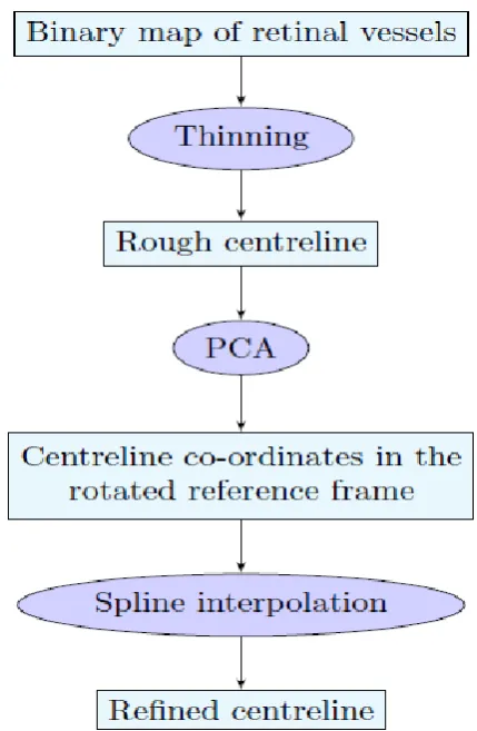
Fig 2: Flowchart representing the main steps of the algorithm for vessel centreline extraction and re_nement
This section presents an algorithm extracting and re_ning the centreline of each vessel. A owchart, representing the main steps of this procedure, is shown above for the sake of clarity.
(a)Thinning
A _rst, rough set of centrelines can be easily obtained from a vessel binary map using a morphological thinning algorithm. This method iteratively erode exterior pixels from the detected vessel structure, until no more erasable pixels exist. The resulting image is a binary mask of the vessel skeleton, i.e. a
connected chain of pixels 1 pixel thin. It is useful for further processing to separate this binary structure into individual vessel segments by removing branching points from the vessel skeleton. These points are pixels belonging to the thinned centre line that have more than two neighbours. After that, segments that are less than 15 pixels long are removed, as they are considered insigni_cant for later analysis. Before proceeding with centreline re_nement, a _rst estimate of vessel width can be computed using the distance transform of the binary vessel mask. The result of this operation is a graylevel image in which pixel intensity values are the Euclidean distances from the considered pixel and the closest background pixel. By doubling the distance values along the thinned centreline, a coarse estimate of vessel diameter in these points is obtained.
(b)PCA:
The scatter matrix H is a 2-by-2 positive-de_nite and symmetric matrix, hence its eigenvalues are always real valued and positive. The eigenvector e1 with the largest eigenvalue is the direction of greatest data variation; the other eigenvector e2 is orthogonal to e1 given the above properties of H. Thus, the x and y axes are centred in p and rotated into e1 and e2 directions. The co-ordinates of pj 2 P in the new reference frame are
where _ is the matrix that has e1 and e2 as columns.
3.4 Vessel edge extraction:
The next step is to extract vessel border points from the binary retinal maps starting from the spline-smoothed centreline and the preliminary vessel widths already computed. The goal is to identify vessel edges using the information given by pixel intensity pro_le along vessel cross-sections. The re_ned centerline is smooth enough to compute reliably, most of the times, orthogonal segments that do not intersect each other. Hence, for each centreline pixel Cj , the perpendicular dj is computed. To ensure that segment dj will be long enough to pass even through the widest vessels, its length is set to wj , where wj is the preliminary vessel width at Cj , estimated from the distance transform described
above.
The image pixel intensity pro_le along dj is computed using linear interpolation. Since the image is binary, vessel edge points are those pixels where intensity pro_le changes value.
Table 1 reports the performance of our method and its comparison with algorithms reported in the literature: Extraction of Segment Profiles (ESP) procedure [6] and Xu’s graph based method [7]. The accuracy achieved with simple double spine fit with parallelism constraint at knots is comparable accuracy to that of specialized, sophisticated width estimation
algorithm. The proposed method has a performance comparable to the observers in HRIS dataset: = 0:760 pixels (2.75 times the mean of observers’). Nevertheless, Xu’s graph-based method and ESP algorithm perform slightly better. On the contrary, in CLRIS
dataset, the spine-based
5. Conclusion
This thesis has proposed a novel algorithm re_ning vessel boundaries in retinal binary vessel maps. Since these black-and-white vessel maps often present jagged edges that are not suitable for width estimations, the spline-based algorithm provides an improved version of the input binary image, in which vessel contours are smoothed and re_ned. However, this procedure does not perform vessel extraction; rather, it re_nes the boundaries of vessels that have been already located by previous vascular segmentation methods. 6. References
[1] Soares J.V.B, Leandro J.J.G, Caesar Jr.R.M, Jelinek H.F, Cree M.J, "Reti- nal Vessel Segmentation Using the 2-D Gabor Wavelet and Supervised Classi_cation", IEEE
Trans on Med Imaging, Vol 25, No. 9, September 2006, pp 1214-1222.
[2] Lupas,cu C.A, Tegolo D, Trucco E, "FABC: Retinal Vessel Segmentation Using AdaBoost", IEEE Trans on Info Tech in Biomedicine, Vol 14, No. 5, September 2010, pp 1267-1274.
[3] Chutatape O, Zheng L, Krishnan S, "Retinal blood vessel detection and tracking by matched Gaussian and Kalman _lters", Proc IEEE Int Conf Emg Bio Soc, Vol 20, No.6, 1998, pp 3144-3149.
Location Re_nement", PLoS ONE, Vol 7, Issue 3, March 2012, e32435, pp 1-12.
[5] Vermeer K.A, Vos F.M, Lemij H.G, Vossepoel A.M, "A model based method for retinal blood vessel detection", Comput Biol Med, Vol 34, 2004, pp 209- 219.
[6] Chaudhuri S, Chatterjee S, Katz N, Nelson M, Goldbaum M, "Detection of blood vessels in retinal images using two-dimensional matched _lters", IEEE Trans on Med Imaging, Vol 8, No. 3, September 1989, pp 263-269. [7] Curtis F.G, Wheatley P.O (1970), Applied numerical analysis, Addison- Wesley, Reading, 1989.
[8] Al-Diri B, Hunter A, Steel D, Habib M, Hudaib T, Berry S, "REVIEW – A Reference Data Set for Retinal Vessel Pro_les", 30th Annual International Conference of the IEEE Engineering in Medicine and Biology, August 2008, pp 2262-2265.
[9] Al-Diri B, Hunter A, Steel D, "An active contour model for segmenting and measuring retinal vessels", IEEE Trans on Med Imaging, Vol 28, No. 9, March 2009, pp 1488-1497.
[10] Gregson P.H, Shen Z, Scott R.C, Kozousek V, "Automated grading of venous
beading", Computers and Biomedical
Research, Vol 28, No. 4, 1995, pp 291-304.
[11] Brinchmann-Hansen O, Heier H, "Theoretical relations between light streak characteristics and optical properties on retinal vessels", Acta Ophthalmologica supplement,
Trans on Med Imaging, Vol 13, No. 4, December 1994, pp 619-626.
[13] Lowell J, Hunter A, Steel D, Basu A, Ryder R, Kennedy R.L, "Measurement of retinal vessel widths from fundus images based on 2-D modeling", IEEE Trans on Med Imaging, Vol 23, No. 10, October 2004, pp 1196-1204.
1st author
G.Shivakumari pursuing her M.Tech, from Vijaya Engineering College, Telangana, India. Email: [email protected]
2nd Author
N.VEERAIAH completed his M.Techand working as a Associate Professor, from Vijaya Engineering college, Telangana,India, Email: [email protected]
3rd Author