COMPUTER ASSISTED
DECISION MAKING FOR IMAGE
UNDERSTANDING IN MEDICINE
by
Paul Martin Taylor B.Sc. M.Sc.
Dept of Medical Physics and Bioengineering University College London
Gower Street London W C IE 6BT
Advanced Computation Laboratory Imperial Cancer Research Fund
61 Lincoln’s Inn Fields London W C2A 3PX
Submitted for
ProQuest Number: 10016126
All rights reserved
INFORMATION TO ALL USERS
The quality of this reproduction is dependent upon the quality of the copy submitted. In the unlikely event that the author did not send a complete manuscript and there are missing pages, these will be noted. Also, if material had to be removed,
a note will indicate the deletion.
uest.
ProQuest 10016126
Published by ProQuest LLC(2016). Copyright of the Dissertation is held by the Author. All rights reserved.
This work is protected against unauthorized copying under Title 17, United States Code. Microform Edition © ProQuest LLC.
ProQuest LLC
789 East Eisenhower Parkway P.O. Box 1346
Leis na bha dhomh de bhreannachadh gun d'rinn mifaileas strV,
gun d'rinneadh gleachd le m' che'ill
(With all I had o f apprehension I put up a shadow o f a fight; my reason struggled)
ACKNOWLEDGEMENTS
H eartfelt thanks are due:
to my supervisors John Fox and Andrew Todd-Pokropek; to D avid Ingram who gave me space to finish;
to Claire Dicks-Mireaux, John Pritchard and Carol Young o f Great Ormond Street Hospital;
to Regina Pauli and the radiographers at the Jarvis Breast Screening Centre;
to all the radiologists who contributed including N igel Barratt, Beatrice Barreau, Julie Cook, Jackie Davis, M arie-H élène Dilhuydy, Ruth English, M ike King,
Caroline Kissen, M ike Michel, N ick Perry, Basil Shepstone, Kate Stoner and Kate Wharmsley; to Lida Graupner, Saki Hajnal, Paul Krause, Simon Parsons, A li Ram enzadeh and Jeremy Wyatt, at the Im perial Cancer Research Fund;
Abstract
We are all familiar with the after-the-fact tone — weary, self-justifi catory, aggrieved, apologetic - shared by ship captains appearing before boards o f inquiry to explain how they came to run their ships aground, and by authors composing forewords.
John Lanchester
This thesis considers the different kinds of computer system which assist in the interpretation of medical images. These systems contain information represented in two very different ways: as images and as symbolic knowledge. If a decision aid is to provide access to all the information that might assist a radiologist, it must be able to employ information represented in images and as symbolic knowledge. Image processing is needed to provide the descriptions required by knowledge-based systems. Symbolic representations are needed to relate image data to the decisions radiologists take. This thesis sets out a design for decision aids which combine image data and symbolic representations.
The approach is based on a model of decision making, a symbolic decision procedure which constructs arguments, or lines of reasoning, about possible solutions to a problem. An extension to the decision procedure provides a model of three generic tasks in image interpretation: detection, classification and measurement. The extended decision procedure is implemented as a program which allows the symbolic decison procedure to draw on information obtained from processing images.
A generic architecture, based on the extended decision procedure, has been used in the implementation of two prototypes: one to assist in the interpretation of breast X-rays or mammograms and one to assist in the use of CT for the management of abdominal tumours.
Contents
Chapter One: Radiology and Computers
1.1 Introduction
1.2 The Medical Context
1.2.1 Establishing the need for the examination 1.2.2 Performing the examination
1.2.3 Interpreting the image 1.2.4 Reporting the findings
1.3 Computer Support for Radiological Tasks 1.4 Summary
1.5 Chapter Outlines
12
12 15 15 18 26 30 33 36 37Chapter Two: Decision Aids for Radiology
39
2.1 Introduction 39
2.2 Decision Support 40
2.3 Computers and Medical Image Interpretation 46
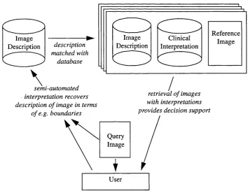
2.3.1 Image databases 46
2.3.2 Decision systems based on numerical methods 52
2.3.3 Expert systems 57
2.3.4 Image-processing systems 63
2.4 Conclusion 67
Chapter Three: The Signal/Symbol Problem
71
3.1 Introduction 71
3.2 Signal and Symbol Information 72
3.2.1 The signal representation of information 73 3.2.2 The symbolic representation of information 74 3.3 Systems Combining Signal and Symbol Information 76
3.3.1 M ultimedia systems 77
3.4 Discussion 99 3.4.1 Mapping between signals and symbols 99 3.4.2 The representation of signal data 106
3.5 Conclusion 109
Chapter Four: A Framework for Decision Support
111
4.1 Introduction 111
4.2 The Symbolic Decision Procedure 113
4.3 Decision M aking in Radiology 121
4.4 The Extended Decision Procedure 123 4.4.1 Rules for proposing candidates and arguments 123
4.4.2 The interpretation rules 127
4.5 Supporting Image Interpretation Tasks 134 4.5.1 The detection of calcifications 134 4.5.2 The classification of calcifications 137 4.5.3 The measurement of tissue density 139
4.6 Discussion 142
Chapter Five: A Generic Architecture
147
5.1 Introduction 147
5.2 Principles for the Design of a Radiologist’s Workstation 148 5.2.1 Integration of functions in a single environment 148 5.2.2 Combination of image processing and reasoning 149 5.2.3 A task-oriented interface 150 5.3 Components of the Architecture 150
5.3.1 User interface 152
5.3.2 Information sources 154
5.3.3 Information-processing components 159
5.4 Functional Components 161
5.5 Conclusion 164
Chapter Six: Decision Support for Mammography
167
6.1 Introduction 167
6.2 The Medical Context 167
6.3 Decision-Making in Mammography 170 6.3.1 The analysis of breast masses 171 6.3.2 The classification of microcalcifications 176 6.3.3 Assessment of tissue density 188
6.3.4 Conclusions 189
6.4 Image Processing for the Detection of Calcifications 192 6.5 Image Processing for the Classification of Calcifications 193
6.5.1 Magnin [M agninl989] 194
6.5.2 Lefebvre [Lefebvrel991] 196
6.5.3 Patrick [Patrick 1991] 198
6.5.6 Chitre [Chitrel994] 203 6.5.7 Nishikawa [Nishikawal993, Nishikawal994] 206
6.5.8 L o [L ol9 9 5] 208
6.5.9 Parker [Parker 1995] 209
6.5.10 Discussion 212
6.6 Discussion 216
Chapter Seven: The Mammography Workstation
218
7.1 Introduction 218
7.2 Image Processing Operators 219
7.2.1 Detection of calcifications 219 7.2.2 The classification of calcifications 222 7.3 The Representation of Medical Knowledge 224 7.4 The Representation of the Protocol 230
7.5 Overview of the Prototype 232
7.6 Conclusions 236
Chapter Eight: Evaluation
237
8.1 Introduction 237
8.2 Accuracy of the Knowledge Base 237 8.3 Association between Knowledge and Image Processing 241 8.3.1 An informal evaluation of the image-processing 242 8.3.2 Agreement between radiologists and processing 246 8.4 Value of the Tool as an Aid to Decision Making 252
Chapter Nine: Decision Support for Abdominal CT
258
9.1 Introduction 258
9.2 The Medical Context 258
9.3 Decision Making in Investigations of Neuroblastoma 259
9.3.1 Diagnosis 259
9.3.2 Staging 260
9.3.3 Response to Treatment 262
9.3.4 Conclusions 263
9.4 Image Processing for the Management of Neuroblastoma 265 9.4.1 Detection of the Midline 265 9.4.2 M easurement of tumour volume 267 9.5 The Representation of Medical Knowledge 272 9.6 The Representation of the Protocol 275
9.7 Overview of the Prototype 277
9.8 Conclusion 278
Chapter Ten: Discussion
280
10.1 Introduction 280
10.2 The Potential for Decision Support in Radiology 280
10.3 The Signal-Symbol Question 283
10.5 The Generic Architecture 289 10.6 The Choice of Clinical Problem 292
10.7 The Mammography System 294
10.7.1 The construction of the knowledge base 295
10.7.2 Image processing 298
10.7.3 The evaluation of the mammography system 299
10.8 The Neuroblastoma System 300
10.8.1 The measurement of tumour volume 301 10.8.2 Decision support for staging and assessment 302
10.9 Conclusions 302
10.10 Future W ork 304
Appendices
306
11.1 Image Processing Calculations 306
11.1.1 Compactness 306
11.1.2 H u’s Invariant Moments 306
11.1.3 Eccentricity 308
11.1.4 Elongation 308
11.2 Knowledge base used in the mammography system 310
11.2.1 Hierarchy of terms 310
11.3 Properties of calcifications 313
11.4 Positive signs for the possible diagnoses 314
11.5 The Mammography Protocol 317
11.5.1 Graphical Representation 317
11.5.2 Knowledge Base 320
List of Tables
Table 1 : kinds of medical image referred to in this thesis 25
Table 2: the toplevel of the Symbolic Decision Procedure 126
Table 3; knowledge required for the example of a breast lump 127
Table 4: the three different image interpretation tasks 128
Table 5: a model to identify a region of interest 131
Table 6: rules describing the three image interpretation tasks 133
Table 7: rules and facts used to represent the detection task 137
Table 8: rules and facts used to represent the classification task 139
Table 9: rules and facts used to describe the measurement task 141
Table 10: a classification of circumscribed lesions [T abari983] 171
Table 11: spectrum of Breast Mass Appearances [Feigl992] 175
Table 12: characteristics of ductal and lobular calcifications 178
Table 13: microcalcifications seen in 1044 mammograms[Lanyi 1987] 180
Table 14: classification of the American College of Radiology 182
Table 15: the predictive value of microcalcifications 185
Table 16: measures used in the classification of calcifications 214
Table 17: results from testing the four individual segmentors 221
Table 18: results from testing the three combined segmentors 222
Table 19 decision specifications for the mammography workstation 225
Table 20: the mean scores for the radiologists’ ratings 241
Table 21: values for five calcifications 243
Table 22: weighted kappa values 248
Table 23: true and false positives, true and false negatives 256
Table 24: the INSS criteria for the staging of neuroblastoma 261
Table 25: the INSS criteria for the assessment of neuroblastoma 262
Table 26: results for the connectivity and threshold algorithms 271
Table 27: decision specifications for the neuroblastoma workstation 273
List of Figures
Figure 1 : a planar X-ray of the compressed breast 19
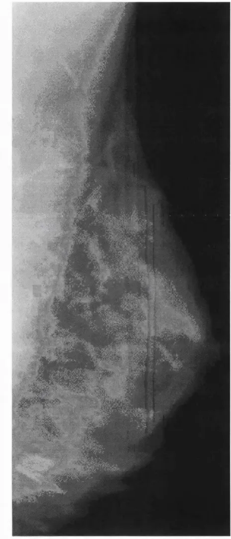
Figure 2: an example of a breast X-ray or mammogram 20
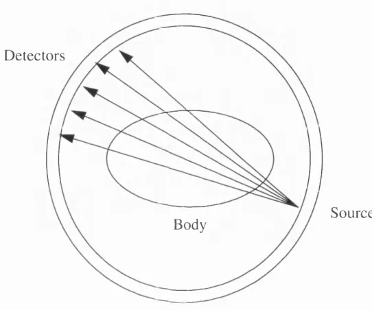
Figure 3: a fan beam of X-rays is used to generate a 2-D image 22
Figure 4: an example of a X-ray CT slice 23
Figure 5: a model of static human vision, after [Pizerl990] 28
Figure 6: schematic outline of the examination of a visual display 29
Figure 7: Image Indexing by Content 50
Figure 8: one of the rules in Smets et al.’s system. 88
Figure 9: image data are combined with symbolic knowledge 100
Figure 10: AXON 101
Figure 11: I^C 102
Figure 12: the Collins et al. system 104
Figure 13: AutoMEX 105
Figure 14: the decision procedure 117
Figure 15: the domino model 118
Figure 16: procedural interpretation of the rule describing detection. 130
Figure 17: the method used to measure the density of tissue 140
Figure 18: the operation of the rule describing the detection task 143
Figure 19: the components of the generic architecture 151
Figure 20: the image reporting function 161
Figure 21 : the decision support function 162
Figure 22: the task-management function 163
Figure 23: the information retrieval function 164
Figure 24: the anatomy of the female breast 168
Figure 25: the five types of microcalcifications 177
Figure 26: a protocol [Dilhuydy 1994] 184
Figure 27: a protocol [M onseesl995] 187
Figure 28: the knowledge model 226
Figure 29: the interface of the mammography workstation 233
Figure 30: the system used to display mammograms 234
Figure 31: the display of decision support 235
Figure 34: values of eccentricity and elongation 245
Figure 35: plots of mean radiologists’ rating 250
Figure 36: plots of mean radiologists’ rating 251
Figure 37: the decision support advice used in the evaluation 254
Figure 38: pooled ROC curves for four trained radiographers 255
Figure 39: an example of a CT scan for a patient with neuroblastoma 265
Figure 40: the output from the spinefinder program 266
CHAPTER ONE
RADIOLOGY AND
COMPUTERS
Whilst my Physitians, by their love are growne Cosmographers, and I their Mapp
John Donne
1.1
Introduction
Consider the sequence of events through which a woman who has detected a
lump in her breast is diagnosed as having cancer and given access to treatment. First
she will see her GP, who will refer her to a breast clinic. There she will see first a
surgeon and then a radiologist. A number of investigations will be performed. Almost
certainly one of them will be a mammogram - a breast X-ray - performed by a trained
radiographer. A sample of tumour cells will be taken and inspected by a pathologist.
All of these experts - GP, surgeon, radiographer, radiologist, pathologist - will have
undergone years of education and training and their expertise represents a scarce
resource which must be used efficiently. Each encounter, therefore, is organised by
the most informed decision to be made at every stage. This requires that, at each stage,
the experts have access not just to the information supplied by their colleagues but to
other data about the patient and her family, as well as about the latest medical research
and the locally available investigations and treatments.
The effective provision of medical care requires the careful management of
different kinds of information: to ensure sound administration, permit clear communi
cation and guarantee informed decision making. An obvious component of the required
infrastructure is an appropriate computer system. Consider the role of the radiologist.
Having met the patient, he or she must decide on the appropriate investigations. If these
are to include a mammogram, the patient must be seen by the radiographer who will
take the mammogram. The radiologist will then inspect it and provide a report for the
surgeon. Advances in digital mammography mean that in the future the mammogram
may exist only as an array of data in a computer. The trend towards electronic patient
records and advanced hospital information systems suggests that a patient’s history will
exist only on computer. Medical databases will allow radiologists to obtain information
about the available investigations and treatment options via computer. In such a
scenario the computer will underpin much of the activity of the radiologist. W hat kind
of computer system would provide him or her with the best possible tool?
A radiologist interpreting a mammogram would want a single computer system
which combined the ability to display images with access to information about the
patient and access to databases of medical knowledge and to recommended care
pathways. He or she would want to use this computer to assist in interpreting the
mammogram, processing the image in order to help detect abnormalities and classify
certain features. This thesis is concerned with the design of a computer system that
Systems to help in the interpretation of medical images have been developed in
a number of different areas of computer science, areas such as image databases, image
processing, expert systems and numerical methods. One of the aims of the thesis is to
evaluate this diversity of svstems from a unifving perspective, one in which radio
logists are viewed as decision-makers and computer systems are assessed for the
contribution thev can make to improving radiologists’ decision making. The argument
which will be made is that each of the different kinds of system has advantages and
limitations, and there is therefore a strong case for exploring the extent to which
different techniques can be used to complement each other. This, then, is the second
aim of the thesis: to consider how the technologies developed within these different
fields can be combined to provide a system within which different forms of decision
support, based on different kinds of information, would be available for use as
required. In particular, the question is asked: how can information represented in
symbolic knowledge bases be combined with information represented in images?
Numerous papers have been published reporting optimistic results in tests of
decision support systems, including 29 rigorous studies of the effects of such systems
on clinical practice [Johnston 1994]. Few have entered widespread clinical use. Recent
papers have discussed the failure of research into decision support to change clinical
practice and a number of explanations have been proposed. Researchers responding to
the perceived failures of early decision support systems suggest that both effective
performance and user acceptability require a co-operative model of decision making in
which the abilities of user and machine are matched [M iller1990]. A third aim o f this
thesis is to develop a conceptual framework within which diverse forms o f information
can be made available to a radiologist as he or she requires. The final aim is to demon
strate the practicalitv of these ideas through the implementation of two prototype
decision support systems aimed at quite different areas of medicine: the use of
mammography in the investigation of breast cancer and the use of abdominal CT in the
The next section gives a brief introduction to the sequence of activities which
are involved in carrying out a radiological investigation. This provides the background
for a discussion of the role to be played by computers in assisting radiology.
1.2
The Medical Context
M uch o f the work that is done on the application of computers to the analysis of
medical images is carried out in isolation from clinical practice. An attempt to provide
practical assistance for radiologists making decisions about medical images ought to be
based on a more thorough analysis of the tasks radiologists perform and how they
perform them. The next four sub-sections consider four stages in the radiological
process: establishing the need for an investigation, performing the investigation, inter
preting the results and communicating the findings.
1.2.1
Establishing the need for the examination
The process of radiology begins when additional information is needed to make
a decision about a patient. The need may be identified by a clinician, by the patient or,
in the case of population screening, by some other authority. It is frequently assumed in
discussions of radiological practice that the decision involved is a diagnostic one,
although radiological investigations are also used to inform decisions taken in the
management of patients with known diseases. Establishing the need for the exam
ination is a key part of the interface between radiology and clinical activity, it involves
focusing on the clinical problem, identifying possible procedures, determining their
appropriateness and appraising their impact on subsequent strategy. In the past the
radiologist would often not have played any part in this process, his or her role was
simply to interpret the examinations requested by the referring physician. There is now
need to minimise unnecessary investigations and radiologists may help in the selection
of an appropriate procedure.
A number of papers in medical decision making, e.g. [Kuhns 1989] and
[Chang 1990] have considered how the need for radiological investigations is assessed
and have argued for the use of mathematical models in appraising their value. The
concern here is to minimise the number of unnecessary investigations by considering
the impact on diagnosis of the information obtained from an investigation. Medical
diagnosis is a ‘hypothetico-deductive’ process, in which the clinician uses some initial
information to make a tentative assessment of the probability of disease and this
assessment then guides decisions about what information to gather, which investiga
tions to request, and the new information in turn allows the initial assessment to be
updated. It is important to note that the clinician is dealing in probabilities: his or
deductions are, in most cases, not being made from principles that have the status of
natural laws but from knowledge gained through collective experience and from
imperfect clinical data.
One model o f radiological decision making [Kuhns 1989] requires clinicians
first to calculate the degree of certainty they should have in a diagnosis before selecting
a treatment - thus setting a threshold above which an investigation is superfluous
because the already available evidence provides an adequate basis for recommending
treatment - and second to determine the impact on that certainty which a positive test
result would have - setting a second threshold below which an investigation is unnec
essary since even a positive result would not provide sufficient evidence to warrant
treatment. This approach assumes both that the decision can be clearly structured, with
the set of possible diseases and their indications laid out, and that reliable data will be
available on the prior probability of diseases and of the probabilities of the diseases
given a particular test result. Often things will not be so straightforward, and when they
interest in this thesis. It is assumed that there will be an increasing move towards the
establishment and use of consensus guidelines for clinical practice [Audetl990,
Grim shaw l993] and that this will be supported by - and create a demand for - decision
support tools [Renaud 1994].
Mendelson [M endelsonl995] describes how the American College of
Radiology is drawing up appropriateness guidelines for radiological investigations.
The aim of these guidelines is to provide an orderly sequence of studies most likely to
assist in the diagnosis and management of a clinical condition. Guidelines are produced
only for clinical conditions meeting a set of criteria, to do with the importance o f the
condition, the variability of practice and outcomes in the absence of guidelines and the
existence of evidence on which guidelines may be based. A panel of experts, drawn
from all the relevant professional specialities, is initially asked to consider a table of
evidence based on 15-20 studies, classified according to the type of study, the number
of subjects, the purpose of the study and the strength of the finding. Successive
questionnaires are then sent to each member of the panel, to elicit information about the
appropriateness of possible imaging techniques. The process is terminated when 80%
of the panel agree, the guideline is then written to incorporate abstracted recommenda
tions and indicate areas of uncertainty. The guideline, which must meet standards of
validity, reproducibility, clarity, flexibility and applicability is then disseminated in
different forms (as a report, a reference guide and a patient guide) through government,
healthcare and other agencies. Guidelines for the appropriateness of radiological inves
tigations are being drawn up in other countries and by other organisations. Sometimes
these will be national guidelines, others will be drawn up by a particular institution for
internal use.
The research described in this thesis seeks to combine support for radiological
guidelines with support for image interpretation and, where appropriate, to structure
assistance for decisions around models of ‘best practice’ which embody the same kind
explore the value of combining support for protocol or guideline based care with
support for image interpretation. The work has not included a rigorous attempt to
identify or validate a particular guideline or a set of guidelines, but rather to develop an
approach which can accommodate various kinds of guideline.
1.2.2
Performing the examination
Once the need for an examination has been established, a request has to be
made to the imaging facility where the investigation is to be performed and the patient
has to be invited to attend. The investigation itself will normally be performed by a
technician or radiographer and the radiologist will only be present in emergencies.
The underlying principle in all radiological techniques is the measurement of
radiation to which the body is semi-transparent. Different techniques are characterised
in part by the radiation they use (X-rays, gamma radiation, ultrasound, perturbations of
magnetic field), in part by the relative positions of camera, anatomy and radiation
source (whether the detected radiation has passed through the body, been emitted from
it or reflected by it) and in part by how the image is constructed from the detected
radiation (whether a single image is taken, or images taken over a period of time and
amalgamated, or an image is created from a set of different projections).
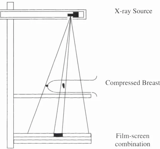
The oldest form of medical imaging is planar X-ray imaging, or radiography, in
which emitted photons pass through a patient and create a two-dimensional image,
which is a projection of the three-dimensional distribution of the X-ray attenuating
properties of a section of anatomy. In traditional film-based radiography, the photons
are detected by a screen-film combination in which the photons are absorbed by a
phosphor coating which emits light on absorption, the light exposing the emulsion of
example, using a photo-stimulable phosphor plate that stores a latent image which can
by read out by a scanning laser beam.
X-ray Source
Compressed Breast
Film-screen combination
Figure 1: traditional m amm ography involves obtaining a pla n a r X-ray o f the com pressed breast
One of the application areas considered in this thesis is that of planar X-rays of
the compressed breast, known as mammograms. Breast cancer is the most common
cancer in women in the UK and there are nearly 32,500 cases every year with about
14,500 deaths. Mammography is the investigation of preference throughout the
detection, diagnosis and management of the disease.
4
.It is a particularly demanding technique for a number of reasons. First, the
difference between the X-ray attenuation of cancer and normal tissues is relatively
small, which means that images must he created with photons emitted at relatively low
energies, in order to optimise the absorption. Second, the range of transmitted
exposures is relatively high, owing to the mix of tissue and radiolucent fat in the breast,
which is a problem since higher exposure leads to higher film contrast only within a
limited range of exposure values. Third, the objects which must be distinguished can be
relatively small and spatial resolution must be traded against detector efficiency in
determining the thickness of the film screen combination. A proportion of the photons
arriving at the detector will not have been transmitted undisturbed through the breast
but will have been scattered, the noise generated by this scattered radiation can, in part,
be removed by a collimating grid, but this has the effect o f increasing the dose required
to expose the film adequately.
The difficulty of producing the best possible film mammogram, given these
constraints, is in part due to the fact that the film is at once the medium o f acquisition,
o f storage and of display. In digital mammography these processes can be separated
and each one optimised. Digital mammography is the subject of much research,
although at the time of writing little used in practice [Feigl995]. M uch research is also
being carried out into computer aids for mammography, since digital mammograms
can conveniently be processed by computers, although the scarcity of digital mammo
graphy machines means that most of the research in this topic uses traditionally
captured images which have then been digitised using a light-box and a CCD ( ‘charge-
coupled device’) camera or by scanning a laser across the film.
The other class of images considered in this thesis is that of X-ray computed
tomography (CT), in which anatomy is viewed slice by slice, forming a stack of two-
dimensional images which can be used to create a three-dimensional representation.
through the body. In modern CT scanners a fan beam is rotated around the body to
generate projection data from which a two-dimensional image of the slice is computed.
Detectors
Body Source
Figure 3: in X-ray CT a fan beam o f X-rays is used to generate a 2-D im age o f a slice through the patient
If the slice is viewed as a grid of cuboids (known as voxels) then the value of
detected radiation at any point is determined by the absorption properties of the tissue
in every voxel through which is passes. If sufficient number of measurements are taken
for different lines through the grid, then a value can be calculated for the absorption
properties of the voxel. This value is called the Hounsfield number. A CT data set
consists of a set of arrays of Hounsfield numbers. This is how CT data are stored, but
generally not how they are displayed. The display of a CT scan usually involves
altering the function used to map Hounsfield numbers into screen values so that the
available contrast on the display medium is concentrated in the range of interest for the
childhood; the interesting contrast is therefore between the tumour and the tissue
making up the surrounding organs.
Mammography and X-ray CT are the two image modalities discussed in detail
in later chapters. A number of other techniques are discussed in considering previous
research in decision support for medical imaging. These are briefly introduced in the
remainder of this section.
Figure 4: an example o f a X -ray CT slice
X-rays are generated externally to the body and passed through it to create an
image of anatomy. In another class of medical images, radiation is generated inside the
body and detected externally to create an image of biological function. The distribution
around the body of an injected radiopharmaceutical will depend on blood flow, blood
volume and a variety of metabolic processes, so radiation detected using a gamma or
scintigraphic camera, can provide information about these processes. In these images.
unlike X-ray images, the point of emission is not known, so some form o f collimation
is required to provide information about direction.
This technique, known as Nuclear Medicine or Scintigraphy is used to create
both planar and tomographic images. Planar scintigraphic images may also be
considered in two categories: static images, 2D single view images of the distribution
of activity at a moment in time, and dynamic images, in which multiple images are
taken over a period of time that may be milliseconds or a few hours. Important classes
o f dynamic planar scintigraphic images include those used to measure cardiac and
renal function. Emission computed tomography is usually considered as two separate
modalities, SPECT (single photon emission computed tomography), using radio
isotopes where a single gamma ray is emitted per disintegration, and PET (positron
emitted tomography) where two gamma rays are emitted simultaneously when a
positron from a nuclear disintegration annihilates in tissue. Owing to their high cost,
PET scanners are found only in major research institutions and have found few applica
tions as yet in routine clinical practice.
A third kind of radiation is ultrasound. Like X-rays ultrasound is generated
outside the body and attenuated differently by different tissue types. Unlike X-rays, it is
usually the reflected rather than the transmitted signal which is measured. Since the
speed of sound is slow enough to allow the time between pulse and echo to be
measured for distances travelled in the body, the doppler shift can also be measured and
ultrasound used as a functional imaging modality in the measurement of blood flow.
Equally the speed allows all the data for an image to be collected in time to present, for
example, a real time image of the moving heart. Ultrasound has the further advantages
o f being relatively cheap and low-risk. The principal uses of ultrasound are in
Name Radiation Configuration Construction
X-ray X-ray Transmitted Planar
Angiogram X-ray Transmitted Difference Image
X-ray CT X-ray Transmitted Computed Tomography
Ultrasound Ultrasound Reflected Planar
Doppler Ultra sound
Ultrasound Reflected Time of flight calculation
Nuclear Medi cine
Gamma Rays Emitted Planar or Difference Image
SPECT /PET Gamma Rays Emitted Computed Tomography MRI Radio Waves Emitted Computed
Tomography
Table 1: kinds o f medical image referred to in this thesis
The other major imaging modality which should be discussed here is magnetic
resonance imaging or MRI. Like X-ray CT, MRI is used to create sets of images
providing a 3D representation. However, while X-ray images provide a map of electron
density, which relates to physical density, MRI can be used to obtain information about
proton density (or other relevant nuclei), and about tissue characteristics, such as the
freedom of hydrogen containing molecules and the proportion of water in different
regions of the body. MRI can also be used to obtain images of function. The basis for
the radiation used here is the magnetic field created by orbiting protons in the
molecules of the body. The net magnetic moment of a sample of nuclei is shifted when
placed in a rotating magnetic field. This shift can be detected with a suitable coil. In
fact different pulse sequences can be generated and measured to give information of
different kinds. Saturation recovery, inversion recovery, spin/echo, longitudinal relax
images. Spin/echo, T l and T2 images are particularly important; the latter two provide
information about vibrational motion in the lattice of molecules.
There are other methods of imaging the human body, such as the use of infra
red light to trans-illuminate, a technique which is used in the investigation o f skin
melanoma, and techniques which use visible light detected by miniature cameras
inserted into the body, but the above represent the principal methods for which
computer decision aids have been developed.
These different classes of image have quite different characteristics in terms of
spatial resolution, dynamic range and signal-to-noise ratio and are used for very
different purposes. In each, however, some form of radiation is used to create a signal
which carries information about the body and stores it in a form which the human eye
can read. This ‘reading off’ of information stored in images is so effortless a task for
human beings that some imagination is required to understand what it must involve.
This process is discussed in the next sub-section.
1.2.3
Interpreting the image
Designing appropriate tools to assist in the interpretation of images requires
some understanding of how images are interpreted by radiologists. Perception, at the
most basic level, involves a response to luminance values. At a more abstract level it
involves the detection of significant form in these responses. The process of image
interpretation is also an active one, and there are important questions concerned with
how the image is searched for potential abnormalities.
Pizer [Pizerl990] describes a model of image perception which, although not
high-level vision. Perception starts with the spatial distribution of luminance values
detected on the retina with a logarithmic sensitivity. The next column consists of
samplings over fields within the retina. M ost of these receptive fields signal the result
of a comparison of the amount of light on a field of the retina with the average amount
falling on the surround. The information transmitted from the receptive fields is
therefore about intensity changes in the image and not about absolute intensity values.
There are many different overlapping receptive fields and receptive fields o f many
different sizes. The result is that the early vision system provides information about
changes at different spatial scales. The system also responds to higher order changes
and so transmits information about orientation and curvature. The local measurements
of features such as edges, bars and corners are combined in a way which allows edges
corresponding to sharp luminance changes to be linked by ‘subjective’ edges which do
not correspond to intensity changes in the image. The edges and corners perceived at
different spatial scales are thought to combine in some way with a hypothesis, which
may be generated by a tentative decision or by expectation.
A radiological investigation is performed with the aim of answering a particular
question. The nature of this question determines the task which the radiologist
performs in interpreting the image. He or she may be attempting to detect any o f a class
of known possible abnormalities, assessing the likelihood that a particular feature has a
malignant cause or measuring the change in size of a tumour following treatment.
These different tasks define sets of hypotheses which guide the radiologist in his or her
reading o f the image.
The human visual system is able to adapt to an extraordinary range of
luminance values. However, for any given level of adaption it can make distinctions
only within a relatively narrow range of luminance values. The properties which give
the system its range of sensitivity to objects of different scales and luminance, also
make it an unreliable indicator of absolute luminance. Judgements about the relative
reliable. The role of hypotheses means that what is detected depends, to an extent, on
what is looked for in the image. A faint or subtle finding to which a radiologist has been
directed in some way is therefore much more likely to be perceived.
edge
singularities m
texture
edge/ shape strength measure
ment
m motion
intensity diffusion
depth
colour
Intermediate vision High-level vision Front-end vision
Figure 5: a m odel o f static human vision, after [P ize r1990]
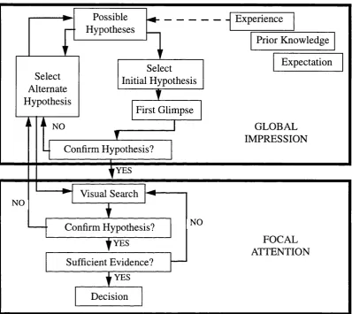
Greenes [Greenes 1989] distinguishes two tasks in the interpretation of images:
the detection and classihcation of features. This distinction is accepted by a number of
authors [Gale 1993a, Swettl993]. Gale presents a detailed account of how images are
searched by radiologists, distinguishing between the ‘somewhat parallel’ initial
processing of the image within the first glimpse and a second phase in which the
display is serially examined using eye-movements. The model also provides a role for
experience in furnishing radiologists with hypotheses. Kundel [Kundell972] showed
which may be due to the richer set of stored expectations available to experienced radiologists. Possible Hypotheses Select Alternate Hypothesis Select Initial Hypothesis
' ' f
NO First Glimpse
I
Confirm Hypothesis? Experience Prior Knowledge Expectation GLOBAL IMPRESSION YES NO— ► Visual Search
----Confirm Hypothesis?
y YES
Sufficient Evidence?
| y e s
Decision
NO
FOCAL ATTENTION
Figure 6: schematic outline o f the examination o f a visual display fo r a possible target. Factors such as experience provide initial hypotheses. The first glimpse provides a global impression which is fo llo w ed by a fo c a l attention stage in which active visual search
takes place. A dapted from [G a lel9 9 3 a ].
Kundel also found evidence to support the distinction between detection and
classification [Kundel 1990] by tracking eye-movements in order to study the errors
made by radiologists examining chest X-rays for lung nodules. He found that approxi
mately 30% of nodules were missed because the fovea did not pass over the appro
on the appropriate section of the image and in approximately 30% of cases the nodule
received prolonged visual attention. Thus roughly one third of errors were errors of
search, one third of detection and one third of classification. These findings have been
used by researchers in computer aided radiology to justify work in enhancing images,
providing prompts and in providing decision aids for classification, e.g. [Swettl993].
The roughly equal distribution of the errors across the three classes of error is a strong
argument for providing a decision support tool which is capable of assisting in search,
detection and classification. The role played by hypotheses suggests a potential role for
a system which can provide information about possible diseases and diagnoses.
1.2.4
Reporting the findings
The next task in the radiological process concerns the reporting of radiological
findings. Many authors identify this as a common source of problems. Robertson
[Robertson 1989] describes a study of communication problems in a mammography
screening programme. In the course of the 11-week study 1,404 screening mammo
grams were taken, of which 63 required additional evaluation or biopsy. Written reports
were sent to the referring physician and, in addition, the physician’s office was notified
by telephone of the need for additional investigation or biopsy. An average o f 2.5
months after the initial examination was performed, the 63 cases were followed up;
computers in radiology and pathology departments were searched for information
about additional mammography studies or pathology results. Where no results were
found, the relevant physicians’ offices were contacted by telephone. These steps were
repeated at fortnightly intervals. In some cases the patient was contacted directly. W hen
the cases were first followed up, at 2.5 months, no action had been taken in 40 out of
the 63 recommendations. After 3.5 months no action had been taken in 10 out of 63
studies and at 4.5 months four had still not undergone the recommended additional
Robertson lists the various explanations offered for the breakdown in communi
cation but gives no indication as to their relative frequency. It seems that problems can
occur at every stage in the process. Some letters were sent to the wrong address. In
some cases the referring physician had moved on. In some cases the letter was misfiled.
In other cases it was misunderstood by the referring physician and in others the diffi
culty was in getting in touch with the patient or with persuading them to comply with
the recommendation. Robertson notes that the particular problem with screening is that
‘no news is good news’ and therefore when a letter fails to reach its intended recipient
the failure is not noticed unless the sender follows up the communication.
D ’Orsi [D’Orsil995] describes in detail the communication issues associated
with mammography. Reporting occurs at two levels: the communication of results and
recommendations in lay language to the patient and the technical medical report to the
patient’s healthcare provider. Robertson cites a number of studies which have shown
the importance of providing a report directly to the patient, and have shown that this
results in improved compliance with recommendations, fewer delays in diagnosis and
reduced confusion over later treatment options. Problems surrounding the sharing and
communication of information with patients have been identified as one of the most
influential factors in patient decisions to sue for malpractice.
The technical report to the patient’s healthcare provider has also been the
subject o f study. D ’Orsi describes how the American College of Radiology has
supported the development of a standard approach to reporting which includes a
statement of which elements should be included in a report and a lexicon of appropriate
terms to be used in describing mammographie features. The aim is to encourage
concise, understandable reports. The report should begin with a brief initial statement
o f the reason for the examination, the breast composition, significant findings,
comparison with previous images (if applicable) and an overall assessment and recom
statement of the communication that has taken place with the patient and the healthcare
provider.
D ’Orsi lists the statements that can be used to describe composition, the
different abnormalities which can be described, the properties which may be attributed
to them and the manner in which these properties should be expressed. Some authors
e.g. [Heilbrunl994] have taken exception to this lexicon. Heilbrun states that the job of
a mammographer is to recognise three kinds of lesion and classify them as benign,
probably benign, suspicious and highly suspicious. Any findings which cannot be so
classified require further analysis. Experienced mammographers have no difficulty in
doing this, and in reporting succinctly that they have done so, inexperienced mammo
graphers attempt to cover up their lack of confidence with wordy equivocations. It is
argued that terminology is not therefore the root problem. Another difficulty is that the
terms in the terminology concentrate on morphology and this inhibits the development
o f thinking about the underlying processes which cause abnormalities.
Coding schemes, such as that proposed by the American College of Radiology
are becoming more widely used throughout the medical community. This is an
important issue for the many computer based decision aids which require that relevant
information about a patient be encoded in a form that is intelligible to the computer.
The question whether the contents of radiological reports can be represented in this
form is of crucial importance to this thesis. Coiera [Coieral995] argues that coding
systems can provide a practical basis for managing the language of medicine so long as
it is understood that they define a limited and consensual language. The approach taken
in this thesis has been to assume that codified medical terminologies can be developed
for restricted tasks where the intended users and the context of the intended use are
understood. A standardised reporting language for particular classes of medical investi
gation is therefore assumed not to be an impractical prerequisite for a radiological
1.3
Computer Support for Radiological Tasks
The different tasks involved in diagnostic radiology have different information
requirements and are amenable to different forms of computer support. A radiology
department will normally exist within a hospital which will have its own information
system providing for the storage, capture and transmission of patient-related and other
information (a hospital information system or HIS). There will often be a separate
computer system providing these functions within the radiology department (a
radiology information system or RIS). There may be, in addition to these systems,
some form of computerised patient record or electronic healthcare record (EHCR).
A number of the imaging modalities will involve digital capture, storage and
display of images. There may be another system for the archiving and communication
of images independent of the individual imaging systems (a picture archiving and
communication or PACS system). There are clearly considerable benefits to be
obtained from the integration of these different systems. Such integration or ‘inter
operability’ is now a major topic of research in medical informatics and many groups
are working on the development of standards and formal models that will facilitate the
development of systems which can share information. Two directly relevant projects
are the development of the DICOM standard and the MIMOSA approach to modelling
PACS systems.
The American College of Radiology and the National Electrical Manufacturers
Association jointly developed a standard for Digital Imaging and Communication
(DICOM) which was published in 1993 [DICOM 1993]. Continuing work on this
standard has involved working groups from European and Japanese standardisation
bodies and has been adopted by vendors of imaging devices and clinical workstations.
The standard covers such topics as the encoding of image data and file transfer
representation of concepts such as ‘patient’ or ‘visit’ and the specification of operations
performed on Information Objects as well as the expected behaviour of the user and
provider of the operation.
The MIMOSA^ project [Garfagnil994] developed an approach to modelling
medical image management systems which tackles the issue of inter-operability
between PACS and HIS/RIS. The project, which was supported by the EU as a research
rather than a standardisation initiative, attempts to reconcile two different views of
medical imaging: the HIS view which considers medical images as a subset of medical
information and the PACS view which is concerned with the acquisition, processing
and storage of images without being concerned with why they are produced or how the
information they contain is used. The MIMOSA model consists of three loosely
coupled models: the data model describes the structure and relationships of the data
represented in the system, the functional model describes what the system does, the
dynamic model describes the behaviour of the system over time.
It is worth reflecting on the ultimate goal of this work. It is theoretically
possible to have a filmless, paperless hospital in which all information, image-based
and otherwise, exists only on computer. This is not necessarily desirable. Indeed, CT
images which can only be created by computer and which must be viewed on a
computer screen in order to establish the appropriate display parameters, are routinely
printed on film to be seen by the radiologist. Film offers many advantages to the radio
logist: they can pick it up, view it next to other films, scan large numbers o f images and
carry them from office to office.
The development of networks of communicating computer systems supporting
gained by standardisation and computerisation with the flexibility and familiarity of
traditional ways of working. Consider the four tasks described in the previous section.
• Establishing the need for a radiological examination requires communi
cation between a referring clinician and a radiologist. This might be
supported by a computer, if both clinicians regularly use email or video
conferencing, but it may be more conveniently done some other way. It may
involve reference to guidelines, it could involve reference to research
material. Again, this kind of information can be conveniently accessed by
computer, but it doesn’t have to be. It could involve calculations or infer
ences made by a computer on the basis of some form of stored knowledge.
• Once the need for an examination is established the examination must be
performed. The efficient use of imaging devices and the attendant personnel
requires the use of computerised scheduling systems. Such systems are
generally provided with radiology information systems and the basic
concept, that of the worklist, is being defined in standards to allow inter
operability of PACS and RIS systems. Many image generation techniques
are now digital and require computers. Others may become computerised.
• The interpretation of images, if at some point they exist in digital form, may
be facilitated by computer systems which either enhance, analyse or interpret
the image or which provide information that the radiologist can use in
making a decision.
• The generation of radiological reports can be assisted by systems of menus
which provide access to standard lexicons, or by systems which provide
access to reporting standards. Voice recognition and word-processing
software can ease the creation of a written report and information systems
can be used to manage the sending out of reports and appropriately timed
The research described in this thesis focuses on the kind of computer system
which would be used by a radiologist to help make a decision about an image. The
work, however, is also guided by a desire to consider this as part of a process of care
and to bear in mind that a computer system designed to support part of a process must
sit within a network of inter-operable systems supporting the whole process.
1.4
Summary
The opening section of this chapter set out four aims for this thesis:
• to consider the range of systems which could potentially improve medical
decision making from a unifying perspective
• to consider how different kinds of system could be integrated into a single
decision aid
• to propose a generic model for decision support tools which allows users to
access relevant information
• to develop prototype systems which demonstrate the practicality o f these
ideas
The following section described four different activities involved in carrying
out a radiological investigation: establishing the need for the investigation, performing
the examination, interpreting the results and providing a report. Each of these tasks can
be supported by the different computer systems which are currently in place in most
radiology departments. A number of projects are now working on the integration and
inter-operability of these different kinds of system. That work provides the context
1.5
Chapter Outlines
Chapter Two reviews existing work on computer aids for decision making in
diagnostic radiology. Systems developed in a number of different areas of computer
science (image databases, numerical decisions aids, expert systems, image processing
systems) are identified and reviewed. The value of the different approaches is assessed
and it is argued that a system capable of drawing on different kinds o f information
source is desirable.
Chapter Three considers the problems involved in designing a decision aid to
handle different kinds of information. Examples of existing systems which attempt to
combine image processing and symbolic reasoning are considered.
Chapter Four sets out an approach intended to combine image processing and
symbolic reasoning. The approach is based on a logical model of the processes
involved in making decisions about images. In the model, decision making is viewed as
involving the proposal of candidate solutions and then the consideration of arguments
for and against these solutions. In the case of decisions made on the basis of the inter
pretation of images, some of the arguments may rely on information about what has
been detected on the image, about how it can be classified or on measurements o f the
contents of the image. These different processes can all be described using logical
rules, rules which can be implemented as a logic program and used as the basis for a
decision support system.
Chapter Five gives details of a generic architecture for decision aids drawing on
different information sources. The architecture sets out three components of a design: a
set o f displays which together make up the user interface, the set of information sources
model described in Chapter Four - which draw on the information in the different
sources to generate new information, which is then used to provide decision support.
Chapter Six describes the background to the prototype decision support system
developed to assist in the differential diagnosis of calcifications on mammograms. The
radiological literature on decisions made in the interpretation of mammograms is
reviewed, concentrating on the problem of the differential diagnosis of microcalcifica
tions. The application of image processing to this problem is considered and work in
the area reviewed. The potential for a decision aid for the differential diagnosis of
microcalcifications drawing on both image processing and symbolic reasoning, is
assessed.
Chapter Seven gives a detailed account of the implementation of the prototype
mammography system, concentrating on the development of the knowledge base and
the selection of image processing measures. Chapter Eight describes the evaluation of
the prototype mammography system. Chapter Nine describes a prototype system
developed to assist in the staging of neuroblastoma, a tumour of childhood, on X-ray
CT scans. Chapter Ten discusses the conclusions of the thesis, and highlights some
CHAPTER TWO
DECISION AIDS FOR
RADIOLOGY
The nurse conducts the exercise in lim ited extinction;
the p late prepared, the source exposed with no more than a glib nictation.
I am told to g et dressed and go home. Later, in my absence
the doctors make their guesses from the holes left by my bones.
Don Paterson
2.1
Introduction
This chapter reviews research into computer aids for radiological image inter
pretation. The aim is to bring together work from quite disparate areas of computer
science and to consider the contribution it could make to improving the quality and
efficiency of radiological decision making. Attention is therefore restricted to systems
which would come into play when or after the image is displayed, and which would be
computers can be used to create new kinds of image, the creation of tomographic
images, the reconstruction of three-dimensional images, the enhancement o f digital
radiographs, the segmentation of MRI images and the registration of images of
different modalities are not covered. Electronic information sources such as hypertext
systems and medical databases are also excluded since, although they could assist in
making decisions, they are not specifically designed to do so.
2.2
Decision Support
Shortliffe [Shortliffel991] includes all systems designed to help health profes
sionals make decisions within the definition of clinical decision support systems. He
notes that “in a sense any computer system which deals with clinical data or medical
knowledge is intended to provide decision support” and suggests that it is accordingly
useful to consider three types of decision support function:
• tools for information management, which would include hospital inform
ation systems and bibliographic retrieval systems which make information
and knowledge available to clinicians but do not help them apply it to
particular cases
• tools for focusing attention, which would include systems which provide
reminders or detect abnormal values, hence which provide advice on the
basis of general rules covering different situations
• tools for a patient-specific consultation, which includes only those systems
which provide tailored advice on the basis of patient-specific information
In practice, systems in the first of these categories are rarely considered in
accounts of decision support systems and where a distinction is drawn between systems
medical decision support systems as “active knowledge systems which use two or more
items of patient data to generate case-specific advice”.
The term decision support system indicates a development in the conception of
the role of knowledge-based systems, so that they are viewed as tools which can offer
advice, but are designed to do so in a supporting role. An early definition [Keen 1978]
of decision support systems states that:
D ecision Support Systems (DSS) represent a p o in t o f view on the role o f the computer in the management decision-making process. D ecision support implies the use o f computers to:
1. Help managers in their decision processes in semi-structured tasks.
2. Support, rather than replace, managerial judgement.
3. Improve the effectiveness o f decision making rather than its effi ciency.
The notion of ‘semi-structured’ tasks is crucial to this definition. These systems
are not designed for tasks where a clear and rigid structure makes a completely compu
terised system a practicality, nor for those where there is no structure on which to base
the design of such a tool, but rather for tasks between these two extremes. Johnston et
al. [Johnston 1994] found that decision support systems in medicine were more
frequently shown to be successful when applied to tasks such as prescribing than when
applied to diagnosis. It could be argued that prescribing is a better exemplar o f the
notion of semi-structured task than diagnosis, since - at least for a GP - diagnosis is a
confrontation with all the variety, complexity and ambiguity of human life whereas
prescribing is only attempted once the available data has been rendered in medical


![Figure 5: a model of static human vision, after [Pizer1990]](https://thumb-us.123doks.com/thumbv2/123dok_us/8623991.1413699/29.595.29.529.180.466/figure-model-static-human-vision-pizer.webp)