1556-6811/10/$12.00
doi:10.1128/CVI.00204-10
Copyright © 2010, American Society for Microbiology. All Rights Reserved.
Recombinant Diabody-Based Immunocapture Enzyme-Linked
Immunosorbent Assay for Quantification of Rabies
Virus Glycoprotein
䌤
Sridevi V. Nimmagadda, Shukra M. Aavula, Neelakantam Biradhar, Varaprasada Sankarasetty Rao,
Rajalakshmi Shanmugham, Dev Chandran, Nagarajan Thirumeni,
Nagendrakumar Balasubramanian Singanallur,
and Srinivasan Alwar Villuppanoor*
Research and Development Centre, Indian Immunologicals Limited, Rakshapuram, Gachibowli, Hyderabad 500032, India
Received 20 May 2010/Returned for modification 10 June 2010/Accepted 14 June 2010
The potency of rabies vaccines, determined using the NIH mouse protection test, can be directly correlated
to the amount of rabies virus glycoprotein (RV GP) present in the vaccine. In an effort to develop a simple and
sensitive enzyme-linked immunosorbent assay (ELISA) using recombinant diabody for quantification of RV
GP, the variable heavy (V
H) and light chain (V
L) domains of an RV GP-specific human monoclonal antibody
(MAb) secreted by a human
ⴛ
mouse heterohybridoma (human MAb R16E5) was amplified, linked using
splicing by overlap extension PCR (SOE PCR), and expressed as a recombinant diabody (D06) in the pET28a
bacterial expression system. The diabody D06 was purified by immobilized metal affinity chromatography on
a nickel-nitrilotriacetic acid (NTA) agarose column and characterized. The purified diabody was used in
combination with a well-characterized RV GP-specific mouse MAb, M5B4, to develop an immunocapture
ELISA (IC-ELISA) for the quantification of RV GP in human rabies vaccine preparations. The maximum
detection limit of the IC-ELISA using the M5B4-D06 combination was up to 31.25 ng/ml of RV GP. The
specificity of the diabody was established by its nonreactivity toward other human viral antigens as determined
by ELISA and toward RV GP as determined by immunoblot transfer assay and competitive ELISA with the
parent human MAb R16E5 and MAb M5B4. The adjusted
r
2value obtained by the regression through the
origin model was 0.902, and the equation for predicted potency values for M5B4-D06-based IC-ELISA and
MAb M5B4 IC-ELISA were 0.5651
x
and 0.8044
x
, respectively, where
x
is the estimate of RV GP from the
IC-ELISA in micrograms. Analysis of variance (ANOVA) results showed the estimates of the two methods
differed significantly (
P
< 0.001), while the predicted potencies by the two tests did not differ significantly (
P
>
0.05). The IC-ELISA can be readily adapted to measure the RV GP content in purified antigen, and a vaccine
can be formulated based on the estimated GP.
Rabies is a fatal viral infection of the nervous system
affect-ing all mammals, includaffect-ing humans through bite wounds from
a rabid animal, which can be prevented by vaccination coupled
with administration of anti-rabies virus serum (6, 11). Rabies
transmission from nonbite exposures is rare. Scratches,
abra-sions, open wounds, or mucous membranes contaminated with
saliva or other potentially infectious material (such as brain
tissue) from a rabid animal constitute nonbite exposures.
Oc-casionally reports of nonbite exposure are such that
postexpo-sure prophylaxis is given. Inhalation of aerosolized rabies virus
is also a potential nonbite route of exposure, but with the
exception of laboratory workers, most people are unlikely to
encounter an aerosol version of the rabies virus (5). Organ
transplantations have also been credited with nonbite
trans-mission of rabies from human to human (3). Despite significant
scientific progress, rabies remains an important zoonotic
dis-ease globally. Annually, 20,000 deaths are reported in India,
making rabies one of the major causes of human mortality
(21). Vaccination is therefore considered one of the most
via-ble and important methods for the prevention of rabies by way
of preexposure prophylaxis in high-risk groups, postexposure
prophylaxis in contact groups, and preexposure prophylaxis in
pet animals that are at risk due to possible contacts with rabid
animals. The most cost-effective means of prevention and
con-trol of rabies in humans is by eliminating rabies in dogs and
other susceptible animals through vaccination.
The NIH mouse protection test is an
in vivo
potency test that
has been used widely by all manufacturers of rabies vaccines.
The role of different immunological parameters and the
pres-ence of virus-neutralizing antibodies are not well established
because of a weak correlation between the NIH potency test
results and immunogenicity when vaccines containing different
strains of rabies virus were tested (2). Furthermore, this
method is time-consuming and expensive, requires a large
number of animals, and involves the use of live rabies virus. As
a result, there is increased exposure in human beings to live
and virulent rabies strains. The NIH test also requires a
secure biosafety level 3 (BSL-3) facility for housing and
challenging the experimental animals. Therefore, for both
practical and ethical reasons, replacement of this test by
more rapid and reliable
in vitro
methods is highly desirable.
Based on the fact that the rabies virus glycoprotein (RV GP)
is the antigen responsible for inducing virus-neutralizing
* Corresponding author. Mailing address: Indian Immunologicals
Limited, Rakshapuram, Gachibowli, Hyderabad 500032, India. Phone:
91-40-23000894. Fax: 91-40-23005958. E-mail: [email protected].
䌤
Published ahead of print on 23 June 2010.
1261
on August 17, 2020 by guest
http://cvi.asm.org/
antibodies and conferring protection against a lethal
intra-cerebral challenge, it has been suggested that the
antigenic-ity of the rabies vaccines could be evaluated by titration of
the RV GP (17).
Though some laboratories have used enzyme-linked
immu-nosorbent assay (ELISA) to assess RV GP content for
deter-mination of the potencies of inactivated vaccines, variable
cor-relation between ELISA and the NIH test (7, 8, 9, 13, 14, 17,
18, 20) has been reported. Essentially, all these ELISAs
incor-porate the use of either polyclonal antibodies or
hybridoma-derived monoclonal antibodies (MAbs). Although MAbs offer
substantial advantages with respect to potency, reproducibility,
and freedom from contaminants (4), they are difficult to
pre-pare in a quality-assured manner.
Recombinant DNA technology has been used to a great
extent in the expression of antibodies/antibody fragments (12).
Antibody fragments can be readily produced from the genes
encoding antibody variable domains, which can be derived
either from hybridomas (19) or from bacteriophage displaying
antibody fragments (16). Diabodies are bivalent or bispecific
antibody fragments generated by the dimerization of variable
heavy (V
H)-light chain (V
L) fragments (10) as a result of
re-duction in the size of the linker between variable light and
variable heavy chains (1), and these antibodies have many
practical applications, including immunoassay and therapy.
We describe for the first time the use of a recombinant
diabody in the development of an ELISA for quantification of
RV GP content in human rabies vaccines incorporating the PV
strain of rabies virus and its comparison with the NIH mouse
protection test.
MATERIALS AND METHODS
Cells and heterohybridoma.The human⫻mouse heterohybridoma cell line formed by fusion between primary immune peripheral blood B cells from a donor immunized with a human rabies vaccine (PV strain; Abhayrab) and a heteromyeloma cell line, K6H6/B5, secreting an RV GP-specific human MAb of the IgG1 isotype (human MAb R16E5) obtained from the hybridoma laboratory Indian Immunologicals Limited (IIL), Hyderabad, India, was used for amplifi-cation of variable light and variable heavy chain genes. A mouse neuroblastoma (Neuro-2a) cell line procured from ATCC and maintained in the cell culture laboratory IIL, Hyderabad, was used to carry out the indirect fluorescent anti-body test (IFAT).
Bacterial strains, vectors, and chemicals.The bacterial strainEscherichia coli
BL21(DE3) (Invitrogen) was used for the propagation of plasmids and overex-pression of protein. The bacterial exoverex-pression vector pET28a, used for cloning and expression of the diabody, was procured from Novagen (Madison, WI). Nickel-nitrilotriacetic acid (Ni-NTA)-agarose (Qiagen, Germany) was used for the purification of 6⫻His-tagged proteins.
Isolation of total RNA and cDNA synthesis.Total RNA isolated from the heterohybridoma cell line (1⫻106cells) using the TRIzol reagent (Invitrogen) was resuspended in diethyl pyrocarbonate (DEPC)-treated water and quantified using a Biophotometer instrument (Eppendorf, Germany). The cDNA was syn-thesized using random hexamers and a Thermoscript reverse transcriptase (RT)-PCR kit (Invitrogen) according to the manufacturer’s instructions. The cDNA was stored at⫺20°C until further use.
Amplification of variable domains, assembly, and cloning of diabody.The cDNAs encoding the antibody variable domains (VHand VL) were PCR ampli-fied using universal primers (15). The variable regions were assembled using splicing by overlap extension (SOE) PCR (Table 1). The 711-bp-long PCR product was purified and cloned into the pCR2.1 TOPO TA vector to obtain pCRdia. pCRdia was transformed intoE. coliTOP 10 chemically competent cells and plated on Luria-Bertani broth medium supplemented with 100g/ml of ampicillin (LB-Amp) and incubated overnight at 37°
C. Selected clones were grown overnight in LB-Amp medium, and pure plasmid DNA was isolated using the Qiagen Miniprep kit (Qiagen, Germany) according to the manufacturer’s
instructions. pCRdia was purified, and the sequence was verified by automated cycle sequencing. pCRdia plasmids containing sequences with no stop codons were selected and stored at⫺20°C until further use.
pCRdia was subjected to PCR with the VLforward 1 and VHreverse 1 primer pairs to incorporate the EcoRI and NotI restriction sites at the 5⬘and 3⬘end of the PCR product, respectively (Table 1). The PCR product and pET28a were digested with EcoRI and NotI and gel purified before being subjected to ligation using T4 DNA ligase to obtain pET28aRD, which was transformed into XL-blue
E. colicompetent cells and plated on LB agar supplemented with 50g/ml kanamycin (LB-Kan). The plates were incubated overnight at 37°C. Selected clones were grown overnight in LB-Kan medium, and pure plasmid DNA was isolated using the Qiagen (Germany) Miniprep kit according to the manufac-turer’s instructions. The plasmid DNA was purified, the sequence was verified by automated cycle sequencing, and the plasmid was stored at⫺20°C until further use.
Expression and purification of the diabody.The pET28aRD was transformed intoE. coliBL21(DE3), plated on LB-Kan, and incubated overnight at 37°C. A single colony ofE. coliBL21(DE3) containing pET28aRD was inoculated in LB-Kan and grown overnight in an orbital shaker at 30°C at 200 rpm. The overnight culture was diluted 40 times in fresh LB-Kan and grown at 37°C at 200 rpm until the culture reached an optical density (OD) of 0.8 to 0.9 at 600 nm. The culture was induced with 1 mM isopropylthio--D-galactopyranoside (IPTG) by incubation at 28°C for 4 h. The bacterial pellet was collected by centrifugation at 5,000⫻gfor 20 min at 4°C.
Purification of diabody by IMAC.The bacterial pellet was resuspended in lysis buffer (50 mM Tris–HCl, 155 mM NaCl, pH 7.6) to prepare a 10% (wt/vol) suspension. Lysozyme was added to a final concentration of 50g/10 ml of lysate and incubated overnight at⫺20°C. The sample was subjected to sonication and centrifuged at 9,200⫻gfor 30 min at 4°C. The pellet was discarded, and the supernatant was subjected to immobilized metal affinity chromatography (IMAC).
An IMAC column (5-ml volume) was equilibrated with 10 column volumes of 50 mM Tris–HCl, 155 mM NaCl, pH 7.6 (equilibration buffer). The su-pernatant was loaded onto the column at a flow rate of 1 ml/min and washed with 20 column volumes of washing buffer (equilibration buffer with 30 mM imidazole, pH 7.6). Bound diabody was eluted with 5 column volumes of elution buffer containing equilibration buffer with 300 mM imidazole, pH 7.6, as 1-ml fractions. All the eluted fractions were analyzed by SDS–PAGE and immunoblotting. Fractions containing the recombinant diabody were pooled and dialyzed against phosphate-buffered saline (PBS), and the protein con-centration was determined by the bicinchoninic acid (BCA) method before storage at⫺20°
C until further use.
Characterization of the diabody. (i) Detection of diabody by SDS-PAGE and immunoblot analysis.The purified diabody was electrophoresed by SDS-PAGE (12) using a 12% gel and electroblotted onto a polyvinylidene difluoride (PVDF) membrane (Hybond-C; GE Health care), using a transblot apparatus (Bio-Rad), following the manufacturer’s instructions. The blot was probed with an anti-His MAb probe (Pierce) and developed using 0.05% 3,3⬘-diaminobenzidine tetrahy-drochloride (DAB) (Sigma) and 0.03% hydrogen peroxide in PBS.
(ii) Immunocapture ELISA for determination of sensitivity of the diabody against RV.Titration of diabody D06 (450g/ml) at different concentrations (1:200 to 1:2,400) was performed against different concentrations of RV (1g/ml to 1.95 ng/ml) in a sandwich ELISA as described previously by Nagarajan et al. (18) with a few modifications wherein the diabody D06 at different concentration was used for detection. The binding of the diabody with RV GP was detected by addition of anti-His probe followed by 3,3⬘,5,5⬘-tetramethylbenzidine (TMB). The plate was incubated at 37°C for 10 min, and the reaction was stopped by addition of 1.25 M H2SO4.The absorbance was measured at 450 nm using a microplate reader (Bio-Tek).
(iii) Demonstration of RV GP-specific activity of the diabody. (a) Immunoblot transfer assay with rabies virus antigen.Zonal purified whole virus (PV strain) antigen (4g) was fractionated by SDS-PAGE using a 10% gel under native condition. The resolved proteins were transferred onto a PVDF membrane (Hybond-C; GE Health care) and blocked with 2% nonfat milk powder (Difco) in PBS for 1 h at room temperature. The blot was washed thrice with PBS containing 0.05% Tween 20 (PBS-T), followed by probing of the membrane with diabody (0.5 mg/ml) for 1 h at room temperature. A rabies virus glycoprotein (RV GP)-specific mouse MAb, M5B4 (18), and the parent human MAb, R16E5, were used as a positive control, and a rabies virus nucleoprotein (NP)-specific MAb, N5G4, was used as a negative control. The immunoreactivity with RV GP was detected by probing the blot with a His probe (Pierce) at a dilution of 1:5,000 followed by staining with DAB.
on August 17, 2020 by guest
http://cvi.asm.org/
(b) Competitive ELISA.A competitive ELISA was performed to identify the specificity of the diabody D06 for RV GP. A microtiter plate was coated with 100 ng/well of purified RV (PV) antigen in 50 mM carbonate-bicarbonate buffer (pH 9.6) and incubated overnight at 4°C. The plate was washed thrice with PBS-T and blocked with 1% bovine gelatin (Sigma) in PBS-T, followed by washing with PBS-T to remove the excess gelatin. Diabody (500 ng/100l) was added by serial dilution and incubated at 37°C for 1 h.E. colilysate (vol/vol) was used as a negative control. An RV GP-specific MAb, M5B4 (156 ng/well), and the human MAb R16E5 (200 ng/well) were added to each well containing diabody and incubated at 37°C for 1 h. The plate was washed with PBS-T and dried by flicking. Goat anti-mouse IgG horseradish peroxidase (HRP) conjugate (1:5,000) was added to each well, and the plates were incubated at 37°C for 1 h. The plate was washed five times with PBS-T, and 100l of TMB (Sigma) was added. The reaction was stopped by addition of 100l of 1.25 M H2SO4to each well, and absorbance was read at 450 nm using a microplate reader (Bio-Tek). The spec-ificity of the diabody toward antigenic site III of PV GP was determined, fol-lowing a method described by Nagarajan et al. (18).
(iv) IFAT.The diabody (500 ng/100l) was used to probe the unfixed RV (PV strain)-infected and uninfected mouse neuroblastoma (Neuro-2a) monolayer (6⫻104
/well) in a 96-well tissue culture plate (Nunc, Denmark) and incubated at 37°C for 45 min in a CO2incubator. The plate was washed with PBS, pH 7.4, and incubated with mouse anti-His IgG-fluorescein isothiocyanate (FITC) con-jugate at a concentration of 1g/5 ml (AnaSpec) at 37°C for 45 min in a CO2 incubator.The plate was washed with PBS to remove the excess conjugate and observed under a fluorescence microscope (Olympus, Japan). The RV GP-specific MAb M5B4 was used as a positive control.
(v) Determination of binding specificity of the diabody by IC-ELISA.The binding specificity of the diabody for RV-GP alone and not toward other unre-lated viruses was evaluated by testing its reactivity with hepatitis B surface antigen (HBsAg), hepatitis A virus (HAV), and Chikungunya virus (CHIKV)
using sandwich ELISA. Maxisorp plates (Nunc, Denmark) were coated with monoclonal antibodies M5B4, specific for RV-GP, and 1F6, specific for HBsAg (unpublished), and polyclonal mice sera raised against HAV and CHIKV at concentrations of 156 ng/well, 140 ng/well, 300 ng/well, and 300 ng/well, respec-tively, and incubated overnight at 4°C. The wells were washed thrice with PBS-T, and the unreacted sites were blocked with 1% bovine gelatin in PBS-T by incubation at 37°C for 1 h. The plate was washed thrice with PBS-T and incu-bated with doubly diluted RV (50g/1 ml), and HBsAg (32g/ml), HAV (100 IU/ml) and log diluted CHIKV (10750% tissue culture infective doses [TCID
50]/ ml), followed by incubation at 37°C for 1 h. The plates were washed thrice with PBS-T and dried. The diabody at a concentration of 450 ng/100l was added to each well. The plates were incubated at 37°C for 1 h, washed with PBS-T thrice, and dried by flicking. Anti-His probe was added, followed by incubation for 1 h at 37°C. The plate was developed using hydrogen peroxide-activated TMB. The reaction was stopped by addition of 1.25 M H2SO4, and the absorbance was measured at the 450-nm wavelength using a microplate reader (Bio-Tek). The experiment was performed in triplicate.
Immunocapture ELISA for quantification of rabies virus glycoprotein in hu-man rabies vaccine.IC-ELISA was performed to quantify the RV GP content in rabies vaccine formulations, according to the method described by Nagarajan et al. (18), with a few modifications wherein the diabody (450 ng/well) was used for detection. Briefly, the ELISA plate was coated with MAb M5B4 overnight at ⫹4°C, and the unreacted sites were blocked with 1% bovine gelatin. The test vaccines and an internal reference standard (IRS) vaccine of known RV GP were subjected to 8 serial 2-fold dilutions in PBS-T. The RV GP trapped by MAb M5B4 was detected using the diabody followed by the addition of anti-His probe. The plate was developed with TMB at room temperature for 10 min. The reaction was stopped by addition of 1.25 M H2SO4, and the absorbance was measured at the 450-nm wavelength using a microtiter plate reader (Bio-Tek). The assay was performed in triplicate. The RV GP content was also
TABLE 1. Primers used for PCR of V
Hand V
Lregions and SOE PCR for construction of diabody
Primer ID Sequence (5⬘–3⬘)a
Human variable heavy chain forward primers
HuVH1a ...GGCGGCGGCGGCTCCGGTGGTGGTCAGGTGCAGCTGGTGCAGTCTGG
HuVH2a ...GGCGGCGGCGGCTCCGGTGGTGGTCAGGTCAACTTAAGGGAGTCTGG
HuVH3a ...GGCGGCGGCGCCTCCGGTGGTGGTGAGGTGCAGCTGGTGGAGTCTGG
HuVH4a ...GGCGGCGGCGGCTCCGGTGGTGGTCAGGTGCAGCTGCAGGAGTCGGG
HuVH5a ...GGCGGCGGCGGCTCCGGTGGTGGTGAGGTGCAGCTGTTGCAGTCTGC
HuVH6a ...GGCGGCGGCGGCTCCGGTGGTGGTCAGGTACAGCTGCAGCAGTCAGG
Human variable heavy chain reverse primers
HuJH1-2 ...GGAATTCTGAGGAGACGGTGACCAGGGTGCC
HuJH3...GGAATTCTGAGGAGACGGTGACCATTGTCCC
HuJH4-5 ...GGAATTCTGAGGAGACGGTGACCAGGGTTCC
HuJH6...GGAATTCTGAGGAGACGGTGACCGTGGTTCC
Human variable light chain forward primers
HuLAM1 ...GCCATGGCGCAGTCTGTGTTGACGCAGCCGCC
HuLAM2 ...GCCATGGCGCAGTCTGCCCTGACTCAGCCTGC
HuLAM3a ...GCCATGGCGTCCTATGTGCTGACTCAGCCACC
HuLAM3b ...GCCATGGCGTCTTCTGAGCTGACTCAGGACCC
HuLAM4 ...GCCATGGCGCACGTTATACTGACTCAACCGCC
HuLAM5 ...GCCATGGCGCAGGCTGTGCTCACTCAGCCGTC
HuLAM6 ...GCCATGGCGAATTTTATGCTGACTCAGCCCCA
Human variable light chain reverse primers
HuJLAM1 ...GGAGCCGCCGCCGCCAGAACCACCACCACCAGAACCACCACCACCACC
TAGGACGGTGACCTTGGTCCC
HuJLAM2-3 ...GGAGCCGCCGCCGCCAGAACCACCACCACCAGAACCACCACCACCACC
TAGGACGGTCAGCTTGGTCCC
HuJLAM4-5 ...GGAGCCGCCGCCGCCAGAACCACCACCACCAGAACCACCACCACCACC
TAAAACGGTGAGCTGGGTCCC
Primers used for construction of diabody
Variable heavy forward primer...GGTGGTGGTGGTTCTGGTGGTGGTCAGGGTCAGCTGGTGCAG
Variable light chain reverse ...ACCACCACCAGAACCACCACCACCTAGGACGGTCAGCTTGGT-3’
Variable light chain forward primer ...ATGCATGAATTCTCAGATTGCCATGGCGTC
Variable heavy chain reverse primer ...ATGCGCGGCCGCCGCATCCTGCAGACGCGT
a
Restriction sites are underlined.
on August 17, 2020 by guest
http://cvi.asm.org/
estimated by MAb M5B4 IC-ELISA, previously described by Nagarajan et al. (18), using the reference standard vaccine. The RV GP content was estimated using the formula RV GP estimate (micrograms/dose)⫽(X⫻Z⫻A⫻10)/Y, whereXis the optical density of the sample,Yis the optical density of the IRS equivalent to twice the mean of the optical density of the negative control,Z
is the reciprocal of the endpoint dilution, andAis the GP estimate ofYin nanograms.
NIH potency test on rabies vaccine formulations.The NIH potency test was carried out on the different rabies vaccine formulations in mice using standard procedures (23).
Statistical analysis.The estimates of RV GP antigen and predicted potency derived from the IC-ELISA using the MAb M5B4 (18) and diabody D06 were compared with thein vivoNIH potency results using ANOVA with the regres-sion through the origin (RTO) model (22).
RESULTS
Assembly, cloning, and expression of diabody.
The V
Hand
V
Ldomains of the antibody were PCR amplified from a
hu-man
⫻
mouse heterohybridoma secreting RV GP-specific
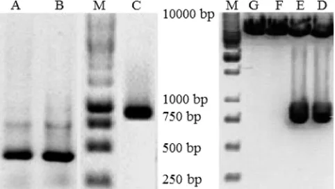
hu-man MAb (Fig. 1, lanes A and B). The amplified V
Hand V
Lchains were joined together with the 24 -mer polynucleotide
linker using SOE PCR, and the resultant 711-bp-long PCR
product (Fig. 1, lane C) was cloned into the TOPO-TA vector
to yield pCRdia. pCRdia was sequenced, and the entire
se-quence was submitted to the international ImmunoGeneTics
information system (IMGT) for sequence verification.
Se-quence information revealed the presence of 363-bp-long V
H,
324-bp-long V
L, and a 24-bp-long linker region (Fig. 2).
The diabody gene was reamplified using primers containing
EcoRI and NotI sites at the 5
⬘
and 3
⬘
ends, respectively, from
pCRdia, gel purified, and cloned into pET28a to obtain
pET28aRD (Fig. 1, lanes D and E). pET28aRD was
trans-formed into BL21(DE3), and the diabody was expressed by
induction with 1 mM IPTG. The cell pellet was lysed, and
the cytoplasmic fraction was purified by IMAC. Analysis of
the purified recombinant diabody by immunoblotting
indi-cated the presence of an
⬃
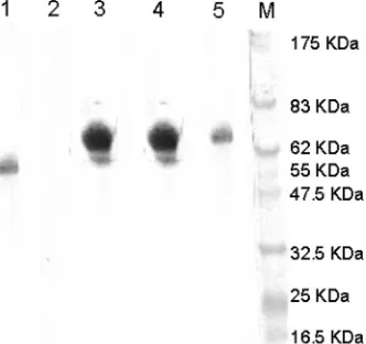
30-kDa band (Fig. 3). The yield
of diabody was
⬃
5 mg per 10-liter culture.
Characterization of diabody. (i) Determination of sensitivity
of the diabody using IC-ELISA.
A checkerboard titration was
performed to determine the optimal concentration of diabody
D06 to be used in IC-ELISA for quantification of RV GP (Fig.
4). The highest and lowest detection limits for RV GP under a
linear detection range were determined using purified RV (PV
strain). The optimal dilution of D06 required was 450 ng.
(ii) Reactivity of diabody with rabies virus glycoprotein.
Immunoblotting of the diabody D06 against the Pasteur rabies
virus (PV) structural proteins resolved in a 10% gel by
nonre-ducing SDS-PAGE clearly indicated the binding of the
dia-body, parent human MAb R16E5, and MAb M5B4 to an
⬃
66-kDa protein which corresponded to PV GP (Fig. 5). The
RV NP-specific MAb N5G4 was used as a negative control,
and it bound to an
⬃
55-kDa protein corresponding to PV NP.
(iii) Competitive ELISA.
Competitive ELISA was
per-formed to determine the competition between diabody D06,
parent MAb R16E5, and MAb M5B4 for the binding site on
PV GP. Competition could be seen when the constant amount
of MAb M5B4 was allowed to compete with various amount of
FIG. 1. Agarose gel electrophoresis analysis of PCR-amplified
products from human
⫻
mouse heterohybridoma. Lanes M show the
DNA ladder, and lanes A, B, and C show the variable heavy and
variable light chain genes and assembled PCR products. Lanes D to G
show the recombinant expression cassette after EcoRI and NotI
di-gestion, wherein lanes D and E show release of
⬃
711-bp product.
FIG. 2. Amino acid sequence of anti-rabies virus human diabody containing V
L, linker peptide, and V
H. The linker peptide is marked in italics.
The restriction enzyme sites for cloning of the diabody gene are underlined.
FIG. 3. Detection of recombinant diabody by Immunoblotting.
SDS-PAGE was performed with a 12% gel. Lane M shows the protein
molecular size standard (New England Biolabs), and lane 1 shows
purified diabody. The blot was transferred onto a PVDF membrane,
probed with anti-His probe, and developed by using the DAB
sub-strate. Lane M shows prestained protein molecular size markers; lane
1 shows a soluble fraction. The diabody (30 kDa) is denoted by an
arrow.
on August 17, 2020 by guest
http://cvi.asm.org/
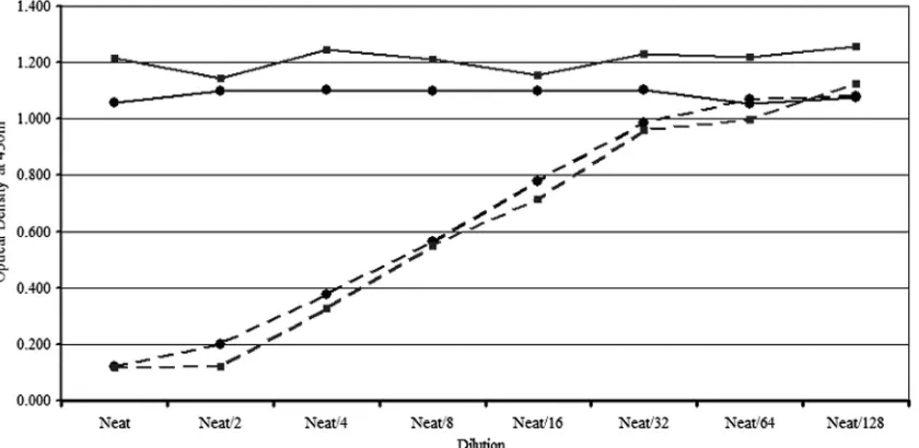
the diabody. A gradual increase in OD values following the
dilution of the diabody indicated that the diabody competed
with MAb M5B4 for antigenic site III on RV GP (Fig. 6).
(iv) IFAT using diabody.
The binding specificity of diabody
for RV GP was determined by IFAT using unfixed,
nonin-fected control and PV-innonin-fected Neuro-2a cells. Typical
mem-brane fluorescence could be seen both with the diabody and
with MAb M5B4 (Fig. 7). No fluorescence could be seen in
uninfected control Neuro-2a cells.
(v) Specificity of diabody.
Sandwich ELISA performed to
determine the binding specificity of diabody D06 clearly
showed reactivity with RV and not with other viruses, such as
HAV, hepatitis B virus (HBV), and CHIKV (Table 2),
indi-cating that diabody D06 can be used to detect RV without any
cross-reaction with other virus antigens.
Estimation of PV-GP in various vaccine preparations.
PV
GP content was estimated in 65 batches of experimental
hu-man rabies vaccine preparations using the IC-ELISA, and the
estimates were compared to the NIH potency values of those
respective batches. ANOVA with the regression through the
origin (RTO) model was performed to compare the potency
estimates derived by M5B4-D06 IC-ELISA and MAb M5B4
IC-ELISA previously described by Nagarajan et al. (18).
Regression analysis was performed using the data analysis
pro-gram in Microsoft Excel 2003 to compare the potency
esti-mates derived by M5B4D06 ELISA and MAb M5B4
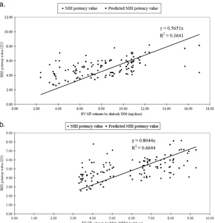
IC-ELISA with the NIH estimate. The adjusted
r
2value obtained
was 0.902, and the equation for predicted potency values for
M5B4-D06 based IC-ELISA and MAb M5B4 IC-ELISA were
0.5651
x
and 0.8044
x
, respectively, where
x
is the estimate of RV
GP determined by the IC-ELISA in
g (Fig. 8). ANOVA
results showed that the estimates by the two methods
com-pared differed highly significantly (
P
⬍
0.001), while the
pre-dicted potencies determined by the two tests did not differ
significantly (
P
⬎
0.05).
DISCUSSION
Rabies endemicity in many developing countries is
respon-sible for human deaths. The annual number of human deaths
worldwide caused by rabies is estimated to be between 40,000
and 70,000 in Africa and Asia, where rabies is endemic (25),
and India ranks among the highest with 20,000 human deaths
(21). The disease can be prevented by the timely
administra-tion of vaccine or a combinaadministra-tion of vaccine and RV
immuno-globulins. Vaccine manufacturers around the world determine
the potency of the rabies vaccines using the
in vivo
mouse
protection test (23), which is time-consuming and expensive
and requires the use of a large number of mice and virulent
rabies virus for challenge in a biosafety level 3 (BSL-3) facility.
The test, apart from being labor-intensive, time-consuming,
and expensive, also suffers from poor intra- and interlaboratory
FIG. 4. Immunocapture ELISA for titration of diabody D06 with different concentrations of rabies virus antigen.
FIG. 5. Reactivity of the diabody with the rabies virus glycoprotein
in immunotransfer blot analysis.
on August 17, 2020 by guest
http://cvi.asm.org/
reproducibility, thus making it a prime target for replacement
with easier
in vitro
tests that allow the accurate quantification
of the rabies glycoprotein (20). In this article, we describe the
expression, purification, and immunological characterization
of a recombinant diabody followed by its application in
devel-opment of an IC-ELISA for the quantification of RV GP in
human rabies vaccines.
The study describes the construction and characterization of
a recombinant diabody and its further development into a
reagent for use in an IC-ELISA format to quantify RV-GP.
Total RNA was isolated from a heterohybridoma, and the V
Hand V
Lfragments were amplified using universal primers and
assembled into a diabody using a reduced peptide linker. The
resultant PCR product was cloned into a bacterial expression
vector, pET28A, and expressed in
E. coli
under the control of
a T7 RNA polymerase promoter as an
⬃
30-kDa soluble
mol-ecule. The diabody was purified by IMAC. Purification of the
bacterial lysate resulted in a yield of
⬃
0.5 mg of purified
diabody/liter of culture with a homogeneity of
⬃
85%. The ease
of purification reiterated the fact that expression of a
func-tional recombinant antibody in bacteria offered many
advan-tages over the maintenance of a hybridoma cell line, which
included minimal batch-to-batch variation, ease of scale-up,
etc., at a reasonable cost.
The RV GP has been shown to induce neutralizing
antibod-ies and confer protection against lethal rabantibod-ies virus challenge.
Thus, much of the effort has been directed toward the
devel-opment of
in vitro
methods like ELISA, which is capable of
estimating GP content in rabies vaccine preparations. The
conventional/recombinant antibodies used in the assay should
be able to recognize the highly immunogenic, natively folded
RV GP present on the virus particle and should circumvent
problems associated with the estimation of the poorly
immu-nogenic soluble form of RV GP that could lead to the
over-estimation of the antigen, resulting in lower vaccine potency.
In order to demonstrate the binding of the diabody to the
FIG. 6. Competitive ELISA using the diabody and the RV GP-specific parent human MAb R16E5 and mouse MAb M5B4. Complete or broken
lines with squares show the result of competitive ELISA with diabody D06 and parent R16E5 with purified rabies virus antigen or
E. coli
lysate,
respectively. Complete or broken lines with circles show the result of competitive ELISA with diabody D06 and MAb M5B4 with purified rabies
virus antigen or
E. coli
lysate, respectively.
FIG. 7. Demonstration of the specificity of recombinant human diabody expressed in
E. coli
cells for RV GP by IFAT. (a) Detection with
anti-mouse IgG–FITC conjugate. (b) Detection with mouse anti-His IgG-FITC conjugate. (c) Uninfected-cell control.
on August 17, 2020 by guest
http://cvi.asm.org/
native RV GP, rabies-infected Neuro-2a cells were allowed to
react with the diabody. The appearance of specific membrane
fluorescence indicated that the antibody bound to the native
form of RV GP. The diabody also bound well to the RV GP,
as demonstrated by Western blot analysis. Further, the diabody
competed with MAb M5B4 for binding to RV GP in a
con-centration-dependent manner, suggesting that diabody and
M5B4 bind to the same epitope. A specificity study showed
that the diabody reacted only with RV but not with other
viruses, as indicated by sandwich ELISA. Diabody holds
prom-ise in quantification of the RV GP in vaccine preparations,
which correlated well with the NIH mouse potency. Various
vaccine batches tested by diabody-based IC-ELISA showed a
good correlation with the NIH mouse potency studies, as seen
with MAb M5B4-based IC-ELISA (18, 24).
TABLE 2. Reactivities of different polyclonal and monoclonal
antibodies and diabody D06 against different viruses
Antibody
Reactivity againsta
:
Rabies virus
Hepatitis A virus
Hepatitis B virus
Chikungunya virus
Diabody D06
⫹
⫺
⫺
⫺
MAb M5B4
⫹
⫺
⫺
⫺
Parent human MAb R16E5
⫹
⫺
⫺
⫺
MAb 1F6
⫺
⫺
⫹
⫺
Mouse serum HAV
⫺
⫹
⫺
⫺
Mouse serum CHIKV
⫺
⫺
⫺
⫹
a⫹, positive reactivity;⫺, no reactivity.
FIG. 8. (a) Line fit plot for regression through origin analysis for GP content estimated by MAb M5B4-D06 IC-ELISA and the actual NIH
potency value (IU). (b) Line fit plot for regression through origin analysis for GP content estimated by MAb M5B4 IC-ELISA and the actual NIH
potency value (IU).
on August 17, 2020 by guest
http://cvi.asm.org/
Quantification of RV GP using diabody-based IC-ELISA
provides exact information on the natively folded RV GP
an-tigen in vaccine preparations and in-process control samples,
which enables reliable estimation of RV GP. The method
described in this article, using diabody, provides a simple,
novel, and efficient option for quantification of RV GP antigen
in vaccine preparations without a loss of antigen and aids in the
manufacture of good-quality vaccines, which can reduce the
cost and make the vaccine affordable in developing countries,
where rabies prevention and control are a challenge. The
dia-body-based ELISA could replace the MAb-based
IC-ELISA due to better reagent stability and ease of production.
Conclusions.
A diabody D06 consisting of the V
Hand V
Lportions of a parent human
⫻
mouse heterohybridoma human
MAb, R16E5, was constructed using standard procedures. The
sensitivity and specificity of the diabody were established for
rabies virus glycoprotein. An immunocapture ELISA was
stan-dardized using the diabody D06 to estimate the glycoprotein
content of the human rabies vaccine containing Pasteur virus.
The IC-ELISA was compared with another procedure,
de-scribed earlier by Nagarajan et al. (18), and with an
in vivo
NIH
mouse protection test. Correlation could be established
be-tween the IC-ELISA using M5B4-D06 and the mouse
protec-tion test. This diabody-based ELISA can be used as an
alter-native for quantification of RV GP in purified RV antigens or
vaccines before or after blending, respectively.
ACKNOWLEDGMENTS
We thank the department of Quality Control, Human Biologicals
Institute, Ootacamund, India, for providing the rabies vaccine batches
used in the present study. We also thank L. Rajendra and R. Ramya
for providing technical assistance for this study.
REFERENCES
1.Alfthan, K., K. Takkinen, D. Sizmann, H. So¨derlund, and T. T. Teeri.1995. Properties of a single-chain antibody containing different linker peptides. Protein Eng.8(7):725–731.
2.Blancou, J., M. F. Aubert, E. Cain, M. Selve, O. Thraenhart, and L. Bruck-ner.1989. Effect of strain differences on the potency testing of rabies vac-cines in mice. J. Biol. Stand.17:259–266.
3.Bronnert, J., H. Wilde, V. Tepsumethanon, B. Lumlertdacha, and T. Hema-chudha. 2007. Organ transplantations and rabies transmission. J. Travel Med.14:177–180.
4.Cheung, S. C., B. Dietzschold, H. Koproswski, A. L. Notkins, and R. F. Rando.1992. A recombinant human Fab expressed in Escherichia coli neu-tralizes rabies virus. J. Virol.66:6714–6720.
5.Constantine, D. L.1962. Rabies transmission by the non-bite route. Public Health Rep.77:287–289.
6.Favoretto, S. R., C. C. de Mattos, N. B. de Morais, M. L. Carrieri, B. N. Rolim, L. M. Silva, C. E. Rupprecht, E. L. Durigon, and C. A. de Mattos.
2006. Rabies virus maintained by dogs in humans and terrestrial wildlife, Ceara State, Brazil. Emerg. Infect. Dis.12:1978–1981.
7.Ferguson, M., and A. Heath.1992. Report of a collaborative study to assess the determination of glycoprotein antigen content of rabies vaccines for human use. Biologicals20:143–154.
8.Fournier-Caruana, J., B. Poirier, G. Haond, C. Jallet, F. Fuchs, N. Tordo, and P. Perrin.2003. Inactivated rabies vaccine control and release: use of an ELISA method. Biologicals31(1):9–16.
9.Gamoh, K., M. Senda, O. Itoh, M. Muramatsu, N. Hirayama, R. Koike, Y. S. Endoh, and N. Minamoto.1996. Use of ELISA for in vitro potency test of rabies vaccines for animal use. Biologicals24(2):95–101.
10.Holliger, P., T. Prospero, and G. Winter.1993. Diabodies: small bivalent and bispecific antibody fragments. Proc. Natl. Acad. Sci. U. S. A.90(14):6444– 6448.
11.Knobel, D. L., S. Cleaveland, P. G. Coleman, E. M. Fe`vre, M. I. Meltzer, M. E. Miranda, A. Shaw, J. Zinsstag, and F. X. Meslin.2005. Re-evaluating the burden of rabies in Africa and Asia. Bull. World Health Organ.83:360– 368.
12.Laemmli, U. K.1970. Cleavage of structural proteins during the assembly of the head of bacteriophage T4. Nature227:680–685.
13.Lafon, M., P. Perrin, P. Versmisse, and P. Sureau.1985. Use of a monoclo-nal antibody for quantification of rabies vaccine glycoprotein by enzyme immunoassay. J. Biol. Stand.13:295–301.
14.Lindbladh, C., K. Mosbach, and L. Bu¨low.1993. Use of genetically prepared enzyme conjugates in enzyme immunoassay. Trends Biochem. Sci.18(8): 279–283.
15.Marks, J. D., H. R. Hoogenboom, T. P. Bonnert, J. McCafferty, A. D. Grif-fiths, and G. Winter.1991. By-passing immunization: human antibodies from V-gene libraries displayed on phage. J. Mol. Biol.222:581–597.
16.McCafferty, J., A. D. Griffiths, G. Winter, and D. J. Chiswell.1990. Phage antibodies: filamentous phage displaying antibody variable domains. Nature 348:552–554.
17.Mousli, M., I. Turki, H. Kharmachi, M. Saadi, and K. Dellagi.2007. Re-combinant single-chain Fv antibody fragment-alkaline phosphatase conju-gate: a novel in vitro tool to estimate rabies viral glycoprotein antigen in vaccine manufacture. J. Virol. Methods146(1–2):246–256.
18.Nagarajan, T., G. S. Reddy, B. Mohana Subramanian, S. Rajalakshmi, D. Thiagarajan, N. Tordo, C. Jallet, and V. A. Srinivasan. 2006. A simple immuno-capture ELISA to estimate rabies viral glycoprotein antigen in vaccine manufacture. Biologicals34:21–27.
19.Orlandi, R., D. H. Gu¨ssow, P. T. Jones, and G. Winter.1989. Cloning immunoglobulin variable domains for expression by the polymerase chain reaction. Proc. Natl. Acad. Sci. U. S. A.86(10):3833–3837.
20.Perrin, P., S. Morgeaux, and P. Sureau.1990. In vitro rabies vaccine potency appraisal by ELISA: advantages of the immunocapture method with a neu-tralizing anti-glycoprotein monoclonal antibody. Biologicals18:321–330. 21.Sudarshan, M. K., S. N. Madhusudana, B. J. Mahendra, N. S. Rao, D. H.
Ashwathnarayana, S. Abdul Rahman, F. X. Meslin, D. Lobo, K. Ravikumar, and Gangaboraiah.2007. Assessing the burden of human rabies in India: results of a national multi-center epidemiological survey. Int. J. Infect. Dis. 11:29–35.
22.Snedecor, G. W., and W. G. Cochran.1989. Statistical methods applied to experiments in agriculture and biology, 8th ed. Iowa State University Press, Ames, IA.
23.Wilber, L. A., and M. F. A. Aubert.1996. The NIH test for potency, p. 360–368.InF. X. Meslin, M. M. Kaplan, and H. Koprowski (ed.), Laboratory techniques in rabies, 4th ed. WHO, Geneva, Switzerland.
24.WHO.1988. Potency and stability of various modern rabies vaccines for veterinary use. http://whqlibdoc.who.int/hq/1988/WHO_Rab.Res._88.28.pdf. WHO, Geneva, Switzerland.
25.WHO.2001. Rabies. Fact sheet no. 99. WHO, Geneva, Switzerland.