Western University Western University
Scholarship@Western
Scholarship@Western
Electronic Thesis and Dissertation Repository
1-10-2017 12:00 AM
Maternal Nutrient Restriction in Pregnant Guinea Pigs and the
Maternal Nutrient Restriction in Pregnant Guinea Pigs and the
Impact on Fetal Growth and Brain Development
Impact on Fetal Growth and Brain Development
Andrew Ghaly
The University of Western Ontario
Supervisor
Dr. Bryan Richardson
The University of Western Ontario
Graduate Program in Physiology and Pharmacology
A thesis submitted in partial fulfillment of the requirements for the degree in Master of Science © Andrew Ghaly 2017
Follow this and additional works at: https://ir.lib.uwo.ca/etd Part of the Developmental Neuroscience Commons
Recommended Citation Recommended Citation
Ghaly, Andrew, "Maternal Nutrient Restriction in Pregnant Guinea Pigs and the Impact on Fetal Growth and Brain Development" (2017). Electronic Thesis and Dissertation Repository. 4359.
https://ir.lib.uwo.ca/etd/4359
This Dissertation/Thesis is brought to you for free and open access by Scholarship@Western. It has been accepted for inclusion in Electronic Thesis and Dissertation Repository by an authorized administrator of
ABSTRACT
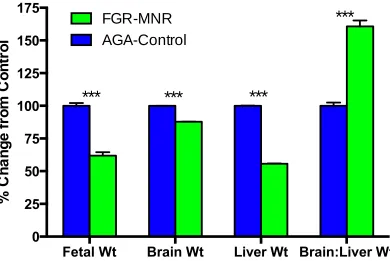
Maternal nutrient restriction (MNR) in guinea pigs results in placental structural abnormalities that reduce nutrient transport contributing to fetal growth restriction (FGR). However, whether brain weights are similarly reduced, or preserved by “brain sparing” mechanisms, and whether energy levels are depleted leading to membrane failure and overt injury remains unknown. Guinea pig sows were fed ad libitum (Controls) or 70% of the control diet pre-pregnant switching to 90% at mid-pregnancy (MNR). Animals were necropsied near term for fetal growth measures and fetal brains were assessed for markers of necrotic cell injury, apoptotic cell injury, endoplasmic reticulum stress, and altered development proteins. MNR resulted in FGR with brains that are large relative to body weight and livers that are small relative to body weight, which suggests a degree of blood flow redistribution. These fetuses have reduced brain weights, but with substantial brain sparing, and with no increased necrotic cell injury and no changes in synaptic development, indicating that the threshold for membrane failure or aberrant development with energy depletion has likely not been reached. However, apoptotic indices were increased in FGR-MNR cohort compared to appropriate for gestational age (AGA)-control cohort and more so in males than females. Changes in apoptosis were primarily in hippocampal regions and were not accompanied by significant changes of protein levels of investigated pro-apoptotic factors.
CO-AUTHORSHIP STATEMENT
TABLE OF CONTENTS
Abstract….…………...i
Co-Authorship Statement...ii
Table of Contents………...iv
List of Figures...vii
List of Tables...viii
List of Abbreviations………..………..………...ix
Chapter 1- Introduction………...1
1.1 Clinical Relevance ...2
. 1.1.1 Incidence and Classification of FGR...2
. 1.1.2 Symmetrical vs. Asymmetrical Growth...3
. 1.1.3 Etiology of FGR...5
. 1.1.4 Placental Insufficiency...6
. 1.1.5 Maternal Undernourishment………...7
. 1.1.6 FGR and Long-Term Health...………...9
. 1.1.7 Developmental Programming……….11
1.2 FGR and the Brain...13
. 1.2.1 FGR and Neurological Outcome...13
. 1.2.2 Fetal Brain Growth and Development………14
. 1.2.3 Synaptogenesis……….15
. 1.2.4 Myelination……….16
. 1.2.5 Apoptosis……….17
. 1.2.6 Mechanisms of Damage and Aberrant Development………18
. 1.2.7 Mechanisms of Adaptation………20
1.3 Animal Models of FGR and Adverse Development………..22
. 1.3.2 Animal Studies of FGR with Placental Insufficiency………..23
. 1.3.3 Animal Studies of FGR with Maternal Undernourishment………24
. 1.3.4 Animal Studies of FGR and Altered Brain Development……….25
1.4 Summary………...26
1.5 References..………..28
Chapter 2- Rationale, Hypothesis, and Research Objectives……….42
2.1Rationale ...43
2.2 Hypothesis...45
2.3 Objectives………..46
2.4 References………48
Chapter 3- Maternal Nutrient Restriction in Guinea Pigs as an Animal Model for Inducing Fetal Growth Restriction………51
3.1 Introduction………52
3.2 Materials and Methods……….55
. 3.2.1 Animal Feeding, Breeding and Pregnancy………..55
. 3.2.2 Necropsy, Tissue Collection and Blood Analytes……….56
. 3.2.3 Data Acquisition and Statistical Analysis……….58
3.3 Results……….58
. 3.3.1 Breeding and Pregnancy Outcomes...58
. 3.3.2 Maternal and Fetal Population Characteristics………59
. 3.3.3 Blood Analytes………..62
3.4 Discussion……….66
Chapter 4- Maternal Nutrient Restriction in Guinea Pigs with Fetal Growth Restriction
Leads to Altered Brain Development………...…………80
4.1 Introduction………81
4.2 Materials and Methods……….85
. 4.2.1 Animal Cohorts and Tissue Collection…..……….85
. 4.2.2 Necrosis Analysis with Haematoxylin and Eosin (H&E) Stain….………...87
. 4.2.3 Apoptosis Analysis with ApopTag….………...89
. 4.2.4 Markers of ER Stress and Apoptosis via Western Blot………91
. 4.2.5 Synaptic Development with Synaptophysin Immunohistochemistry. ………..92
. 4.2.6 Data Acquisition and Statistical Analysis….………..………..94
4.3 Results ………104
. 4.3.1 Fetal Population Characteristics……….………...104
. 4.3.2 Quantification of Necrotic Appearing Cells………..105
. 4.3.3 Quantification of Apoptotic Appearing Cells………...105
. 4.3.4 Western Blot Analysis….………..106
. 4.3.5 Quantification of SYN IR.. ………...106
4.4 Discussion.. ………..114
4.5 References……….………119
Chapter 5- General Discussion….……….127
5.1 General Discussion………...128
5.2 Future Studies……….133
5.3 Conclusions…..……….135
5.4 References…..………...137
LIST OF FIGURES Chapter 3
Figure 3.1 Fetal weights of all liveborn fetuses from MNR and control groups displaying 50th and 10th percentile
Chapter 4
Figure 4.1 Gross picture of the fetal guinea pig brain microdissection
Figure 4.2 High power section from FGR-MNR fetus of necrotic appearing cells
Figure 4.3 High power section from FGR-MNR fetus of TUNEL positive cells.
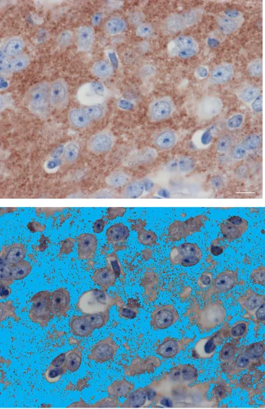
Figure 4.4 High power section from AGA-control fetus of SYN IR
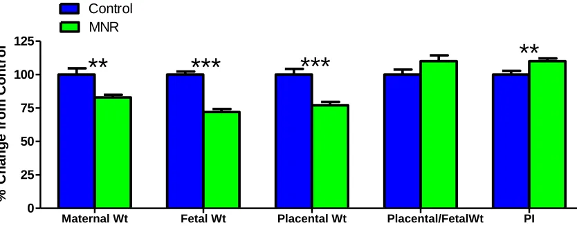
Figure 4.5 Growth measures for all control and MNR animals
Figure 4.6 Growth measures for select AGA-control and FGR-MNR animals
Figure 4.7 Bar chart of necrotic-appearing cells
Figure 4.8 Bar chart of TUNEL positive cells
Figure 4.9 Bar chart of TUNEL positive cells with sex comparison
Figure 4.10 Quantification of Western Blots with Representative immunoblots
LIST OF TABLES Chapter 3
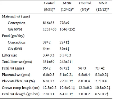
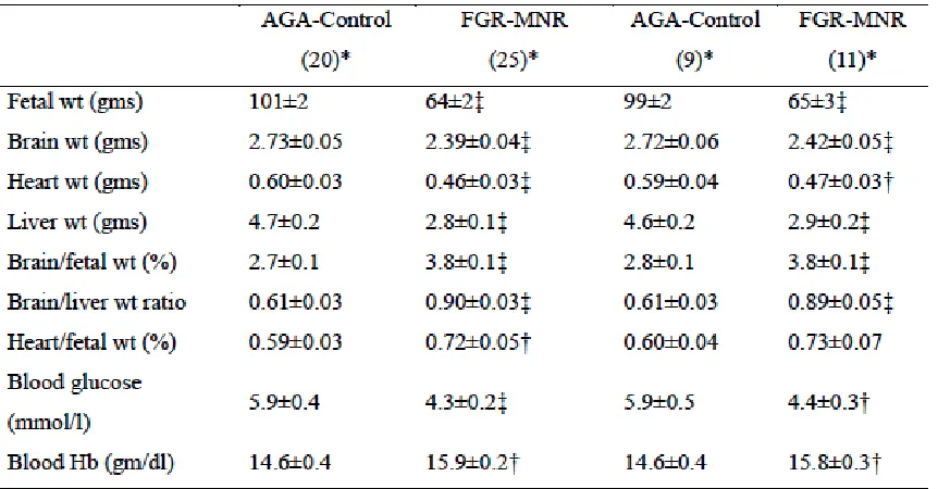
Table 3.1 Overall Maternal and Fetal Population Characteristics Table 3.2 Select Fetal Population Characteristics
Chapter 4
Table 4.1 Western Blot primary antibodies, dilutions used in experiments and company and catalogue information
Table 4.2 Western Blot secondary antibodies, dilutions used in experiments and company and catalogue information
LIST OF ABBREVIATIONS
aFGR Asymmetrical Fetal Growth Restriction AGA Appropriate for Gestational Age
AIF Apoptosis-Inducing Factor BAX Bcl-2 Associated X Protein BCL2 B-Cell Lymphoma 2 CV Coefficient of Variation DAB Diaminobenzidine ER Endoplasmic Reticulum FGR Fetal Growth Restriction Grp78 G-protein Coupled Receptor 78 H&E Haematoxylin and Eosin HPF High-Power Field
IGF Insulin-like Growth Factor IR Immunoreactivity
IQ Intelligence Quotient
IUGR Intrauterine Growth Restriction MNR Maternal Nutrient Restriction MRI Magnetic Resonance Imaging PARP1 Poly ADP Ribose Polymerase 1
sFGR Symmetrical Fetal Growth Restriction SGA Small for Gestational Age
SYN Synaptophysin
TBST Tris-buffered saline-Tween 20
TDT Terminal Deoxynucleotidyl Transferase
1.1CLINICAL RELEVANCE
1.1.1 Incidence and Classification of FGR
Fetal growth restriction (FGR), also known as intrauterine growth restriction
(IUGR), and formerly as fetal growth retardation, is defined clinically as birth weight
below the 3rd, 5th, or 10th percentile for gestational age and is indicative of a reduction in
growth rate that prevents a newborn from achieving their complete growth potential
(Lackman, Capewell, Richardson, DaSilva, & Gagnon, 2001). FGR pregnancies have up
to a 6-fold increase in fetal mortality and a 5-30 fold increase in neonatal mortality which
correlates with the severity of FGR (Piper et al., 1996; Seeds et al., 1998), as well as
increased neonatal morbidity (Bernstein, Horbar, Badger, Ohlsson, & Golan, 2000;
Kramer, Olivier, McLean, Willis, & Usher, 1990). Approximately 30% of pregnancies
with weights below the 10th percentile have a pathological origin, whereas the remaining
70% are constitutionally small. This incongruity highlights that these measures are not
always able to accurately delineate genuine FGR from infants that are simply small for
gestational age (SGA).
Early differentiation between FGR and constitutionally small fetuses arising from
non-pathological conditions in utero is important, as misdiagnosing small, healthy fetuses
as FGR may waste valuable resources. As such, other measures of fetal growth are used
to provide supplementary criteria for a clearer definition of FGR, rather than
constitutional SGA. Customized growth curves, which account for maternal,
environmental, and socioeconomic status may be beneficial for the identification of
pathological FGR (Arbuckle, Wilkins, & Sherman, 1993; Figueras et al., 2007; Gardosi
& Francis, 2009; Hutcheon, Walker, & Platt, 2011; Kierans et al., 2008; Kramer et al.,
mother would have an expected mean body weight at term that is 140 g heavier than a
child born to an average sized mother (Gardosi & Francis, 2009). Therefore,
identification of FGR would require a higher threshold based on maternal size to avoid
misdiagnosis of FGR. These customized growth curves result in reclassifying 2.7-4.3% of
babies (Figueras et al., 2007).
Other measures of fetal growth are used in conjunction with fetal growth curves
to produce a clearer definition of FGR vs. SGA. Ratios of fetal weight/length, are used as
a measure of leanness and fetal nutrition at birth. Additional studies have found placental
weight and placental function to positively correlate with birth weight and thus relate to
FGR (Lackman, Capewell, Richardson, et al., 2001; Pollack & Divon, 1992). Markers of
fetal size and shape at a particular gestational age are useful predictors of fetal health and
postnatal outcome.
1.1.2 Symmetrical vs. Asymmetrical Growth
FGR presents phenotypically as symmetrical (sFGR) or asymmetrical (aFGR)
fetal growth restriction; this distinction is based primarily on head and body proportions.
An sFGR fetus is typically impacted very early in pregnancy, with all growth parameters
being affected equally. This presents with brain growth inhibition remaining proportional
to reductions in weight, length, and head circumference and with a normal ponderal index
at birth (al Riyami et al., 2011; Halliday, 2009; Pollack & Divon, 1992) In aFGR, growth
restriction onsets later in pregnancy, which tends to occur in a more severe fashion,
however the later onset is associated with better health outcomes compared to the very
early-onsetting growth restriction seen in sFGR cases. aFGR presents with average head
muscle mass, and low weight/length ratios (due to increased leanness reflective of
disproportionate body dimensions) (al Riyami et al., 2011; Campbell & Thoms, 1977;
Halliday, 2009; Jones & Parer, 1983). This “brain sparing” effect occurs as a mechanism
of adaptation when chronically exposed to a hypoxic in utero environment to ensure fetal
survival. There is a redistribution of blood towards essential organs such as the brain, the
heart, and the adrenal glands at the expense of other organs such as the liver, kidneys, and
skeletal muscle (Dubiel, Breborowicz, & Gudmundsson, 2003; Dubiel, Breborowicz,
Marsal, & Gudmundsson, 2000; Poudel, McMillen, Dunn, Zhang, & Morrison, 2015;
Salihagic-Kadic et al., 2006). This redistribution is intended to ensure that the brain
continues to receive sufficient nutrient supply and maintain relative growth. The effects
of this are visible at birth, with the size of the fetal head being relatively large compared
than that of the abdomen, giving rise to the observable asymmetry observed in aFGR
fetuses (Jensen, Storgaard, Madsbad, Richter, & Vaag, 2007).
Unlike aFGR, sFGR can be difficult to distinguish from inherently small
fetuses because they are proportionally similar, it is the presence of pathological
processes that causes an infant to be considered sFGR rather than constitutionally SGA;
therefore, additional tests, such as umbilical artery Doppler assessment or extreme
discrepancy from a growth curve may be necessary to infer pathological sFGR. The
most reliable means of differentiating aFGR and sFGR is by measuring brain-to-liver
weight ratios. An aFGR fetus will have a disproportional decrease in liver weight,
but a relatively normal brain weight and therefore an increased brain-to-liver
weight ratio. Whereas sFGR fetuses will have small livers but with the brain being
appropriate for gestational age (AGA) fetuses (P. Cox & Marton, 2009).
Consequently, the brain-to-liver weight ratio is used in many animal studies to
identify aFGR at the time of necropsy. In clinical settings, head
circumference-to-abdominal circumference ratio measurements can serve as a proxy in a newborn or
human fetus.
There is a higher incidence of aFGR, reportedly occurring in 70% of growth
restricted fetuses (Campbell & Thoms, 1977). Population studies report that
60-80% of growth restricted babies born in developed countries are aFGR whereas
70-80% of growth restricted babies in developing countries are classified as sFGR
(Villar, Altobelli, Kestler, & Belizan, 1986). This indicates that both aFGR and sFGR
do not arise as the result of a single anatomical dysfunction or abnormality during
development; as such, there is a large amount of research dedicated to learning
more about the exact combination of conditions that contribute to FGR.
1.1.3 Etiology of FGR
Fetal growth is dependent on the interplay between maternal, placental, and
genetic factors. Optimal fetal growth is achieved when there is a balance of genetic
growth potential of the fetus, efficiency of the placenta in nutrient and oxygen transport,
and the state of the maternal environment. These maternal-placental-fetal factors work
synergistically to provide a healthy intrauterineenvironment for the development of
the fetus throughout gestation while supporting physiological changes in the mother.
When one or more of these factors is impacted, a sub-optimal intrauterine environment
2004; Han, 1993; Pollack & Divon, 1992). Maternal causes for FGR can arise
pre-pregnancy or during pre-pregnancy; maternal etiologic factors include weight, age, and
parity as well as variables like undernutrition, hypertension, hypoxic conditions,
vascular disease and environmental choices such as cigarette smoking and substance
abuse (Fang, 2005; Pollack & Divon, 1992). Placental factors that can contribute to FGR
and sub-optimal development include abnormal cord insertion, nuchal cord, placental
infarcts, placenta previa, and multiple gestations (Pollack & Divon, 1992). Fetal factors,
though relatively uncommon, are capable of contributing to FGR; fetal etiologic factors
include genetic factors such as fetal aneuploidy, congenital irregularities such as major
heart abnormalities, and infections such as cytomegalovirus (Demirci et al., 2015). Any
one of these factors is sufficient to cause FGR, however it is estimated that 60% of FGR
is idiopathic in nature and likely derived from a combination of factors, the majority of
which involve some form of abnormal placental development, commonly referred to as
placental insufficiency (Ghidini, 1996; Pallotto & Kilbride, 2006; Pollack & Divon, 1992).
1.1.4 Placental Insufficiency
The placenta is responsible for the transfer of nutrients between maternal and
fetal tissue throughout gestation. The human placenta has a complex system of blood
vessels that constitute the main area of fetal-maternal exchange. At approximately 21
days of gestation, there is an increase in fetal-placental blood vessels that allows for
vascularization and transforms the initially hypoxic in utero environment to one that is
capable of supporting fetal growth and organ system development during later
gestation (J. Kingdom, Huppertz, Seaward, & Kaufmann, 2000). This increase in
placental labyrinth, and a decrease in trophoblast thickness, all of which promote
elevated exchange of nutrients, essential substrates, and oxygen between maternal and
fetal tissue (Myatt, 2006).
The majority of conditions compromising fetal growth, especially in developed
countries, involve idiopathic aberrant development of the placenta referred to as
placental insufficiency (Hashimoto et al., 2012; Lumey, 1998). Placental insufficiency is
a term used to describe conditions in which the placenta is unable to transfer
appropriate amounts of nutrients or oxygen for full fetal growth. Clinical studies of
placental insufficiency with FGR have shown aberrant placental vascularization,
involving insufficient or incomplete trophoblastic invasion of spiral arteries in the
placental bed, and decreases in umbilical blood flow (J. Roberts, 1998; Salafia, Charles, &
Maas, 2006; Wang, Walsh, & Kay, 1992). This decrease in umbilical blood flow may
result in inadequate nutrition and oxygen reaching the fetus, resulting in a hypoxic,
nutrient deprived in utero environment (Economides & Nicolaides, 1989; Giussani,
Salinas, Villena, & Blanco, 2007; Romo, Carceller, & Tobajas, 2009; Salafia et al., 2006);
thus resulting in poor fetal development and therefore FGR. Under these conditions, the
fetus adapts by altering metabolic and developmental processes at the expense of
complete growth (T Jansson & Persson, 1990; Jones & Parer, 1983; Myatt, 2006). It
should be noted that independent of reductions in nutrient supply, hypoxia alone has
been shown to have a significant impact on fetal growth, therefore hypoxia may be a
major contributor to impaired fetal growth and FGR (Giussani et al., 2007).
As previously stated, FGR is a prevalent problem worldwide, however there are
differences in FGR etiology between developed and developing countries. While the
majority of conditions negatively affecting fetal growth in developed countries involve
placental insufficiency, in developing countries maternal nutrition plays a more
prominent role (Hashimoto et al., 2012; Lumey, 1998). It is estimated that 11% of
offspring in developing countries fail to reach their full growth potential due to
nutritional, social, and health factors related to poverty (de Onis, Blossner, & Villar,
1998; S. P. Walker et al., 2007a). Early evidence of the impact of maternal nutrition on
fetal health outcomes was observed in population studies during the Dutch famine of
the 1940s; studies of infants whose mothers’ nutrition was compromised at the time of
conception or in early pregnancy showed increases in placental weight but not in birth
weight (Lumey, 1998). These increases in placental weight are possibly an attempt to
compensate for the reduced availability of substrates in the maternal circulation, which
is what is seen in examination of human FGR cases (Lumey, 1998). Clinical study in
populations subjected to food restriction (Lumey, 1998) and in mothers deemed
underweight with low body mass index (Ehrenberg, Dierker, Milluzzi, & Mercer, 2003;
Kalk et al., 2009; Li et al., 2013; Sebastián Manzanares et al., 2012; Sebire, Jolly, Harris,
Regan, & Robinson, 2001) also support the notion that maternal undernourishment is
undoubtedly causative for FGR.
It has been found that nutritional supplementation throughout pregnancy led to
better outcomes in motor development of the child at 8 months, but not at the 5-year
mark. Similar supplementation given during the final trimester of pregnancy and
throughout infancy provided no benefit from 6 months to 3 years (Adair & Pollitt, 1985;
prior to pregnancy are actually better determinants of fetal growth and development
compared to changes in weight during pregnancy (Stevens-Simon, Metlay, &
McAnarney, 1995), reflecting the fact that the majority of women do not significantly
improve lifestyle habits and nutrition over the course of pregnancy (Crozier, Robinson,
Godfrey, Cooper, & Inskip, 2009). These studies show evidence that the placenta adapts
to maternal nutrient supply early on in pregnancy and is not easily altered or rescued in
response to supplementation later on in pregnancy. This shows the importance of
maternal nutrition in fetal programming and development as it has the potential to be a
chronic stressor on the mother, placenta, and developing fetus.
In cases of maternal nutrient restriction (MNR), the mother’s nutrient reserves
are depleted which results in a reduction of nutrient transport to the fetus and the
placenta, increasing the risk of FGR. In addition to this, MNR has been shown to
generate placentas with altered vascular development and increased barrier thickness,
in both animal models (Belkacemi, Nelson, Desai, & Ross, 2010; Redmer, Wallace, &
Reynolds, 2004; C. T. Roberts, Sohlstrom, Kind, Earl, et al., 2001) and human cases
(Aherne & Dunnill, 1966) of maternal undernourishment; which will further diminish
the transport of glucose, amino acids and lipids to the fetus (Belkacemi et al., 2010;
Gaccioli, Lager, Powell, & Jansson, 2012) in a manner similar to that seen in placental
insufficiency induced FGR.
1.1.6 FGR and long-term health
FGR remains the second most common adverse condition, behind preterm birth,
that arises from complications in pregnancy; and studies have shown that FGR is
Capewell, Richardson, et al., 2001). FGR pregnancies have up to a 6-fold increase in fetal
mortality and a 5-30 fold increase in neonatal mortality which correlates with the severity
of FGR (Piper et al., 1996; Seeds et al., 1998), as well as increased neonatal morbidity. In
addition to risk of adverse fetal and neonatal outcomes, FGR is also a recognized risk
factor for long term adverse health outcomes including heart disease, diabetes and
neurodevelopmental disability, with the greatest risk observed in those with severe and
early-onsetting FGR (Barker, 2004; Thomas Jansson & Powell, 2007; Low et al., 1992;
Pryor, Silva, & Brooke, 1995; Zaw, Gagnon, & da Silva, 2003). Studies on sex differences
in FGR offspring found that there is a significantly higher incidence of FGR in females,
which suggests that there is a higher tendency for adaptation to a sub-optimal
intrauterine environment in females relative to males (J. Liu, 2014); however females
tend to have more favourable outcomes and a better likelihood of surviving FGR related
outcomes compared to males (Synnes et al., 2010). Studies of MNR in mice showed a
higher susceptibility to FGR in males, with a higher incidence of impaired cognitive
function (Akitake et al., 2015) and this sex difference persists in adults, as less tissue
damage for an equivalent insult has been reported in global and focal cerebral ischemia
in adult female rodents compared to males (Alkayed et al., 1998; Hall, Pazara, &
Linseman, 1991).
At the time of birth, FGR newborns are at high risk for hypoxia, hypoglycemia,
and hypothermia due to limited nutrient supply and storage, low tissue adiposity,
diminished gluconeogenesis and impaired lipid metabolism (Fang, 2005; Rodríguez et
al., 2011). Interventions in early pregnancy are aimed at promotion of fetal growth and
survival. Mothers often receive glucocorticoid supplementation if they are at risk of
that is necessary for lung development. Another major goal of intervention in postnatal
care is the achievement of catch-up growth, whereby there is rapid growth after birth to
match the growth curves of AGA infants. Previous studies have demonstrated that aFGR
infants are capable of full catch-up growth within 3 years, however sFGR infants have a
reduced rate of weight gain and growth up to 40 weeks, followed by an increased linear
weight gain until approximately 8 months (Strauss & Dietz, 1997). The same study
demonstrated that 70% of sFGR infants had weights greater than that of the 10th
percentile but remained at a lower weight compared to AGA infants up to 3 years old
(Strauss & Dietz, 1997). A related study, which examined growth of sFGR infants, found
that weight, height, and head circumference were still reduced even up to 14 years of
age (Indredavik et al., 2010). While post-natal treatment may aid in catch-up growth, it
does not lessen the long-term morbidity that arises due to sub-optimal in utero
conditions and aberrant development.
1.1.7 Developmental Programming
Fetal programming is the theory that there are critical periods of organ
development, during which the system is highly sensitive to environmental cues, which
are capable of setting the platform for health outcomes in later life (Barker, 2004). This
is known as the “Barker hypothesis” or the “developmental origins of health and
disease” in later life. The adaptations that occur in response to adverse intrauterine
conditions during these critical periods of development are capable of causing
permanent structural and functional changes to several of the body’s systems. The three
primary means by which fetal programming can occur are: 1) direct damage, such as
structure resulting from insult during a critical period of development; or 3)
physiological re-setting by an early stimulus or insult at a critical period with long term
consequences for endocrine/autocrine/paracrine axes (Lucas, 1994). Barker described
these periods as most likely occurring in the fetus during phases of rapid cell division,
and being more impacted by intrauterine environment than genetics (Cosmi, Fanelli,
Visentin, Trevisanuto, & Zanardo, 2011). Early evidence which supported claims that
undernutrition in utero, resulting in FGR, increased the risk of cardiovascular disease in
adult life lead to the popularization of the idea of fetal programming (Barker, 2004).
More recently, studies have found a positive correlation between low birth weight,
caused by either FGR or preterm birth, and increased rates of cardiovascular disease
and type-2 diabetes in adult life (Langley-Evans, 2009). There is growing evidence that
supports Barker’s hypothesis of developmental adaptations in utero having long-term
changes on the “programming” of later life health outcomes (Barker, 2004), and there is
an increasing amount of research that suggest that the etiology of many neurological
disorders can also be attributed to the effects of the suboptimal intrauterine
environment seen in FGR.
Maternal undernutrition gives rise to fetal programming by means of the above
mentioned physiological re-setting. The early stimulus of placental compromises in
nutrient/oxygen delivery causes growth restriction to occur as an adaptive process by
which energy needs are decreased in order to ensure survival (Hochachka, Buck, Doll, &
Land, 1996). However, this has consequences for aberrant growth processes which lead
1.2 FGR AND THE BRAIN
1.2.1 FGR and neurological outcome
The link between FGR and neurological disorders and cognitive deficits is
apparent in all age groups and has been reported in many studies. FGR is associated
with a higher risk of development of a number of neurological disorders that manifest
in early childhood including cerebral palsy, attention deficit hyperactivity disorder, and
autism spectrum disorder (Halliday, 2009; Indredavik et al., 2010; Rodrigues, Mello, &
Fonseca, 2006). The effects of FGR in early childhood can be observed with reductions
in cognitive skills, impaired memory, learning difficulties, difficulties reading, writing,
and with adaptive skills, inattention, reduced psychosocial function, behavioural
problems, sensorineural deterioration, reduced mathematics abilities, and reduced
intelligent quotient (IQ) scores (Geva, Eshel, Leitner, Fattal-Valevski, & Harel, 2008;
Pallotto & Kilbride, 2006; Rodrigues et al., 2006; Synnes et al., 2010; D.-M. Walker &
Marlow, 2008). More severe effects can be seen with altered brain development later in
life as FGR-born adults are at a higher risk of schizophrenia, epilepsy, and psychiatric
hospitalization (M. Cannon, Jones, & Murray, 2002); studies of these neurological
disabilities show that their severity correlates positively with the severity of FGR.
Neurological difficulties in FGR-born children continue through adolescence and
into early adult life. At 10 years of age, late-onset aFGR-born children were found to
have continued deficiencies in verbal short term memory of auditory or visiospatially
presented information and reduced IQ scores (Geva et al., 2008). Studies comparing 13
year-old FGR-born children to age matched controls showed poor everyday memory,
mathematical reasoning and numerical operation skills even after learning-skills
were reported to still have reduced head circumference as well as increased
hyperactivity, inattention, psychiatric diagnosis, autism spectrum disorder scores and
reduced psychosocial function (Indredavik et al., 2010). These studies indicate that
behavioural and cognitive difficulties seem to become more complex and quite possibly
more severe with age for FGR-born offspring.
Regional differences exist in the brain’s development and thus, certain areas can
be affected differently by in utero insults. For example, the hippocampus is the least
genetically regulated region of the brain and is therefore more vulnerable to
environmental and developmental influences compared to areas of the brain under
higher levels of genetic influence (Lodygensky et al., 2008). A study by Lodygensky et al.
showed reductions in hippocampal volume of FGR-born infants at 2 years of age, with a
significant correlation between total hippocampal volume and size at birth, this cohort
was also found to have poor performance in motor, attention-interaction, and
self-regulation behavioural function maturation assessments (Lodygensky et al., 2008).
Hippocampal differences have been observed in other studies as well, with reduced
hippocampal volume and enlarged ventricles appearing on magnetic resonance imaging
(MRI) scans in FGR-born infants compared to age matched controls (Isaacs et al., 2000).
These irregularities in the development of the hippocampus may, in part, explain the
basis for several of the neurological deficits observed in FGR as the hippocampus and
associated areas are critical for learning, memory, and proper cognitive function (Reed
& Squire, 1997).
Brain growth and development are intricate processes that occur throughout the
entirety of gestation and continue to be refined after birth. At term, the developing
human fetal brain is responsible for approximately 20% of body mass and 80% of
energy expenditure (Gilles, 2011). The rate of brain growth peaks around term; this
involves the formation and migration of neurons, and once neurons reach their final
migration point myelination begins and there is a notable decrease in the rate of brain
growth which plateaus at about two years after birth (Gilles, 2011); after which brain
alteration is largely dependent upon experience and environmental factors.
Complete growth of the brain requires neuronal generation and differentiation,
navigation and organization of axonal projections between neurons, and the formation
and maturation of synaptic contacts (Bourgeois, 1997). It is theorized that there is an
initial formation of large amounts of synaptic contacts by means of intrinsic
mechanisms of growth; however, subsequent maturation of specific neuronal
connections occurs by means of selective usage (Bourgeois, 1997). As a result, it is
believed that environmental influences play a major role in the development of these
processes and can have significant impacts on later life neurological health.
1.2.3 Synaptogenesis
Synaptogenesis is a process that occurs during gestation and through to about
puberty in humans (Bourgeois, 1997). The early phases involve neurogenesis, neuronal
migration, individualization of cortical layers and synapse formation that are controlled
by intrinsic mechanisms which are considered experience-independent (Bourgeois,
1997). In later phases synapses are formed at a much faster rate, driven by an
brain and pre-adapts the brain for an experience dependent phase with individual
customization on the basis of experience and environmental cues (Bourgeois, 1997). A
plateau phase exists, in which there is elimination of select synapses and selective
maturation of others, in order to fine tune and establish certain neuronal connections.
(Bourgeois, 1997). In order to mature, a synapse requires an abundant amount of
energy. Areas with newly forming synapses have high levels of mitochondria in order to
supply the large amount of energy necessary for protein synthesis and synapse
formation (Mjaatvedt & Wong-Riley, 1988); as such compromises in nutrient/oxygen
delivery as a result of a poor intrauterine environment are capable of affecting the
earlier phases of synaptogenesis and may predispose the fetal brain to deficiencies in
later life.
Synaptophysin (SYN) is a 38 kDa presynaptic protein marker present in all
presynaptic boutons on presynaptic vesicle membranes in the central nervous system
(Calhoun et al., 1996; Jahn, Schiebler, Ouimet, & Greengard, 1985). Histological staining
using SYN antibodies shows punctate staining localized to presynaptic boutons
(Calhoun et al., 1996; Fletcher, Cameron, De Camilli, & Banker, 1991). This presynaptic
protein marker has been used in animal studies of FGR as a measure of synaptic
numbers in the brain (Camm, Gibbs, Harding, Mulder, & Rees, 2005; Tolcos et al., 2003).
It is also found in immature synapses and small synaptic vesicles, and its protein levels
directly increase with synapse formation and development (Daly & Ziff, 1997; Fletcher
et al., 1991). Using immunohistochemistry, the immunoreactivity (IR) of SYN has been
shown to be an accurate presynaptic marker for the detection of synapse formation.
Myelin is necessary for the insulation of electrochemical signals that are sent
along neurons; proper myelination throughout development ensures neuronal
communication and connectivity between areas of the brain. The presence of neurons
and the electrical activity of axons perpetuates myelination in a specific axon (Barres &
Raff, 1993). The degree to which an axon is myelinated dictates the conduction velocity
of an axonal signal and is critical to the synchronous firing of action potentials, which is
necessary to strengthen neuronal connections (Fields, 2008). As such, the increasing
electrical activity in a maturing neuron aids in myelination which in turn establishes
saltatory conduction and aids in the coordination of synaptogenesis between neuronal
connections.
In humans, the rate of myelination of the brain increases towards the end of
gestation and into the 2nd year of life. Myelin sheath proteins are seen in the brain as
early as the 5th week of gestation and become more abundant with advancing
gestational age (Jakovcevski, Mo, & Zecevic, 2007; Kinney, Brody, Kloman, & Gilles,
1988). The high rate of signaling and protein turnover required for myelination makes
it a process with high energy demand; therefore, extended periods of compromised
nutrient/oxygen delivery may impact on the process leading to poor myelination and
therefore poor synaptogenesis and aberrant development of the brain.
1.2.5 Apoptosis
Apoptosis, or programmed cell death, has a physiological basis during the
fetal/neonatal period of brain development that coincides with neuronal differentiation
and synaptogenesis, and possibly relates to the competition for trophic factors
Staley, & Chun, 1996). The process of apoptosis involves stimulation of intrinsic and/or
extrinsic signaling pathways leading to the downstream cleavage of caspase-3, which
together with effector caspases can target multiple proteins for proteolysis causing a
resultant fragmentation of the cell’s DNA and dismantling of cellular structures
(Banasiak, Xia, & Haddad, 2000; D’Amelio, Cavallucci, & Cecconi, 2010). Apoptosis can
be pathologically activated in the brain in cases of chronic hypoxia and result in
selective neuronal loss (Burke et al., 2006; Yue et al., 1997) with immature neurons
and/or milder insults more likely to result in apoptotic death, while terminally
differentiated neurons and/or severe insults are more likely to result in death by
necrosis ( a D. Edwards et al., 1997; Scott & Hegyi, 1997; Yue et al., 1997).
1.2.6 Mechanisms of Damage and Aberrant Development
The histo-pathological alterations seen in human pregnancies with
undernourishment resulting in FGR (Aherne & Dunnill, 1966), have been shown to
result in reductions in nutrient transport to the fetus for glucose, amino acids, and lipids
(Crozier et al., 2009). Recent studies have also shown evidence for chronic hypoxia with
MNR-FGR in guinea pigs (Elias, Matushewski, Zhao, Regnault, & Richardson, 2013), low
grade oxidative stress may play a mechanistic role here given its known role in the
pathogenesis of chronic disorders (Hracsko, Orvos, Novak, Pal, & Varga, 2008; Mohn et
al., 2007). If these insults occur during critical time points in brain growth and
development, there is a high risk for potential derangements.
As previously stated, in FGR with compromised nutrient/oxygen delivery there is
adaptive programming in which growth restriction occurs and select developmental
of essential processes necessary for survival. (Hochachka et al., 1996). Variable
alterations to brain development and injury may be observed on the basis of the insult.
Selective neuronal loss has been observed in developing brain cells upon exposure to
hypoxia or ischemia; with milder insults likely to impact immature neurons and result
in apoptotic death and more severe insults likely to affect terminally differentiated
neurons and result in death by necrosis (Scott & Hegyi, 1997; Yue et al., 1997).
Sensitivity to insults and brain injury also varies on the basis of energy demand, with
areas that have a higher energy demand being more sensitive; as such hippocampal
cells are expected to see the largest change (Zhao & Flavin, 2000). Additionally, there is
regional variation in susceptibility, with the cortex and striatum being more sensitive
than the thalamus (Dirnagl, Iadecola, & Moskowitz, 1999). In prenatal brain developers
such as guinea pigs, and for humans as perinatal brain developers, the most rapid phase
of synapse formation and neuronal myelination onsets during the latter half of
pregnancy (McIntosh, Baghurst, Potter, & Hetze, 1979; Penn & Shatz, 1999). As such,
FGR with nutrient transport impairment may disrupt synapse formation and neuronal
myelination, as they are energy demanding growth processes (Jiang & Schuman, 2002).
This may lead to alterations in brain development and underlie risk for later cognitive
impairment and neuropathology.
Additionally, hypoxia, a decrease in amino acid levels, and/or oxidative stress can
lead to the accumulation of misfolded or unfolded proteins in the endoplasmic
reticulum (ER), which if prolonged can initiate a pro-apoptotic cascade (Koumenis et al.,
2002; Marciniak & Ron, 2006; Szegezdi, Logue, Gorman, & Samali, 2006). Hypoxia and
low amino acid supply, as seen in the MNR guinea pig model (Elias et al., 2013; C. T.
2001), have been demonstrated to hinder disulfide bond formation – one of the key
processes underlying protein maturation and folding within the ER lumen, thus
augmenting ER stress (Benham, van Lith, Sitia, & Braakman, 2013; Frand & Kaiser,
1999; Yu et al., 2012; Zhang et al., 2014). In cases where ER stress is not sufficiently
alleviated, downstream apoptotic pathways will be activated (D. Liu, Zhang, & Yin,
2013; Matsumoto, Minami, Takeda, Sakao, & Akira, 1996). G-protein coupled receptor
78 (Grp78) is an essential component of the ER translocation machinery and plays a
significant role in retrograde transport of aberrant proteins destined for degradation by
the proteasome; as such, synthesis of Gpr78 is induced under conditions that lead to the
accumulation of unfolded polypeptides in the ER (Hendershot, Valentine, Lee, Morris, &
Shapiro, 1994; Ting & Lee, 1988); Consequently, Grp78 is used as an early marker of ER
stress. Since the ER is the primary site of protein synthesis and maturation within the
cell, prolonged ER stress could negatively impact essential signaling and transport
function necessary for proper development of the fetal brain (Braakman, Hoover-Litty,
Wagner, & Helenius, 1991; Frand & Kaiser, 1999; Kawakami et al., 2014; Red-Horse et
al., 2004).
1.2.7 Mechanisms of Adaptation
Previous studies have shown that there are compensatory mechanisms in action
that deal with prolonged episodes of lowered energy and oxygen that involve
suppression of energy-demanding pathways to regulate the cell at new steady state
levels, even during drastic energy depletion. These mechanisms include channel arrest
of membrane ion pumps, cessation of synaptic transmission, and decreased protein
enough, it is possible that these compensatory mechanisms, which are usually
protective for the brain, may become limited and brain energy levels may be sufficiently
impacted, leading to membrane failure with an increase in necrotic cell injury and/or
changes in apoptotic regulators (Rocha, Hammond, & Richardson, 2004). While these
mechanisms have yet to be elucidated, regional differences in the balance of
pro-apoptotic and anti-pro-apoptotic gene expression and activity-dependent changes in this
balance with the strengthening of incoming afferent activity are likely to be involved
(Anand & Scalzo, 2000).
Some of these pro- and anti-apoptotic factors include Bcl-2 associated X protein
(Bax), B-cell Lymphoma 2 (Bcl-2), poly ADP ribose polymerase 1 (PARP1), and cleaved
caspase-3. Pro-apoptotic Bax and anti-apoptotic Bcl-2 are known to synergistically
regulate apoptosis (D. Liu et al., 2013; McCullough, Martindale, Klotz, Aw, & Holbrook,
2001). PARP1 is required for translocation of apoptosis-inducing factor (AIF) from the
mitochondria into the nucleus, in which it induces large scale DNA fragmentation
(Daugas et al., 2000). Extrinsic and intrinsic apoptotic pathways both converge on
caspase-3, with the downstream event consisting of substrate cleavage. There is
abundant evidence that pathways leading to caspase-3 cleavage/activation are engaged
following neonatal hypoxia-ischemia (Blomgren et al., 2001; Felderhoff-Mueser et al.,
2002; Hu, Liu, Ouyang, Blomgren, & Siesjö, 2000; Northington, Ferriero, Flock, & Martin,
2001). It is of note that caspase-3 activation does not always lead to DNA fragmentation,
in the developing brain caspase-3 appears to also play a non-apoptotic role in cellular
differentiation and remodeling of neuroplasticity through selective removal of existing
1.3 ANIMAL MODELS OF FGR AND ADVERSE DEVELOPMENT
1.3.1 Guinea pig brain development
Guinea pigs and sheep have been used to model human FGR due to their delivery
of precocious young after a relatively long gestational period (Dobbing & Sand, 1970).
These species demonstrate a similar timeline of developmental events in the brain to
those occurring in utero during human pregnancy; therefore, intrauterine insults will
coincide with critical periods of development and the associated long-term outcomes
more accurately parallel that of the human situation. Additionally, both humans and
guinea pigs are thought to be born with the full complement of neuronal and synaptic
numbers (Lennon, Francon, Fellous, & Nunez, 1980). This is in contrast to rodents,
which have a shorter gestation and altricial young; therefore many of their
developmental processes occur in the postnatal period and are less affected by the in
utero environment.
In comparison to humans, the guinea pig brain is larger and at a more developed
stage of functionality at birth. In the human situation brain growth plateaus at
approximately 2 years of age, this plateau occurs just prior to birth in the guinea pig
(Dobbing & Sand, 1970). At birth, guinea pig brain functions include motor, cognitive
and regulatory processes that are comparable to that of a human toddler, this is due to
peak neurogenesis occurring in several brain areas much earlier in guinea pigs than in
humans (Clancy et al., 2007). Therefore, in utero insults may actually be magnified in
the guinea pig brain in comparison to humans; while the impact of post-natal
environmental influences that play a role in ongoing brain development will be lessened
in the mature guinea pig brain. The precocious function and comparable anatomy of the
determination of changes in neurodevelopment and their extrapolation to the human
situation.
1.3.2 Animal Studies of FGR with Placental Insufficiency
Clinical studies have shown that a major contributor to placental insufficiency,
and therefore FGR, is aberrant placental vascularization and the accompanying changes
in umbilical blood flow. As a result of this, research on animal models of placental
insufficiency has been a growing area of research. The guinea pig is a good model of
human placental insufficiency because it has a very similar placenta; both human and
guinea pigs have a discoid haemochorial placenta in which fetal vessels and maternal
blood are in direct contact. Placental insufficiency can be induced by a variety of
interventions at pre-pregnancy, mid-pregnancy, and late pregnancy. Some of these
include: uterine caruncletomy, the removal of endometrial tissue, exposure to
hypothermic environment in pregnant sheep, placental embolization midway through
gestation, or uterine artery ligation/ablation midway through gestation; these
techniques have successfully resulted in decreased fetal weight, increased brain-to-fetal
weight ratios in sheep, and increased markers of hypoxemia and hypoglycemia
(Gagnon, Murotsuki, Challis, Fraher, & Richardson, 1997; Harding, Jones, & Robinson,
1985; Murotsuki, Challis, Han, Fraher, & Gagnon, 1997; Regnault, Orbus, Battaglia,
Wilkening, & Anthony, 1999). Of particular interest to this study, are reports of uterine
artery ligation in rodents and sheep that have successfully induced FGR with
accompanying reductions in fetal brain development and long-term brain function
(Olivier et al., 2007; Rees, Breen, Loeliger, McCrabb, & Harding, 1999). Guinea pig
decreased transfer of oxygen and nutrients (T Jansson & Persson, 1990; Jones & Parer,
1983; Lafeber HN, Rolph TP, 1984), which successfully models human placental
insufficiency. Recent studies have additionally shown that uterine artery ligation is
capable of leading to disruptions in synapse formation, regional synapse maturation,
and myelination (Piorkowska et al., 2014). These animal models that mimic placental
insufficiency are highly effective in producing FGR with variable levels of hypoxemia
and nutrient deficiency, however they are not without their flaws. There have been high
reported rates of mortality in the pups in uterine artery ligation and ablation models ( a.
J. Turner & Trudinger, 2009), and they involve a one-time, invasive procedure, which
doesn’t ideally parallel the human condition of a chronic, non-invasive insult.
1.3.3 Animal Studies of FGR with Maternal Undernourishment
As previously stated, clinical studies have also shown that maternal nutrition is a
major contributor to the health outcomes of a fetus. As a result, there are many animal
models that aim to investigate the impacts of maternal undernourishment on fetal and
placental growth and development. These models use a variety of levels of nutrient
restriction and different time points for restriction relative to gestation resulting in a
large range of different levels of growth restriction and altered development. These
models can range from “moderate” restrictions, whereby animals are fed 70-85% of a
control ad libitum diet, to “severe” restriction studies, whereby animals are provided
with 50% of a control ad libitum diet to mimic cases of extreme undernourishment.
Animal models of MNR have been used to study developmental outcomes with induced
FGR (Belkacemi et al., 2010; Thomas Jansson & Powell, 2007; Redmer et al., 2004; C. T.
shown to generate placentas with altered vascular development and increased barrier
thickness, in both animal models (Belkacemi et al., 2010; Redmer et al., 2004; C. T.
Roberts, Sohlstrom, Kind, Earl, et al., 2001) and human cases (Aherne & Dunnill, 1966)
of maternal undernourishment; recent studies have also shown evidence for chronic
hypoxia with MNR-FGR in guinea pigs (Elias et al., 2013).
Studies conducted in sheep and guinea pig animal models, ranging from
moderate to severe nutrient restriction, result in a range of different levels of growth
restriction and altered development. MNR starting pre-conception and lasting over the
entire course of pregnancy in guinea pigs has been shown to be sufficient in causing
altered fetal and placental weights consistent with FGR (C. T. Roberts, Sohlstrom, Kind,
Earl, et al., 2001). Studies that utilized pre-conception moderate MNR showed a
significant impact on fetal-placental growth. MacLaughlin et al. demonstrated that MNR,
applied pre-conception through to gestational day 7 in sheep, was sufficient to impact
fetal and placental weight at birth. Previous studies have used moderate MNR in guinea
pigs, applied pre-conception and through pregnancy, and have observed reduced fetal
weight and altered fetal development, as well as structural and functional changes in
the placenta, including increased barrier thickness, decreased size of the placental
labyrinth, and therefore decreased availability for nutrient exchange between maternal
and fetal tissue (C. T. Roberts, Sohlstrom, Kind, Earl, et al., 2001; C. T. Roberts,
Sohlstrom, Kind, Grant, et al., 2001).
1.3.4 Animal Studies of FGR and Altered Brain Development
Animal studies of aFGR have demonstrated that, despite brain weights being
such as the hippocampus, retina, ventricles, cerebellum, brain stem, and cortex
(Mallard, Loeliger, Copolov, & Rees, 2000; Rees & Bainbridge, 1992; Rees et al., 1999;
Rehn et al., 2004; Tolcos et al., 2003; Tolcos & Rees, 1997). There have been several
studies in FGR guinea pigs showing that synapse formation and myelination are
reduced (Dieni & Rees, 2003; Mallard et al., 2000; Piorkowska et al., 2014). Guinea pig
offspring that utilized a uterine artery ligation model of placental insufficiency were
found to have reduced neuronal numbers in select brain regions at 7 days of age; these
changes significantly correlated with reductions in brain weight (Mallard et al., 2000).
Another study using a uterine artery ligation model in guinea pigs found FGR-born
animals to have increased lateral ventricle size and reduced basal ganglia volume at 8
weeks of age (Rehn et al., 2004). Additional studies in guinea pig models of placental
insufficiency have found reduced myelination and a reduced number of myelinated
fibers in FGR offspring compared to control animals, as well as disproportionally thin
myelin sheath in growth restricted fetuses (Nitsos & Rees, 1990) A number of structural
changes occur in the brain with a great deal of regional variability, and these changes
persist after birth.
1.4 SUMMARY
Fetal growth restriction is a failure to attain full genetic growth potential during
fetal life. It can manifest as asymmetrical or symmetrical growth restriction, based on
the incidence of brain sparing. FGR occurs due to a combination of maternal, placental,
and genetic factors; the most common being the occurrence of placental insufficiency in
developed countries or undernutrition in developing countries. In both cases there is a
either by reduced capacity of the placenta to transport nutrients, and/or by reduced
concentration of nutrients in maternal circulation. FGR leads to an elevated risk of
cognitive deficits and neurological disorders in later life. This includes increased risk of
autism spectrum disorder, attention deficit hyperactivity disorder and schizophrenia.
Cognitive deficits may present as poor academic performance, learning disabilities and
memory deficiencies in childhood and throughout life. Clinical study and research on
potential mechanisms behind FGR have led to the creation and use of many animal
models to mimic the human situation. Previous studies have shown that there is a
connection between FGR and altered growth and development of the fetal brain;
however, there has been little study in models of MNR. As such, determining the effects
on brain growth and development resulting from MNR leading to FGR may lead to a
better understanding of the human situation and may be utilized in extrapolation of
1.5 REFERENCES
Adair, L. S., & Pollitt, E. (1985). Outcome of maternal nutritional supplementation: a comprehensive review of the Bacon Chow study. The American Journal of Clinical Nutrition, 41(5), 948–978. Retrieved from
http://ovidsp.ovid.com/ovidweb.cgi?T=JS&PAGE=reference&D=med2&NEWS= N&AN=3993612
Aherne, W., & Dunnill, M. S. (1966). Morphometry of the human placenta. British Medical Bulletin, 22(1), 5–8.
http://doi.org/10.1001/archinte.1960.03860170112044
Akitake, Y., Katsuragi, S., Hosokawa, M., Mishima, K., Ikeda, T., Miyazato, M., &
Hosoda, H. (2015). Moderate maternal food restriction in mice impairs physical growth, behavior, and neurodevelopment of offspring. Nutrition Research (New York, N.Y.), 35(1), 76–87. http://doi.org/10.1016/j.nutres.2014.10.014
al Riyami, N., Walker, M. G., Proctor, L. K., Yinon, Y., Windrim, R. C., & Kingdom, J. C. P. (2011). Utility of head/abdomen circumference ratio in the evaluation of severe early-onset intrauterine growth restriction. Journal of Obstetrics and
Gynaecology Canada : JOGC = Journal D’obstétrique et Gynécologie Du Canada :
JOGC, 33(7), 715–9. Retrieved from
http://www.ncbi.nlm.nih.gov/pubmed/21749747
Alkayed, N. J., Harukuni, I., Kimes, A. S., London, E. D., Traystman, R. J., & Hurn, P. D. (1998). Gender-linked brain injury in experimental stroke. Stroke, 29(1), 159– 166. http://doi.org/10.1161/01.STR.29.1.159
Anand, K. J., & Scalzo, F. M. (2000). Can adverse neonatal experiences alter brain development and subsequent behavior? Biology of the Neonate, 77(2), 69–82. http://doi.org/10.1159/000014197
Arbuckle, T. E., Wilkins, R., & Sherman, G. J. (1993). Birth weight percentiles by gestational age in Canada. Obstetrics and Gynecology, 81(1), 39–48.
Banasiak, K. J., Xia, Y., & Haddad, G. G. (2000). Mechanisms underlying hypoxia-induced neuronal apoptosis. Progress in Neurobiology, 62(3), 215–49. http://doi.org/10.1016/S0301-0082(00)00011-3
Barker, D. J. P. (2004). The developmental origins of adult disease. Journal of the American College of Nutrition, 23(6 Suppl), 588S–595S.
http://doi.org/10.1159/000273066
Barres, B. A., & Raff, M. C. (1993). Proliferation of oligodendrocyte precursor cells depends on electrical activity in axons. Nature, 361(6409), 258–60.
http://doi.org/10.1038/361258a0
Benham, A. M., van Lith, M., Sitia, R., & Braakman, I. (2013). Ero1-PDI interactions, the response to redox flux and the implications for disulfide bond formation in the mammalian endoplasmic reticulum. Philosophical Transactions of the Royal Society of London. Series B, Biological Sciences, 368(1617), 20110403.
http://doi.org/10.1098/rstb.2011.0403
Bernstein, I. M., Horbar, J. D., Badger, G. J., Ohlsson, A., & Golan, A. (2000). Morbidity and mortality among very-low-birth-weight neonates with intrauterine growth restriction. The Vermont Oxford Network. American Journal of Obstetrics and Gynecology, 182(1 Pt 1), 198–206. http://doi.org/S0002937800406873 [pii] Blaschke, a J., Staley, K., & Chun, J. (1996). Widespread programmed cell death in proliferative and postmitotic regions of the fetal cerebral cortex. Development (Cambridge, England), 122(4), 1165–1174.
Blomgren, K., Zhu, C., Wang, X., Karlsson, J. O., Leverin, A. L., Bahr, B. A., … Hagberg, H. (2001). Synergistic activation of caspase-3 by m-calpain after neonatal hypoxia-ischemia: A mechanism of “pathological apoptosis”?Journal of Biological Chemistry, 276(13), 10191–10198.
http://doi.org/10.1074/jbc.M007807200
Bourgeois, J. P. (1997). Synaptogenesis, heterochrony and epigenesis in the
mammalian neocortex. Acta Paediatrica (Oslo, Norway : 1992). Supplement, 422, 27–33. Retrieved from http://www.ncbi.nlm.nih.gov/pubmed/9298788
Braakman, I., Hoover-Litty, H., Wagner, K. R., & Helenius, A. (1991). Folding of influenza hemagglutinin in the endoplasmic reticulum. Journal of Cell Biology, 114(3), 401–411. http://doi.org/10.1083/jcb.114.3.401
Burke, C., Sinclair, K., Cowin, G., Rose, S., Pat, B., Gobe, G., & Colditz, P. (2006). Intrauterine growth restriction due to uteroplacental vascular insufficiency leads to increased hypoxia-induced cerebral apoptosis in newborn piglets. Brain Research, 1098(1), 19–25. http://doi.org/10.1016/j.brainres.2006.04.129 Calhoun, M. E., Jucker, M., Martin, L. J., Thinakaran, G., Price, D. L., & Mouton, P. R.
(1996). Comparative evaluation of synaptophysin-based methods for quantification of synapses. Journal of Neurocytology, 25(12), 821–828. http://doi.org/10.1007/BF02284844
Camm, E. J., Gibbs, M. E., Harding, R., Mulder, T., & Rees, S. M. (2005). Prenatal hypoxia impairs memory function but does not result in overt structural alterations in the postnatal chick brain. Developmental Brain Research, 160(1), 9–18. http://doi.org/10.1016/j.devbrainres.2005.07.015
Campbell, S., & Thoms, a. (1977). Ultrasound measurement of the fetal head to abdomen circumference ratio in the assessment of growth retardation. British Journal of Obstetrics and Gynaecology, 84(3), 165–74.
http://doi.org/10.1111/j.1471-0528.1977.tb12550.x
Psychiatry. http://doi.org/10.1176/appi.ajp.159.7.1080
Cetin, I., Foidart, J.-M., Miozzo, M., Raun, T., Jansson, T., Tsatsaris, V., … Huppertz, B. (2004). Fetal growth restriction: a workshop report. Placenta, 25(8-9), 753– 757. http://doi.org/10.1016/j.placenta.2004.02.004
Clancy, B., Kersh, B., Hyde, J., Darlington, R. B., Anand, K. J. S., & Finlay, B. L. (2007). Web-based method for translating neurodevelopment from laboratory species to humans. Neuroinformatics, 5(1), 79–94. http://doi.org/10.1385/NI:5:1:1 Cosmi, E., Fanelli, T., Visentin, S., Trevisanuto, D., & Zanardo, V. (2011).
Consequences in infants that were intrauterine growth restricted. Journal of Pregnancy, 364–381. http://doi.org/10.1155/2011/364381
Cox, P., & Marton, T. (2009). Pathological assessment of intrauterine growth restriction. Best Practice and Research: Clinical Obstetrics and Gynaecology, 23(6), 751–764. http://doi.org/10.1016/j.bpobgyn.2009.06.006
Crozier, S. R., Robinson, S. M., Godfrey, K. M., Cooper, C., & Inskip, H. M. (2009). Women’s dietary patterns change little from before to during pregnancy. The Journal of Nutrition, 139(10), 1956–1963.
http://doi.org/10.3945/jn.109.109579
D’Amelio, M., Cavallucci, V., & Cecconi, F. (2010). Neuronal caspase-3 signaling: not only cell death. Cell Death and Differentiation, 17(7), 1104–1114.
http://doi.org/10.1038/cdd.2009.180
Daly, C., & Ziff, E. B. (1997). Post-transcriptional regulation of synaptic vesicle protein expression and the developmental control of synaptic vesicle
formation. The Journal of Neuroscience : The Official Journal of the Society for Neuroscience, 17(7), 2365–75. Retrieved from
http://www.ncbi.nlm.nih.gov/pubmed/9065497
Daugas, E., Susin, S. a, Zamzami, N., Ferri, K. F., Irinopoulou, T., Larochette, N., … Kroemer, G. (2000). Mitochondrio-nuclear translocation of AIF in apoptosis and necrosis. The FASEB Journal : Official Publication of the Federation of American
Societies for Experimental Biology, 14(5), 729–739. http://doi.org/10.1096/fj.00-0388com
de Onis, M., Blossner, M., & Villar, J. (1998). Levels and patterns of intrauterine growth retardation in developing countries. Eur J Clin Nutr, 52 Suppl 1, S5–15. Retrieved from http://www.ncbi.nlm.nih.gov/pubmed/9511014
Demirci, O., Selçuk, S., Kumru, P., Asoğlu, M., Mahmutoğlu, D., Boza, B., … Tandoğan, B. (2015). Maternal and fetal risk factors affecting perinatal mortality in early and late fetal growth restriction. Taiwan J Obstet Gynecol., 54(6), 700–4.
Dieni, S., & Rees, S. (2003). Dendritic morphology is altered in hippocampal neurons following prenatal compromise. Journal of Neurobiology, 55(1), 41–52.
http://doi.org/10.1002/neu.10194
stroke: An integrated view. Trends in Neurosciences. http://doi.org/10.1016/S0166-2236(99)01401-0
Dobbing, J., & Sand, J. (1970). Growth and development of the brain and spinal cord of the guinea pig. Brain Res., 17(1), 115–23.
Dubiel, M., Breborowicz, G. H., & Gudmundsson, S. (2003). Evaluation of fetal
circulation redistribution in pregnancies with absent or reversed diastolic flow in the umbilical artery. Early Hum Dev, 71(2), 149–156.
http://doi.org/10.1016/S0378-3782(03)00006-9
Dubiel, M., Breborowicz, G. H., Marsal, K., & Gudmundsson, S. (2000). Fetal adrenal and middle cerebral artery Doppler velocimetry in high-risk pregnancy. Ultrasound in Obstetrics & Gynecology : The Official Journal of the International
Society of Ultrasound in Obstetrics and Gynecology, 16(5), 414–418. http://doi.org/10.1046/j.1469-0705.2000.00278.x
Economides, D. L., & Nicolaides, K. H. (1989). Blood glucose and oxygen tension levels in small-for-gestational-age fetuses. American Journal of Obstetrics and Gynecology, 160(2), 385–9. http://doi.org/10.1097/00132582-198910000-00032
Edwards, a D., Yue, X., Cox, P., Hope, P. L., Azzopardi, D. V, Squier, M. V, & Mehmet, H. (1997). Apoptosis in the brains of infants suffering intrauterine cerebral injury. Pediatric Research, 42(5), 684–9.
http://doi.org/10.1203/00006450-199711000-00022
Ehrenberg, H. M., Dierker, L., Milluzzi, C., & Mercer, B. M. (2003). Low maternal weight, failure to thrive in pregnancy, and adverse pregnancy outcomes. American Journal of Obstetrics and Gynecology, 189(6), 1726–1730. http://doi.org/10.1016/S0002-9378(03)00860-3
Elias, A., Matushewski, B., Zhao, L., Regnault, T. R. H., & Richardson, B. S. (2013). Maternal nutrient restriction (MNR) in pregnant guinea pigs impacts fetal-placental growth and erythropoietin (EPO): Implications for regulatory mechanisms.
Fang, S. (2005). Management of preterm infants with intrauterine growth restriction. Early Human Development, 81(11), 889–900.
http://doi.org/10.1016/j.earlhumdev.2005.09.004
Felderhoff-Mueser, U., Sifringer, M., Pesditschek, S., Kuckuck, H., Moysich, A., Bittigau, P., & Ikonomidou, C. (2002). Pathways leading to apoptotic
neurodegeneration following trauma to the developing rat brain. Neurobiology of Disease, 11(2), 231–245. http://doi.org/10.1006/nbdi.2002.0521
Fields, R. D. (2008). White matter in learning, cognition and psychiatric disorders. Trends in Neurosciences, 31(7), 361–70.
http://doi.org/10.1016/j.tins.2008.04.001