1556-6811/11/$12.00 doi:10.1128/CVI.00329-10
Copyright © 2011, American Society for Microbiology. All Rights Reserved.
Characterization of Infecting Strains and Superantigen-Neutralizing
Antibodies in
Staphylococcus aureus
Bacteremia
䌤
Dorothee Grumann,
1* Eeva Ruotsalainen,
2Julia Kolata,
1Pentti Kuusela,
3,4Asko Ja
¨rvinen,
2Vesa P. Kontinen,
4Barbara M. Bro
¨ker,
1and Silva Holtfreter
1Institute of Immunology and Transfusion Medicine, University of Greifswald, Greifswald, Germany1; Division of
Infectious Diseases, Department of Medicine, Helsinki University Central Hospital, Helsinki, Finland2; Division of
Clinical Microbiology (HUSLAB), Helsinki University Central Hospital, Helsinki, Finland3; and
Department of Bacteriology and Immunology, the Haartman Institute,
University of Helsinki, Helsinki, Finland4
Received 4 August 2010/Returned for modification 29 September 2010/Accepted 12 January 2011
Staphylococcus aureus superantigens (SAgs) are highly potent T cell mitogens. Antibodies against
non-enterotoxin gene cluster (non-egc) SAgs are common in healthy adults, whereas neutralizing antibodies against
egcSAgs are rare. We investigated the infectingS. aureusstrains and the anti-SAg antibody response during
S. aureusbacteremia (SAB). This prospective clinical study (www.clinicaltrials.gov, NCT00548002) included 43
injection drug users (IDUs) and 44 group-matched nonaddicts with SAB.spagenotypes and SAg gene patterns
(multiplex PCR) of theS. aureusisolates were determined. The neutralizing capacities of sera obtained at the
acute phase and the convalescent phase of SAB were tested against the SAg cocktail of the respective infecting strain and a panel of recombinant SAgs. The lineages CC59 and CC30 were more prevalent among bacteremia strains from IDUs than among strains from nonaddicts. SAg gene patterns in isolates from IDUs and nonaddicts were similar. At the acute phase of bacteremia, IDUs had more neutralizing antibodies against
non-egcSAgs than did nonaddicts. Antibody titers frequently increased during infection. In contrast, there
were no neutralizing antibodies againstegcSAgs at disease onset and such antibodies were not induced by SAB.
SAB triggers an antibody response only against non-egcSAgs. Preimmunization in IDU patients is probably
due to previous exposure to the infecting strain.
Staphylococcus aureus is a major human pathogen that
causes a wide spectrum of infections, such as toxin-mediated diseases and systemic infections, for instance, bacteremia and endocarditis. At the same time,S. aureusis a commensal that colonizes approximately 35% of the healthy population in the nose (46, 48).
Among the numerous toxins ofS. aureusare the 21 known staphylococcal superantigens (SAgs): the toxic shock syndrome toxin (TSST-1), the staphylococcal enterotoxins (SEA to SEE and SEG to SEJ), and the staphylococcal enterotoxin-like tox-ins (SElK to SElU) (13, 24, 33, 35, 38). They are encoded on mobile genetic elements, like phages and pathogenicity islands (25). SAgs are the causative agents of food poisoning and toxic shock syndrome, but their role in bacteremia is not well de-fined (13, 28). They can activate a large fraction of T lympho-cytes by directly cross-linking certain T cell receptor V do-mains with conserved structures on major histocompatibility complex class II (MHC II) molecules. This results in a poly-clonal T cell activation and massive cytokine release.
The more recently described enterotoxin gene cluster (egc) harbors five or six SAg genes (seg, sei, selm, seln, selo, and sometimesselu), which cluster on a staphylococcal pathogenic-ity island (Sa) (17, 23). In contrast to the non-egcSAgs, the
egcis organized as an operon, and its genes are transcribed into a polycistronic mRNA (17). Theegcgenes are the most prev-alent SAg genes in commensal and invasiveS. aureusisolates, with frequencies ranging between 52 and 66% (4, 9, 14).
We previously reported that SAg genes are not randomly distributed but rather strongly associated with the clonal lin-eages (14). Thus, each lineage is characterized by a typical SAg gene profile. However, within each lineage, most SAg genes are mobile (except foregcSAgs). Therefore, several SAg ge-notypes can occur within one clonal complex (CC).
In addition to their superantigenicity, SAgs, like other pro-teins, also act as conventional antigens and induce a specific antibody response. Antibodies against non-egc SAgs (e.g., TSST-1, SEA, SEB, and SEC) are common in the healthy population (12, 21, 39, 41). InS. aureus carriers, these anti-bodies are highly specific for the SAgs of the colonizing strain and they effectively neutralize their mitogenic effects (15). Sur-prisingly, neutralizing antibodies against egc SAgs are very rare, even among carriers ofegc-positiveS. aureusstrains (ref-erence 12 and unpublished observations). This “egcgap” in the antibody response of healthy individuals was unexpected be-cause of the high prevalence of egcSAg genes in clinical S.
aureusisolates (4, 9, 14). A comparison of recombinantegc
and non-egcSAgs revealed that they do not differ in any of the studied aspects of T cell activation, including gene reg-ulation, cytokine secretion, or induction of T cell prolifera-tion (10). Remarkably,egc SAgs are secreted by S. aureus
during exponential growthin vitro, whereas non-egcSAgs— like most virulence factors—are expressed during stationary growth (10, 31).
* Corresponding author. Mailing address: Institute of Immunol-ogy and Transfusion Medicine, University of Greifswald, Interims-gebaeude Q05b, Sauerbruchstrasse, D-17487 Greifswald, Ger-many. Phone: 49-3834-865462. Fax: 49-3834-865490. E-mail: dodo [email protected].
䌤Published ahead of print on 19 January 2011.
487
on August 17, 2020 by guest
http://cvi.asm.org/
The aim of our study was to determine SAg gene patterns of
S. aureusbacteremia (SAB) strains and to test whether the
differentially regulated egc SAgs and non-egc SAgs elicit an antibody response during systemic infection. In particular, we wanted to investigate the role of egc SAgs in SAB among nonaddicts previously less exposed to S. aureus and among injection drug users (IDUs) with more frequent contact with it. In a prospective clinical study, we (i) determined the geno-type and SAg gene patterns in bacteremia isolates from IDUs and matched nonaddicts and (ii) compared the SAg-neutraliz-ing capacities of sera obtained at the acute phase of bacteremia and in the convalescent phase.
MATERIALS AND METHODS
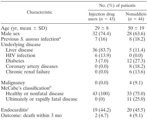
Patient population.We prospectively collected 430 adult patients with blood cultures positive for methicillin-sensitiveS. aureus(MSSA). Twelve university or central hospitals in Finland participated in this study between January 1999 and May 1999 and between January 2000 and August 2002 (36). In this study, 43 IDUs and 44 group-matched nonaddicts as controls were included (Table 1) (37). For each IDU with endocarditis (n⫽19), we chose a nonaddict with preferably definite endocarditis (n⫽20; 16 definite and 4 possible cases). For each IDU without endocarditis (n⫽24), we chose an age (⫾15 years)- and sex-matched nonaddict, whose randomization time was the nearest possible.
Written informed consent was obtained from all patients or their representa-tives. The study was approved by the ethics committees of all study sites and was conducted in accordance with the Declaration of Helsinki.
S. aureusidentification and DNA isolation.Routine bacteriological methods were used to detectS. aureusgrown in blood (36). TotalS. aureusDNA was isolated with the Promega Wizard DNA purification kit (Promega, Mannheim, Germany) according to the manufacturer’s instructions.
Serum samples.After the first positive culture forS. aureus, serum samples were collected at days 2 to 7 (acute phase) and at days 22 to 28 (convalescent phase) (37). Sera were stored at⫺20°C for further analysis. Both samples were available from 27 of 43 IDUs and from 37 of 44 nonaddicts. In the other 16 IDUs and 7 nonaddicts, one or both serum samples were missing.
spagenotyping.PCR for amplification of theS. aureusprotein A (spa) repeat region was performed according to the published protocols (1, 11). PCR prod-ucts were purified with the QIAquick PCR purification kit (Qiagen, Hilden, Germany) and sequenced using both amplification primers by a commercial
recombinant SEB, SEC, SElQ, TSST-1, and SEI which elicited submaximal proliferation were determined in T cell proliferation assays (between 1 and 100 pg/ml). The bacterial supernatants were collected at the stationary growth phase from cultures of the infectingS. aureusisolates grown in tryptone soy broth medium. The recombinant SAgs were produced inEscherichia coliand were efficiently lipopolysaccharide (LPS) depleted as described before (10). T cell proliferation assays were performed in RPMI 1640 supplemented with 10% fetal bovine serum (FBS) using peripheral blood mononuclear cells (PBMCs) from three different, healthy blood donors. Differences in the proliferative responses between the PBMC donors concerned mainly the numbers of responding cells (cpm values) and not their response curves.
Afterwards, four sets of neutralization assays were performed with PBMCs from different donors. The appropriate concentrations of supernatant or recom-binant SAgs were incubated in the presence of heat-inactivated patient sera serially diluted in RPMI 1640-10% FBS. For control, supernatants or recombi-nant SAgs were incubated without human serum and titrated over a broad range. After 20 min, 105PBMCs were added to measure mitogenic potency and
neu-tralizing serum capacity. T cell proliferation was determined by the incorporation of [3H]thymidine after 72 h, quantified by calculating the area under the
prolif-eration curve (AUC), and expressed as a percentage of the control without human serum. All measurements were performed in triplicate. Two IDU patients (T-29359 and T-37698) were excluded because their invasiveS. aureusisolate was SAg negative.
Statistical analysis.Differences in thespagenotype and virulence gene pat-terns between IDUs and nonaddicts were assessed using the chi-square test. The Mann-Whitney test was used to compare the neutralizing capacities of sera from IDUs and nonaddicts. The neutralizing capacities of the acute- and convalescent-phase serum samples from the bacteremic patients were compared with the pairedttest.Pvalues ofⱕ0.05 were considered statistically significant.
RESULTS
Genotypes of SAB isolates from IDUs and nonaddicts.
Among the SAB isolates from 43 IDUs and 44 nonaddicts we found 46 differentspatypes, which were assigned to 12 CCs (CC1, -5, -8, -9, -12, -15, -20, -22, -25, -30, -45, and -59). Moreover, we observed six singletons. Eight isolates could not be clustered by BURP analysis, because clustering parameters excludedspatypes shorter than five repeats, and two strains were nontypeable byspaPCR.
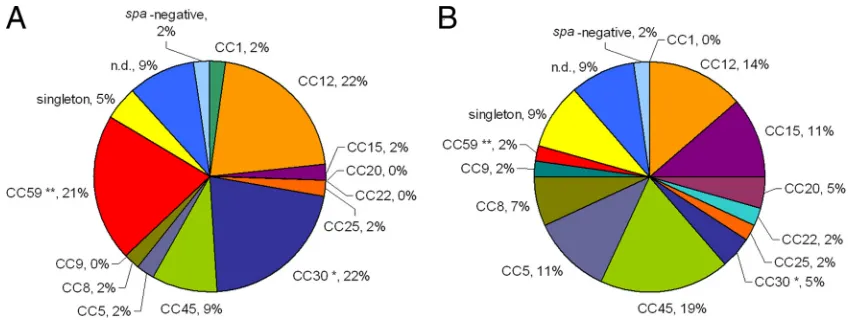
The genetic diversities of SAB strain collections from IDUs and nonaddicts were similar, but CC59 and CC30 were signif-icantly overrepresented among isolates from IDUs in compar-ison to those from nonaddicts (CC59, 20.9% versus 2.3%,Pⱕ
0.01, and CC30, 20.9% versus 4.5%, Pⱕ 0.05, respectively) (Fig. 1). No CC orspatype was associated with endocarditis.
Virulence gene repertoire of SAB isolates from IDUs and
nonaddicts. To test whether SAB isolates from IDUs and
nonaddicts differ in their virulence gene patterns, we next determined the accessory gene regulator (agr) type and SAg, Panton-Valentine leukocidin (PVL), and exfoliative toxin
Malignancy 0 (0.0) 4 (9.1)
McCabe’s classificationb
Healthy or nonfatal disease 43 (100) 33 (75.0) Ultimately or rapidly fatal disease 0 (0) 11 (25.0)
Endocarditisc 19 (44.2) 20 (45.5)
Outcome: death within 3 mo 2 (4.7) 4 (9.1)
aSuperficialS. aureusinfections orS. aureusbacteremia (only IDUs). bPrognosis or severity of underlying diseases classified according to the
crite-ria of McCabe and Jackson.
cClassified as definite or possible by using the modified Duke criteria.
on August 17, 2020 by guest
genes.agris a global regulator of virulence gene expression, and four different agr subgroups, agr 1 to 4, are known. In agreement with previous studies (14, 16, 30, 34, 49), we ob-served a strict linkage ofagrsubgroups with the spa-derived lineages (Fig. 2). PVL and exfoliative toxin genes (etaandetd) occurred only rarely.
Multiplex PCR was applied to detect 19 SAg genes. SAg genes were highly prevalent among SAB isolates from both collections (IDUs, 90.7%; nonaddicts, 84.1%), and SAg gene patterns differed remarkably. As previously reported, SAg genes were linked to staphylococcal lineages (14).
TheegcSAg genes were by far the most prevalent (IDUs, 44.2%; nonaddicts, 61.4%).sebandseawere overrepresented among IDU isolates (seb, 44.2% versus 15.9%,Pⱕ0.01;sea, 30.2% versus 9.1%,P ⱕ 0.05), but this was due to the high prevalence ofsea/seb-positive CC59 andsea-positive CC30 iso-lates among IDU strains. This emphasizes the importance of a simultaneous analysis of virulence genes and genetic back-ground. The comparison of SAg patterns within certain CCs revealed no major differences between isolates from IDUs and those from nonaddicts. Furthermore, we found no association of SAg genes with endocarditis.
Neutralizing serum antibodies in IDUs and nonaddicts.To
test whetheregcSAgs elicit an antibody response during infec-tion, we analyzed the neutralizing antibody responses of SAB patients (i) against the supernatant of their infecting strain and (ii) against representative recombinant SAgs. Supernatants were obtained fromS. aureuscultures in stationary phase and contain both egc SAgs, which are expressed at exponential growth phase and remain in the culture, and non-egcSAgs, which are typically secreted in stationary phase.
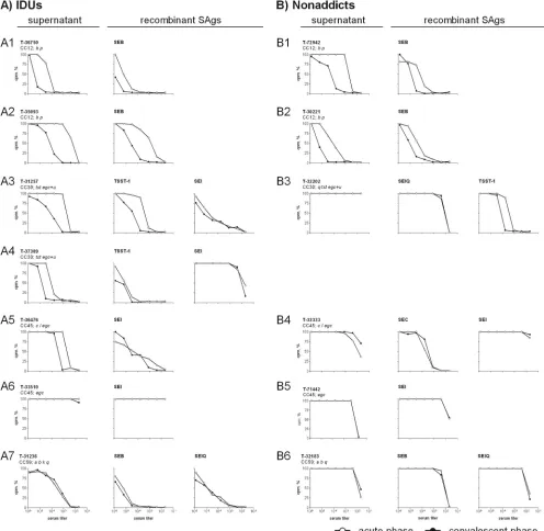
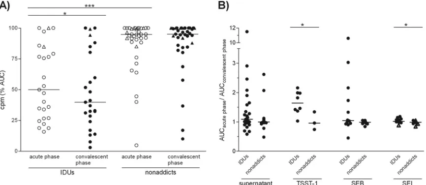
At the onset of bacteremia, many patients already possessed neutralizing serum antibodies against the SAg cocktail pro-duced by their infecting strain (Fig. 3 and 4A). While this neutralizing capacity was mostly low in nonaddicts, IDUs al-ready showed high antibody titers at the acute phase. This suggests that they were preimmunized with the SAgs of their infecting strain.
In several cases, we observed a rise in antibody concentra-tions during SAB, again especially among IDUs (Fig. 3 and 4).
In some individuals, titers increased more than 100-fold (T-30900 and T-35093 [Fig. 3, panel A2]). However, this was different in patients infected with CC59 strains. TheseS. aureus
isolates harbored a number of non-egcSAgs (sea,seb,sek, and
selq), and their supernatants were strongly mitogenic. IDUs infected with these strains had neutralizing antibodies at diag-nosis of SAB, but serum concentrations did not further in-crease thereafter.
The SAg cocktails in theS. aureusculture supernatants may be close to the clinical situation, but their SAg composition is not known. For molecular definition, we complemented anal-ysis by neutralization assays with recombinant SAgs (TSST-1, SEB, SEC, SElQ, and SEI), which in most patients confirmed the results obtained with bacterial supernatants. SEI served as a representative for the coexpressedegcSAgs. Sera from six patients neutralized recombinant SAgs but not the bacterial supernatants (e.g., Fig. 3, panel B3). The neutralizing effect against individual SAgs was probably obscured by the mito-genic effects of others that were also present in the superna-tant.
Notably, neutralizing antibodies againstegcSAgs were rare exceptions at the acute phase of SAB, and such antibodies were not induced during bacteremia. The findings were similar for supernatants from egc-positive strains (Fig. 3, panels A6 and B5; Fig. 4) and for recombinant SEI (Fig. 3, panels A4, A6, B4, and B5; Fig. 4B). High titers of anti-SEI antibodies were present in only 2 of the 62 tested patient sera (T-31257 and T-36476, Fig. 3, panels A3 and A5). Both patients were IDUs, and their anti-SEI antibody serum concentrations did not in-crease during SAB.
DISCUSSION
Neutralizing antibodies against non-egcSAgs are common in healthy adults (21, 39, 41), whereas neutralizing antibodies againstegcSAgs are very rare (12). Remarkably, the regulation of SAg release differs fundamentally between the two groups of SAgs (10, 31). These findings raised the question of how this differential regulation ofegcand non-egcSAgs affects the anti-SAg antibody response during SAB. Comparison of SAB
iso-FIG. 1. Clonal distribution of SAB isolates from IDUs (A) and nonaddicts (B).spatypes were clustered into 12 CCs by BURP analysis. MLST-CC nomenclature was deduced fromspa-CCs using the Ridom SpaServer database. CC59 and CC30 were overrepresented among SAB isolates from IDUs. Significance was determined by the chi-square test (*,Pⱕ0.05;**,Pⱕ0.01). n.d., excluded.
on August 17, 2020 by guest
http://cvi.asm.org/
carriers. Prevalences range from 0% to 4.5% in nasal isolates from the United Kingdom, Poland, Netherlands, and Germany (6, 8, 14, 27, 29). However, in IDUs from the United Kingdom, methicillin-sensitiveS. aureus strains isolated from abscesses and soft-tissue infections frequently belonged to CC59 (30), indicating that this lineage spreads in the internationally con-nected community of IDUs. We found no correlation of cer-tain clonal lineages or SAg gene patterns with endocarditis.
We previously reported that everyS. aureusclonal lineage is characterized by a consensus repertoire ofagrsubgroup and virulence genes (14). The SAB isolates in the present study fit well into this picture. In some CCs, the frequencies of the typical SAg genotypes differed between Finland and Germany (14).
Neutralizing antibodies against non-egcSAgs are frequent in the healthy population (12, 21, 39, 41), and in the present study, this was also found to be the case in SAB patients at the acute phase of bacteremia. However, neutralizing antibody titers were low in most nonaddicts. A possible explanation would be that the majority of them suffered from exogenous infections (15). Unfortunately, the carrier status of our patient cohort is not known. The SAg-neutralizing capacity was much higher in IDUs, suggestive of intensive exposure toS. aureus, probably even to the invasive strain. Repeated bacterial inoc-ulation could be the reason for this, because frequent injec-tions, contaminated drugs, sharing of drug use equipment, and poor hygiene usually characterize the living conditions of IDUs (2, 26). In agreement with this, studies from the 1970s showed higher colonization rates and more frequent endogenous in-fections in IDUs than in the general population (43, 44).
In IDUs but not in nonaddicts, bacteremia increased the neutralizing serum capacity, extending earlier reports about staphylococcal infections (e.g., bacteremia and wound infec-tion) (18, 20). While other groups measured antibody binding, we employed a functional assay, because it has been shown that SAg-neutralizing antibodies can be protective in patients as well as in animal models (3, 22, 32, 45). The high titers and rapid increases of SAg-neutralizing antibodies are indicative of a vigorous antibody response to many S. aureus antigens in IDUs (37). In addition to other factors, such as younger age, lack of preexisting heart disease, and the predominance of right-sided heart involvement (5, 7, 19), an efficient and pro-tective antibody response might contribute to the more favor-able outcome of S. aureus endocarditis in IDUs than in the general population. Only a few nonaddicts responded to bac-teremia with an increase of SAg-neutralizing antibodies. It
FIG. 2. Distribution of virulence genes withinspa-defined CCs among SAB isolates from IDUs (A) and nonaddicts (B). For construction of the phylogenetic tree, several reference strains were included in the BURP clustering (shaded in gray). Virulence genes (SAg genes,agr,eta,etd,
mecA, andpvlgenes) were determined by multiplex PCR. SAB strains from IDUs and nonaddicts did not differ in their virulence gene patterns. Staphylococcal enterotoxins (SEs) are indicated by single letters (a⫽sea, etc.).tst, toxic shock syndrome toxin;egc, enterotoxin gene cluster;etaand
etd, exfoliative toxins a and d;agr, accessory gene regulator;pvl, Panton-Valentine leukocidin (lukPV). a, patients with infective endocarditis; b, serum analyzed in neutralization assays. n.d., excluded.
on August 17, 2020 by guest
appears that bacteremia rarely primes high-affinity antibody responses, which would be required for SAg neutralization, but it seems that this immune stimulus is strong enough to boost preexisting B cell memory.
Overall, the boost of neutralizing antibody titers clearly shows that the immune system is exposed to non-egc SAgs duringS. aureusbloodstream invasion. This remains open for
egc-encoded SAgs, which did not elicit a boost or de novo
generation of specific antibodies, neither against supernatants of egc-positive strains nor against recombinant SEI. As we demonstrated previously, the immune cell-activating proper-ties ofegcand non-egcSAgs are very similar (induction of T cell proliferation, cytokine secretion, and gene regulation) and cannot explain the striking differences in the immune response
FIG. 3. SAg-neutralizing capacities of selected SAB sera from IDUs (A) and nonaddicts (B). The neutralizing capacities of SAB sera (acute phase and convalescent phase) against (i) the supernatant from the infecting strain and (ii) representative recombinant SAgs were determined. Therefore,S. aureusculture supernatants or recombinant SAgs were incubated with serial dilutions of SAB sera. After 20 min, 105PBMCs were added and T cell proliferation was measured after 72 h by [3H]thymidine incorporation. The graph depicts the SAg-induced proliferation in the presence of serum, expressed as percentages of the control without human serum. In contrast to nonaddicts, most IDUs had neutralizing antibodies already at the acute phase of SAB and antibody titers increased more frequently. In general, neutralizing antibodies againstegcSAgs were absent and were not induced. Representative data sets are depicted (IDUs, 7/25; nonaddicts, 6/37).
on August 17, 2020 by guest
http://cvi.asm.org/
to egc and non-egc SAgs (10). Because the amino acid se-quences of theegcSAgs are more closely related to those of individual non-egc SAgs than to each other (17, 42), it also appears unlikely that the two groups of SAgs differ systemat-ically in their immunogenicities. Remarkably, egc SAgs are secreted during exponential growthin vitro, whereas non-egc
SAgs—like most virulence factors—are expressed in stationary growth (10, 31). It remains to be shown whether SAgs are also differentially regulatedin vivoand whether this might explain theegcgap inS. aureuscarriers and noncarriers (12), which is not closed following bloodstream invasion.
ACKNOWLEDGMENTS
This work was supported by the Deutsche Forschungsgemeinschaft (GRK840, “Host-Pathogen Interactions in Generalized Bacterial In-fections,” SFB-TR34) and the Alfried Krupp Wissenschaftskolleg Greifswald (“A Functional Genomics Approach to Infection Biol-ogy”).
We thank the FINTROVA and FINLEVO study group investigators for their invaluable participation in patient collection.
REFERENCES
1.Aires-de-Sousa, M., et al.2006. High interlaboratory reproducibility of DNA sequence-based typing of bacteria in a multicenter study. J. Clin. Microbiol. 44:619–621.
2.Bassetti, S., and M. Battegay.2004.Staphylococcus aureus infections in injection drug users: risk factors and prevention strategies. Infection32:163– 169.
3.Bavari, S., B. Dyas, and R. G. Ulrich.1996. Superantigen vaccines: a com-parative study of genetically attenuated receptor-binding mutants of staph-ylococcal enterotoxin A. J. Infect. Dis.174:338–345.
4.Becker, K., A. W. Friedrich, G. Peters, and C. von Eiff.2004. Systematic survey on the prevalence of genes coding for staphylococcal enterotoxins SElM, SElO, and SElN. Mol. Nutr. Food Res.48:488–495.
5.Chambers, H. F., O. M. Korzeniowski, and M. A. Sande.1983. Staphylococ-cus aureusendocarditis: clinical manifestations in addicts and nonaddicts. Medicine62:170–177.
6.Donker, G. A., et al.2009. The population structure ofStaphylococcus aureus among general practice patients from The Netherlands. Clin. Microbiol. Infect.15:137–143.
7.Fowler, V. G., Jr., et al.2005.Staphylococcus aureusendocarditis: a conse-quence of medical progress. JAMA293:3012–3021.
8.Fowler, V. G., Jr., et al.2007. Potential associations between hematogenous complications and bacterial genotype inStaphylococcus aureusinfection. J. Infect. Dis.196:738–747.
9.Fueyo, J. M., M. C. Mendoza, M. A. Alvarez, and M. C. Martin.2005. Relationships between toxin gene content and genetic background in nasal carried isolates ofStaphylococcus aureusfrom Asturias, Spain. FEMS Mi-crobiol. Lett.243:447–454.
10.Grumann, D., et al.2008. Immune cell activation by enterotoxin gene cluster (egc)-encoded and non-egcsuperantigens fromStaphylococcus aureus. J. Im-munol.181:5054–5061.
11.Harmsen, D., et al.2003. Typing of methicillin-resistantStaphylococcus au-reusin a university hospital setting by using novel software forsparepeat determination and database management. J. Clin. Microbiol.41:5442–5448. 12.Holtfreter, S., et al.2004.egc-encoded superantigens fromStaphylococcus aureusare neutralized by human sera much less efficiently than are classical staphylococcal enterotoxins or toxic shock syndrome toxin. Infect. Immun. 72:4061–4071.
13.Holtfreter, S., and B. M. Broker.2005. Staphylococcal superantigens: do they play a role in sepsis? Arch. Immunol. Ther. Exp. (Warsz)53:13–27. 14.Holtfreter, S., et al.2007. Clonal distribution of superantigen genes in
clin-icalStaphylococcus aureusisolates. J. Clin. Microbiol.45:2669–2680. 15.Holtfreter, S., et al.2006.Staphylococcus aureuscarriers neutralize
superan-tigens by antibodies specific for their colonizing strain: a potential explana-tion for their improved prognosis in severe sepsis. J. Infect. Dis.193:1275– 1278.
16.Jarraud, S., et al.2002. Relationships betweenStaphylococcus aureusgenetic background, virulence factors,agrgroups (alleles), and human disease. In-fect. Immun.70:631–641.
17.Jarraud, S., et al.2001.egc, a highly prevalent operon of enterotoxin gene, forms a putative nursery of superantigens inStaphylococcus aureus. J. Im-munol.166:669–677.
18.Jozefczyk, Z.1974. Specific human antibodies to enterotoxins A, B, and C1 of Staphylococcus: their increased synthesis in staphylococcal infection. J. Infect. Dis.130:1–7.
19.Julander, I.1985. Unfavourable prognostic factors inStaphylococcus aureus septicemia and endocarditis. Scand. J. Infect. Dis.17:179–187.
20.Kanclerski, K., B. Soderquist, M. Kjellgren, H. Homberg, and R. Mollby.
FIG. 4. SAg-neutralizing capacities of SAB sera from IDUs and nonaddicts.S. aureus culture supernatants or recombinant SAgs were incubated with serial dilutions of SAB sera (acute phase and convalescent phase). After 20 min, 105PBMCs were added and T cell proliferation was measured after 72 h by [3H]thymidine incorporation. (A) The neutralizing capacities of SAB sera against the supernatant of the infecting strain. SAg-induced proliferation in the presence of serum, quantified by calculating the area under the proliferation curve (AUC) and expressed as percentages of the control without human serum, for all data sets (IDUs, 25; nonaddicts, 37). In contrast to nonaddicts, most IDUs showed an effective antibody response already at the onset of bacteremia. (B) Fold changes of the neutralizing capacities of SAB sera (acute phase and convalescent phase) against theS. aureusculture supernatants and representative recombinant SAgs. The ratio of the AUCs from both serum samples of one patient was calculated. IDUs frequently showed an increase in neutralizing antibody titers against the SAg cocktail and recombinant non-egcSAgs. In general, neutralizing capacity againstegcSAgs (triangles indicate SAB isolates which were onlyegcpositive) was very low or absent and not triggered during bloodstream invasion.Pvalues were calculated using the pairedttest (acute phase versus convalescent phase) or the Mann-Whitney test (IDUs versus nonaddicts); median values are indicated (*,Pⱕ0.05;***,Pⱕ0.001).
on August 17, 2020 by guest
1996. Serum antibody response toStaphylococcus aureusenterotoxins and TSST-1 in patients with septicaemia. J. Med. Microbiol.44:171–177. 21.Kunstmann, G., E. Schroder, H. Hasbach, and G. Pulverer.1989. Immune
response to toxic-shock-syndrome toxin-1 (TSST-1) and to staphylococcal enterotoxins A, B and C inStaphylococcus aureus infections. Zentralbl. Bakteriol.271:486–492.
22.LeClaire, R. D., R. E. Hunt, and S. Bavari.2002. Protection against bacterial superantigen staphylococcal enterotoxin B by passive vaccination. Infect. Immun.70:2278–2281.
23.Letertre, C., S. Perelle, F. Dilasser, and P. Fach.2003. Identification of a new putative enterotoxin SEU encoded by theegccluster ofStaphylococcus au-reus. J. Appl. Microbiol.95:38–43.
24.Lina, G., et al.2004. Standard nomenclature for the superantigens expressed by Staphylococcus. J. Infect. Dis.189:2334–2336.
25.Lindsay, J. A., and M. T. Holden.2006. Understanding the rise of the superbug: investigation of the evolution and genomic variation of Staphylo-coccus aureus.Funct. Integr. Genomics6:186–201.
26.Lowy, F. D., and M. Miller.2002. New methods to investigate infectious disease transmission and pathogenesis—Staphylococcus aureus disease in drug users. Lancet Infect. Dis.2:605–612.
27.Masiuk, H., et al.2010. Association of recurrent furunculosis with Panton-Valentine leukocidin and the genetic background ofStaphylococcus aureus. J. Clin. Microbiol.48:1527–1535.
28.McCormick, J. K., J. M. Yarwood, and P. M. Schlievert.2001. Toxic shock syndrome and bacterial superantigens: an update. Annu. Rev. Microbiol. 55:77–104.
29.Monecke, S., C. Luedicke, P. Slickers, and R. Ehricht.2009. Molecular epidemiology ofStaphylococcus aureusin asymptomatic carriers. Eur. J. Clin. Microbiol. Infect. Dis.28:1159–1165.
30.Monk, A. B., S. Curtis, J. Paul, and M. C. Enright.2004. Genetic analysis of Staphylococcus aureusfrom intravenous drug user lesions. J. Med. Microbiol. 53:223–227.
31.Munson, S. H., M. T. Tremaine, M. J. Betley, and R. A. Welch.1998. Identification and characterization of staphylococcal enterotoxin types G and I fromStaphylococcus aureus. Infect. Immun.66:3337–3348. 32.Nilsson, I. M., M. Verdrengh, R. G. Ulrich, S. Bavari, and A. Tarkowski.
1999. Protection againstStaphylococcus aureussepsis by vaccination with recombinant staphylococcal enterotoxin A devoid of superantigenicity. J. Infect. Dis.180:1370–1373.
33.Ono, H. K., et al.2008. Identification and characterization of two novel staphylococcal enterotoxins, types S and T. Infect. Immun.76:4999–5005. 34.Peacock, S. J., et al.2002. Virulent combinations of adhesin and toxin genes
in natural populations ofStaphylococcus aureus. Infect. Immun.70:4987– 4996.
35.Proft, T., and J. D. Fraser.2003. Bacterial superantigens. Clin. Exp. Immu-nol.133:299–306.
36.Ruotsalainen, E., et al.2006. Levofloxacin does not decrease mortality in Staphylococcus aureusbacteraemia when added to the standard treatment: a prospective and randomized clinical trial of 381 patients. J. Intern. Med. 259:179–190.
37.Ruotsalainen, E., et al.2008. Methicillin-sensitiveStaphylococcus aureus bacteraemia and endocarditis among injection drug users and nonaddicts: host factors, microbiological and serological characteristics. J. Infect.56:249– 256.
38.Schlievert, P. M., and G. A. Bohach.2007. Staphylococcal and streptococcal superantigens: an update, p. 21–36.InM. Kotb and J. Fraser (ed.), Supe-rantigens: molecular basis for their role in human diseases. ASM Press, Washington, DC.
39.Schroder, E., G. Kunstmann, H. Hasbach, and G. Pulverer.1988. Prevalence of serum antibodies to toxic-shock-syndrom-toxin-1 and to staphylococcal enterotoxins A, B and C in West-Germany. Zentralbl. Bakteriol. Mikrobiol. Hyg. A270:110–114.
40.Strommenger, B., et al. 2006. Assignment of Staphylococcus isolates to groups byspatyping, SmaI macrorestriction analysis, and multilocus se-quence typing. J. Clin. Microbiol.44:2533–2540.
41.Takei, S., Y. K. Arora, and S. M. Walker.1993. Intravenous immunoglobulin contains specific antibodies inhibitory to activation of T cells by staphylo-coccal toxin superantigens. J. Clin. Invest.91:602–607.
42.Thomas, D., S. Chou, O. Dauwalder, and G. Lina.2007. Diversity in Staph-ylococcus aureusenterotoxins. Chem. Immunol. Allergy93:24–41. 43.Tuazon, C. U., and J. N. Sheagren.1974. Increased rate of carriage of
Staphylococcus aureusamong narcotic addicts. J. Infect. Dis.129:725–727. 44.Tuazon, C. U., and J. N. Sheagren.1975. Staphylococcal endocarditis in
parenteral drug abusers: source of the organism. Ann. Intern. Med.82:788– 790.
45.Ulrich, R. G., M. A. Olson, and S. Bavari.1998. Development of engineered vaccines effective against structurally related bacterial superantigens. Vac-cine16:1857–1864.
46.van Belkum, A., et al.2009. Reclassification ofStaphylococcus aureusnasal carriage types. J. Infect. Dis.199:1820–1826.
47.Verkaik, N. J., et al.2009. Anti-staphylococcal humoral immune response in persistent nasal carriers and noncarriers ofStaphylococcus aureus.J. Infect. Dis.199:625–632.
48.Wertheim, H. F., et al.2005. The role of nasal carriage inStaphylococcus aureusinfections. Lancet Infect. Dis.5:751–762.
49.Wright, J. S., III, et al.2005. Theagr radiation: an early event in the evolution of staphylococci. J. Bacteriol.187:5585–5594.