pISSN 2320-1770 | eISSN 2320-1789
Original Research Article
Role of cytology, colposcopy and colposcopic directed biopsy in the
evaluation of unhealthy cervix
Alpana Agrawal*, Amita Sharma, Manisha Gupta, Neelima Agarwal
INTRODUCTION
Cervical cancer is the second most common cancer among women in developing and underdeveloped countries, amounting to 80% of the global burden of this disease.1 India bears over a tenth of the global burden of cancers.2 As per latest data of India from GLOBOCAN 2012, top three cancers in female are breast, cervix uteri and colo-rectum.2
Cervical cancer is a deadly disease once it reaches the invasive stages but out of all the female genital tract cancers, it is the only preventable cancer if detected in its
early stages. It is possible to prevent deaths due to cervical cancer through various strategies that target women >30 yr for screening and treatment.3
The introduction of Papanicolaou test led to significant reduction in mortality and morbidity in developed countries. The screening coverage in Asian countries is low and is 2.6-5 per cent in India.4 In India, unhealthy cervix is a common finding on per speculum examination in gynaecology Out Patient Department (OPD) and it is recommended to do the cervical cytology to detect any epithelial cell abnormality.
Department of Obstetrics and Gynaecology, Santosh Medical College, Ghaziabad, Uttar Pradesh, India
Received: 25 August 2016
Accepted: 24 September 2016
*Correspondence:
Dr.Alpana Agrawal,
E-mail: alpanaishi@gmail.com
Copyright: © the author(s), publisher and licensee Medip Academy. This is an open-access article distributed under the terms of the Creative Commons Attribution Non-Commercial License, which permits unrestricted non-commercial use, distribution, and reproduction in any medium, provided the original work is properly cited.
ABSTRACT
Background: The objective was to find out the magnitude of precancerous lesions and evaluate the performance of colposcopy in estimating the presence and grade of cervical disease vs conventional Pap smear testing of women with unhealthy cervix.
Methods: This was a prospective observational study. All the women were subjected to Pap smear and colposcopy, whereas histopathology was done in patients having abnormal findings on colposcopy. Biopsies were taken from the abnormal areas.
Results: Out of 110 women who completed the study 60 (54.5%) had changes in their cervical epithelium on colposcopy. These 60 women with unhealthy cervix underwent cervical biopsy, and 20 (33.3%) had histologically proven chronic cervicitis, 6 (10%) had chronic cervicitis with condylomatous change followed by 17 (28.3%) with CIN 1 lesion, 8 (13.3%) with CIN 2, 4 (6.7%) with CIN 3 lesion and only one (1.7%) had histologically proven malignancy. Cytology alone identified the population at risk with a high sensitivity [70.00% (95% CI: 50.60 % to 85.24 %)] but low specificity [60.00% (95% CI: 40.61 % to 77.32 %)] rate. Colposcopy showed a high sensitivity rate [93.33 % (95% CI: 77.19 % to 98.99 %)] but a limited specificity [70.00% (95% CI: 50.60% to 85.24%)].
Conclusions: It is very important to diagnose the CIN lesions with accuracy once a woman comes to a tertiary care hospital with symptoms or is referred for a suspicious looking cervix. All these women must be screened by colposcopy and directed biopsy must be taken if indicated in the same sitting.
In a developing country like India, cytology based screening programmes are difficult to organize because of absence of trained manpower, infrastructure, logistics; costs involved and has other limitations like low sensitivity and high false negative rates.5 Colposcopy is a worldwide accepted method for detection of early carcinoma cervix, as it gives faster result and guides the site of biopsy which can be done in a single visit proving itself as a better screening modality for premalignant lesion.
The present study was undertaken to find out the magnitude of precancerous lesions among women in the age group 20-65 years and to identify the various socio-demographic and reproductive risk factors of cervical cancer among those with suspicious cervical lesions presenting in the OPD of a referral hospital. We also tried to evaluate the performance of colposcopy in estimating the presence and grade of cervical disease vs conventional cytology testing.
METHODS
The present prospective, cross sectional study was carried out on 110 patients, attending the outpatient Department of Obstetrics and Gynaecology, Santosh Medical College and Hospital, Ghaziabad for a period of one year. Patients were selected by selective screening (clinically suspicious cervix).
Inclusion criteria
Women in age group of 20-65 years associated with: 1. Persistent vaginal discharge
2. Post coital/ intermenstrual/ perimenopausal/ post menopausal bleeding
3. Cervical hypertrophy
4. Cervical erosion/ ulceration/ growth/ oozing surface
5. Cervix bleed on touch
6. Cervix flushed with petechiae spot
7. Unexplained occasional foul smelling discharge per vaginum.
Exclusion criteria
1. Pregnant or postpartum patients.
2. Patient having any history of treatment for either cervical dysplasia or vulval warts.
3. Patient who had undergone recent endometrial curettage, hysterosalpingography, cervical biopsy or hysterectomy.
4. Immunocompromised patients ( HIV patient on corticosteroids)
Informed written consent was taken from the women, informing them of the background of the study, risks and benefits and voluntary nature of participation. Ethical
clearance was obtained from the institutional ethical committee.
A detailed history was taken and a thorough clinical examination was done in all patients. If there was any evidence of vaginitis or cervicitis, this was first treated and patient was asked to return to OPD after 2 weeks for sample collection.
All the 110 patients were subjected to Pap smear and colposcopy, whereas histopathology was done in patients having abnormal findings on colposcopic examination. Biopsies were taken from the abnormal areas (acetowhite areas and vascular abnormalities like fine punctuations, coarse punctuations, mosaic and areas showing atypical vasculature)
Statistical analysis
It was done by calculating diagnostic efficacy of each test. The Sensitivity, Specificity, Positive Predictive Value (PPV), Negative Predictive Value (NPV), and Accuracy were calculated for Pap smear and Colposcopy with Colposcopy directed biopsies results as gold standard.
Comparison between groups was done by Chi-square test. A P-value <0.05 was considered to indicate statistical significance. The incidence of pre-malignant and malignant lesions was calculated as percentages.
RESULTS
Out of the total of 110 women in the study population highest (43.6%) was from 31 to 40 years of age group, followed by 20-30 years of age group (36.4%). All the women underwent Pap smear and colposcopic examination.
About 50 (45.5%) women with unhealthy cervix had no suspected cervical lesion on colposcopic examination. Only 60 (54.5%) were found to have changes in their cervical epithelium. Colposcopic directed biopsy were taken from these 60 women and the specimen was sent for histopathological examination (Table 2).
In the overall study group of 110 women, 30 (27.3%) of women had cytological diagnosis of low grade squamous intraepithelial lesion (LSIL) followed by inflammatory smear in 18 (16.4%) women. Pap smear was normal in 49 (44.5%) women (Table 1).
Table 1: Distribution of cases under study according to cytological findings.
Pap Smear n =110 (%) Negative 49 (44.5) Inflammatory 18 (16.4)
ASCUS 6 (5.5)
LSIL 30 (27.3)
HSIL 6 (5.5)
Malignant 1 (0.9)
ASCUS: Atypical squamous cells of undetermined significance. LSIL: Low grade squamous intraepithelial lesions’
HSIL: High grade squamous intraepithelial lesion
Figure 1: Results of histopathological examination.
More than half (65 %) of the women with cervical inflammation were in the age group of 31-40 years followed by 20-30 years age group (30 %). More than three-fourth (76.5%) of CIN 1 lesion was seen in 31-40 years. CIN 2 lesions (62.5%) were seen more among the 65 yr age group. All of CIN 3 lesion was seen in 41-65 years age group. The age of the only women with malignancy was 52 years. We compared the pooled data of the cervicitis and cervicitis with condylomatous change with that of CIN1, CIN 2 and CIN 3 lesions regarding age variables; no significant difference was noted among those with or without lesions among women up to 30 years of age with those 31 years and above (P= 0.06).
Cervical inflammation was highest (75%) in >P1 (parity 1) as compared to ≤P1. More than three-fourth (74.5%) of CIN 1 lesion was seen in >P1 as compared to ≤P1. CIN2 lesions (75%) were seen more among the ≥P3. 100% of CIN 3 lesion was seen in women with parity 3 or more. The only women with malignancy was para 5.
Cervical intraepithelial neoplasia was more common among the lower socioeconomic group. We compared the pooled data of the cervicitis and cervicitis with condylomatous change with that of CIN 1, CIN 2 and CIN 3 lesion regarding sociodemographic variables. No significant difference was noted among women of lower socioeconomic status compared to those women of combined middle and upper socioeconomic status (P=1.0).
Vaginal discharge was the most common presenting complaint of patients with dysplasia. Menstrual disturbances and post coital bleeding were frequently found in higher grade of dysplasia. Carcinoma presented with discharge per vaginum, post-menopausal bleeding, backache and post-coital bleeding per vaginum.
Cervicitis (inflammatory lesions) mainly appeared as cervicovaginitis, hypertrophied cervix and erosion. Dysplastic lesions appeared as hypertrophied cervix, erosion and cervico vaginitis. It bled on touch in 47% cases in CIN 1, 75% cases of CIN 2, and 75% of CIN 3. Carcinoma cervix appeared as irregular growth which bled on touch.
[image:3.595.312.546.338.473.2]Cytology alone identified the population at risk with a high sensitivity [70.0% (95% CI: 50.60 % to 85.24 %)] but low specificity [60.0% (95% CI: 40.61 % to 77.32%)] rate. The positive predictive value of Pap test was 63.6% (95% CI: 45.13 % to 79.58 %), negative predictive value was 66.7% (95% CI: 46.04 % to 83.45%) and accuracy was 65.0% (Figure 2).
Figure 2: Correlation between Pap smear and biopsy.
Colposcopy showed a high sensitivity rate [93.3 % (95% CI: 77.19 % to 98.99 %)] but a limited specificity [70.0% (95% CI: 50.60% to 85.24%)]. The positive predictive value of colposcopy was 75.7% (95% CI: 58.80 % to 88.20%), negative predictive value of colposcopy was 91.3% (95% CI: 71.92 % to 98.68 %) and accuracy was 81.7 % (Figure 3).
Figure 3: Correlation between colposcopic finding and biopsy. Normal 7% Cervicitis 33% Cervicitis with condylomat os change 10% CIN1 28% CIN2 13% CIN3 7% Malignancy
2% (n=60)
0 2 4 6 8 10 12 14 N o . o f ca ses
Pap Smear Findings
[image:3.595.311.545.604.729.2]Colposcopy had higher sensitivity (93.33%), higher specificity (70%), higher accuracy (81.66%) and higher PPV (75.68 %) for detecting CIN and cancer than cytological method (Figure 4).
Figure 4: Comparison of diagnostic efficacy of Pap smear and colposcopy.
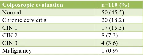
Table 2: Distribution of cases under study according to colposcopic findings.
Colposcopic evaluation n=110 (%)
Normal 50 (45.5)
Chronic cervicitis 20 (18.2)
CIN 1 17 (15.5)
CIN 2 8 (7.3)
CIN 3 4 (3.6)
Malignancy 1 (0.9)
DISCUSSION
Effective screening programme can lead to earlier detection of cancer and its precursor lesions, thus leading to decline in mortality. The incidence of the biopsy-confirmed dysplasia in our study was 27.2% out of which more than half (56.6%) were CIN 1 lesions. We screened women who came to the hospital with various symptoms and unhealthy cervix and hence would belong to a high risk group. That would be a contributory factor to the high incidence of biopsy-confirmed dysplasia in our study.
On correlating Pap smear findings with histology in the present study, the accuracy was found to be 65.0 % which is comparable to a study by Jain et al, (73.2%).6 The accuracy of Pap smear in a study by Bhatla et al, was 89% which is quite high.7
The accuracy of colposcopy in our study was 81.7% which is comparable to the findings of Ashmita and Shakuntala et al,(86.5%) and Mallur et al, (80%).8,9 Colposcopy had higher sensitivity (93.3%), higher specificity (70%), higher accuracy (81.7%) and higher
PPV (75.7 %) for detecting CIN and cancer than cytological method (Pap smear). Colposcopic assessment is a critical stage in the diagnosis of early cervical neoplasia, as the detection of abnormal cervical cytology is dependent on precise visual localisation of micro pathological changes and precise biopsy of such tissue for subsequent histopathologic diagnosis. Colposcopy thus plays a crucial role in the diagnosis of early cervical disease, with management decisions often anchored on the colposcopic assessment.
CONCLUSION
Most of the women who undergo screening with Pap smear in developing countries do not come for follow-up or do not collect their report on time thereby leading to delay in diagnosis and treatment. It is very important to diagnose the CIN lesions with accuracy once a woman comes to a tertiary care hospital with symptoms or is referred for a suspicious looking cervix. All these women must be screened by colposcopy and directed biopsy must be taken if indicated in the same sitting.
Funding: No funding sources Conflict of interest: None declared
Ethical approval: The study was approved by the Institutional Ethics Committee
REFERENCES
1. World Health Organization (WHO). Human
Papillomavirus Infection and Cervical Cancer. 2010.
Available from:
http://www.who.int/vaccine_research/diseases/hpv. (Last accessed on 2016 Feb 10).
2. Ferlay J, Soerjomataram I, Ervik M, Dikshit R, Eser
S, Mathers C, et al. GLOBOCAN 2012;0. Cancer Incidence and Mortality Worldwide: IARC Cancer Base No. 11. Lyon, France: International Agency for Research on Cancer; 2013 (Last accessed on 2016 Jan 27).
3. Goldie SJ, Gaffikin L, Goldhaber-Fiebert JD,
Gordillo-Tobar A, Levin C, Mahe C. Alliance for Cervical Cancer Prevention Cost Working Group. Cost-effectiveness of cervical cancer screening in five developing countries. N Engl J Med. 2005;353:2158-68.
4. Gakidou E, Nordhagen S, Obermeyer Z. Coverage of
cervical cancer screening in 57 countries: low average levels and large inequalities. PloS Med. 2008;5:e132.
5. Dinshaw KA, Shastri SS, Patil SS. Cancer Control Programme in India: challenges for the new Millennium; Health Administrator. Vol: XVII(1):10-13.
6. Jain V, Vyas AS. Cervical
Neoplasia-Cyto-Histological Correlation (Bethesda System) A Study of 276 Cases. J Cytol Histol. 2010;1:106.
7. Bhatla N, Mukhopadhyay A, Kriplani A, Pandey
RM, Gravitt EP, Shah KV. Evaluation of adjunctive 70
60 63.6 66.7 65
93.3
70 75.7
91.3
81.7
Sensitivity Specificity Positive Predictive
value
Negative Predictive value
Accuracy
[image:4.595.52.284.368.458.2]tests for cervical cancer screening in low resource settings. Indian J Cancer. 2007;44:51-5.
8. Ashmita D, Shakuntala PN, Rao SR. Comparison
and Correlation of Pap smear, Colposcopy and Histopathology in Symptomatic Women and Suspicious Looking Cervix in a Tertiary Hospital Care Centre. Int J Health Sci Res. 2013;3(5):50-9.
9. Mallur PR, Desai BR, Anita D, Geeta D, Bhavana S,
Pallav G. Sequential Screening with Cytology and
Colposcopy in Detection of Cervical Neoplasia. J. South Asian Feder Obst Gynae. 2009;1(3):45-8.