David A Sarkar
A thesis submitted for the Degree of Doctor of Philosophy at University College London, University of London
A
UCL
2003
Centre for Clinical Pharmacology & Therapeutics, University College London,
ProQuest Number: U642379
All rights reserved
INFORMATION TO ALL USERS
The quality of this reproduction is dependent upon the quality of the copy submitted.
In the unlikely event that the author did not send a complete manuscript and there are missing pages, these will be noted. Also, if material had to be removed,
a note will indicate the deletion.
uest.
ProQuest U642379
Published by ProQuest LLC(2015). Copyright of the Dissertation is held by the Author.
All rights reserved.
This work is protected against unauthorized copying under Title 17, United States Code. Microform Edition © ProQuest LLC.
ProQuest LLC
789 East Eisenhower Parkway P.O. Box 1346
Previous reports have demonstrated a range of negative and latterly positive inotropic responses in cardiac preparations exposed to NO We set out to investigate the effect of NO donors on isolated myocytes and to elucidate the underlying mechanism and experimental factors governing the observed response.
In isolated guinea-pig ventricular cardiomyocytes newer classes of NO donors including nitrosothiols (GSNO and SNAP) and NONOates (DEANO) induced a positive inotropic response. SNP and GTN showed no positive inotropy. The response was enhanced by co-administration of isoprenaline and reversibly abolished by the free NO scavenger oxyhaemoglobin. ODQ (soluble guanyl cyclase inhibitor) and Rp-cAMPS (protein kinase A inhibitor) did not abolish the effect. Measurement of myocyte cyclic nucleotides demonstrated a rise in cGMP, but not cAMP. Microelectrode recordings of the action potential and steady state la during exposure to DEANO (lO^M) found no change in the action potential, though the la was increased with preservation of the current-voltage relationship.
A faster rate of NO donor decomposition was associated with positive inotropy. Breakdown of nitrosothiols was enhanced by the presence of myocytes. Functionally the fast NO releaser DEANO was more likely to induce an increase in cell shortening compared with the slow releaser detanonoate. Positive inotropy was demonstrated in rabbit and human myocytes (from failing and non-failing hearts) but not in rat. In multi- cellular preparations the inotropic effect was reduced or absent.
First and foremost, I would like to express by gratitude to my supervisors Prof. Patrick Vallance and Prof Sian Harding for their continuous support, encouragement and flow of ideas, which have made my Ph.D. research immensely enjoyable and rewarding.
I wish to acknowledge the British Heart Foundation for their support of my work (PhD Clinical Studentship No FS/97060
I also wish to express my thanks to P O’Gara for assistance with myocyte preparation.
Statement of Contribution
PUBLICATIONS
Work described in this thesis has given rise to the following publications: ABSTRACTS:
ISHR (American section) XXU Annual Scientific Session (poster presentation)
Nitric oxide induced positive inotropy in single cardiomyocytes and multicellular preparations.
Sarkar D, Vallance P and Harding SE. J. Mol Cell Cardiol 2000;32 (5): 36A
The contibution of constitutive NO synthase to the elevated NO production in septic shock: Role of tetrahydrobiopterin. (poster presentation)
Amirmansour C, Bogle RG, Sarkar D, Hingorani A, Hesslinger C, Zeigler I, Heales S, Jones L and Vallance P.
Pteridines 2000.
British Hypertension Society Meeting Sept 1999 Glasgow (oral presentation)
The Positive inotropic effect of NO donors and the Relationship to NO release Kinetics Sarkar D, Vallance P and Harding SE
J Human Hypertension 1999; 13: 1.2
Heart Failure 99, European Society of Cardiology (oral Presentation) NO mediated positive inotropic effect in isolated myocytes.
Sarkar D, Vallance P and Harding SE.
European Journal of Heart Failure 1999:1 Supplement, No5
American College of Cardiology. New Orleans March 1999. (poster presentation) NO mediated positive inotropic effect in isolated myocytes.
Sarkar D, Vallance P and Harding SE.
Positive inotropic effects of NO donors in isolated cardiomyocytes are independent of cGMP and dependent on the rate of NO release. Cardiovasc Res 2000;48: 430-439
Sarkar D, Vallance P and Harding SE Nitric Oxide: Not just a negative inotrope
European Journal of Heart Failure 2001;3:527-534
Sarkar D, Vallance P, Terracciano CMN and Harding SE
Positive inotropy of NO in guinea-pig cardiomyocytes: Mechanism and relevance to integrated cardiac function. (Manuscript under review)
McLean PG, Aston D, Sarkar D and Ahluwalia A.
Protease-activated receptor-2 activation causes EDHF-like coronary vasodilation. Circ Res 2002; 90: 465-472
Chen Z, Ahluwahlia A, Sarkar D, Selwood DL, Vallance P, Hesslinger C and Hingorani AD.
Estrogen modulation of endothelial function involves activation of the pterin pathway. (Manuscript submitted to Circ Res)
Amirmansour C, Sarkar D, Bogle RG, Jones L, Hesslinger C, Zeigler I, Heales S, Hingorani A and Vallance P.
Lipopolysaccharide increases nitric oxide production in mice lacking inducible nitric oxide synthase: Possible role of tetrahydrobiopterin. (Manuscript submitted BBRC)
AWARDS
INDEX TO CONTENTS
Page
TITLE 1
ABSTRACT 2
ACKNOWLEDGEMENT 4
PUBLICATIONS 5
INDEX OF CONTENTS 7
LIST OF FIGURES 14
LIST OF TABLES 16
TABLE OF ABBREVIATIONS 17
CHAPTER ONE: INTRODUCTION
1.1 Background 22
1.2 Structure and function of nitric oxide synthase 23
1.3 Sources of Endogenous Nitric oxide in the Myocardium 24
Endothelial NOS 24
Inducible NOS 24
Induction ofiNOS in heart Failure 27
Neuronal NOS 29
1.4 Paracrine Endothelial NO 29
1.5 The Role of NO in the Human Heart 30
1.6 Sources of Exogenous NO (NO Donors) 30
1.7 Pharmacology of Nitric Oxide Donors 31
Organic Nitrates 32
Sodium Nitroprusside 33
Nitrosothiols 34
“NONOate” Nitric Oxide Donors 37
1.8 Nitric Oxide Species 38
The Nitrosonium Cation 39
The Nitroxyl Ion 39
1.11 Functional effects of NO on the heart 46
Diastolic Function 46
Systolic Function 46
Pharmacological NO donors 48
1.12 Mechanisms to regulate NO synthesis 52
Alteration of NOS activity 52
Substrate availability 52
Enzyme cofactors 53
1.13 Aims of the study 55
CHAPTER TWO: METHODOLOGY
2.1 Isolation of animal ventricular myocytes 58
Preparation of Isolated Guinea-Pig Myocytes 58 Isolation of Rat and Rabbit Myocytes 59 Isolation of Human Ventricular Myocytes 61
2.2 Measurement of Myocyte Contraction 62
Cell Bath 62
Myocyte Selection 62
Video Edge Detection System 64
2.3 Papillary Muscles 65
2.4 Perfused Isolated Heart (Langendorff technique) 66
2.5 Measurement of cAMP 66
2.6 Measurement of cGMP 68
2.7 Measurement of Myocyte Intracellular Calcium 68
2.8 Measurement of Myocyte Calcium Current 69
Simultaneous Measurement of Contraction Amphtude 69
Calcium IV measurement 70
2.9 Preparation of Oxyhaemoglobin 70
2.10 Measurement of NO Release 72
Dual Beam Spectrophotometry 72
NO Electrode 73
Measurement of NOx Levels by Chemiluminescence 73
2.11 Statistical Analysis 76
2.12 Materials 76
CHAPTER THREE: EFFECT OF NO DONORS ON ISOLATED MYOCYTE CONTRACTION
3.1 Introduction 78
3.2 Experimental Protocol 79
Isoprenaline concentration response in different species 79 Reproducibility of isoprenaline concentration response 79
Effect of L-NAME and L-arginine 79
Effect of SNP and GSNO 80
3.3 Results 80
Isoprenaline concentration response in different species 80 Reproducibility of isoprenaline concentration response 81
Effect of L-NAME and L-arginine 82
Isoprenaline Response in the presence of NO donor 83
3.4 Discussion 83
3.5 Section H Protocol 84
3.6 Results 85
Effect of GTN on isoprenaline 0.3 nM 86 Effect of NO donor on a low concentration of isoprenaline 87
GSNO Control Data 88
3.7 Section HI Introduction 88
3.8 Experimental Protocol 88
Oxyhaemoglogin 88
3.9 Results 89
Oxyhaemoglobin 89
Efifect of Low Dose Isoprenaline on Response to NO donors 90 The Role of Soluble Guanylyl Cyclase 90
Efifect of Superoxide Dismutase 92
3.10 Discussion 92
CHAPTER FOUR: MECHANISM OF NO INDUCED POSITIVE INOTROPY
4.1 Introduction 95
4.2 Section I. Rate of NO Release 96
Experimental Protocols 96
DEANO Vs Detanonoate 96
Dual Beam Spectrophotometry 96
Alteration of NOx turnover 97
NO Electrode 97
4.3 Results 97
DEANO Vs Detanonoate 97
Measurement of Spontaneous NO Release 98 NO donors and Myocardial Fragments 99
NO Electrode 99
4.4 Section H. The Role of Cyclic Nucleotides 102
Experimental Protocol 102
Measurement of cAMP 102
Measurement of cGMP 102
Rp-cAMPS treatment 102
4.5 Results
Myocyte cGMP Levels 103
Myocyte cAMP Levels 103
4.6 Section m . The Role of in mediating the observed response
Protocol 104
Thapsigargin 104
Measurement of the Ca^^ Transient 105
4.7 Results 105
Thapsigargin 105
Ca^^ Transient Measurements 105
Microelectrode Studies: Action Potential 107
Calcium Current 107
Current Voltage Relationship 109
4.8 Discussion 110
CHAPTER FIVE: INFLUENCE OF TISSUE PREPARATION ON RESPONSE TO NO DONORS
5.1 Introduction 115
5.2 Protocol 116
Isoprenaline concentration response 116
Effect of NO donor alone 116
Effect of NO donor and Isoprenaline 117
Concentration response relationship to GSNO in the Langendorff 117 Co-administration of DEANO and isoprenaline in the Langendorff 117
5.3 Results 118
Isolated Ventricular Myocytes 118
5.4 Papillary Muscle Preparation 119
Cumulative concentration response to isoprenaline 119 Efifect of isoprenaline on papillary beat parameters 119 Efifect of NO donor on papillary Muscle 121 Efifect of NO donors on papillary beat characteristics 124
5.5 Langendorff Preparation 125
Efifect of NO donors on the guinea-pig Langendorff 126
Effect of DEANO on beat parameters in the Langendorff 129
5.6 Discussion 129
CHAPTER SIX: SPECIES VARIATION IN RESPONSE TO NO DONORS
6.1 Introduction 134
6.2 Protocols 135
6.3 Results: Rat Cardiac preparations 136
6.4 Rat Myocytes and Thapsigargin 136
6.5 Rabbit isolated myocytes 138
6.6 Human isolated Myocytes 140
6.7 Discussion 144
CHAPTER SEVEN: EFFECT OF SEPSIS ON THE RESPONSE TO NO DONORS
7.1 Introduction 145
7.2 Experimental Protocols 145
7.3 Results 146
Response to isoprenaline 146
Effect of L-NAME on LPS treated myocytes 147
Effect of L-NAME on the Response to Isoprenaline 148
Effect of DEANO on septic myocytes 149
7.4 Discussion 149
CHAPTER EIGHT: GENERAL DISCUSSION
8.1 Introduction 152
8.2 Comparison of our results with previous publications 154
8.3 Clinical Implications 158
APPENDIX 161
REFERENCES 165
APPENDIX n 196
Fig 1.1 Conversion of L-arginine to L-citrulline and NO 22 Fig 1.2 Generic structure of groups of NO donors 32 Fig 1.3 Targets for NO in the cardiovascular system 42 Fig 1.4 Cellular mechanisms of Ca^^ handling 45
Fig 1.5 Interaction of cAMP and cGMP 51
Fig 2.1 Schematic of guinea-pig myocyte isolation protocol 60
Fig 2.2 Cell bath apparatus 63
Fig 2.3 cAMP standard curve 67
Fig 2.4 Absorption spectrum of oxyhaemoglobin 71 Fig 2.5 Schematic of chemiluminescence apparatus 74
Fig 2.6 Chemiluminescence standard curve 75
Fig 3.1 Concentration response to isoprenaline in different myocyte species 80 Fig 3.2 Serial concentration response curves to isoprenaline in the guinea-pig 81 Fig 3.3 Effect of L-arginine on isoprenaline concentration response 82 Fig 3.4 Effect of SNP on isoprenaline concentration response 82 Fig 3.5 Effect of GSNO on isoprenaline concentration response 83
Fig 3.6 Effect of SNAP and Papanonoate 85
Fig 3.7 Effect of GTN on low concentration isoprenaline 86
Fig 3.8 Effect of various NO donors 87
Fig 3.9 Trace depicting response to GSNO 87
Fig 3.10 Effect of Oxyhemoglobin on GSNO response 90 Fig 3.11 Concentration response relationship of 8-Br-cGMP 91 Fig 3.12 Effect of ODQ on the response to GSNO 91
Fig 4.3 NO electrode trace with GSNO 101
Fig 4.4 Myocyte cAMP levels 104
Fig 4.5 Calcium current with simultaneous contraction amplitude 106
Fig 4.6 Representative trace of Ca^^ current with DEANO 107
Fig 4.7a Efifect of DEANO on peak /q. 108
Fig 4.7b Simultaneous efifect of DEANO on cell shortening 108
Fig 4.8 Current voltage relationship with DEANO 109
Fig 5.1 Efifect of GSNO and DEANO on myocytes 118
Fig 5.2 Isoprenaline concentration response curve in papillary muscle 119
Fig 5.3 Trace of the papillary response to isoprenaline 120
Fig 5.4a Efifect of GSNO and DEANO on papillary muscle 121
Fig 5.4b Efifect of GSNO with isoprenaline on papillary muscle 122
Fig 5.5 Efifect of DEANO and isoprenaline on papillary muscle 122
Fig 5.6 Trace of Efifect of DEANO and isoprenaline on papillary muscle 123
Fig 5.7a Efifect of GSNO on coronary perfusion pressure 126
Fig 5.7b Efifect of GSNO on spontaneous heart rate 126
Fig 5.7c Efifect of GSNO on developed LV pressure 126
Fig 5.8a Efifect of DEANO and isoprenaline on coronary pressure 128
Fig 5.8b Efifect of DEANO and isoprenaline on spontaneous heart rate 128
Fig 5.8c Efifect of DEANO and isoprenaline on developed pressure 128
Fig 6.1 Efifect of GSNO and Thapsigargin on rat myocytes 137
Fig 6.2 Efifect of GSNO on coronary tone in the rat Langendorfif 138
Fig 6.3 Efifect of GSNO on LV developed pressure in the rat Langendorfif 138
Fig 6.4 Efifect of GSNO on rabbit myocytes 139
Fig 6.5 Efifect of DEANO on rabbit myocytes 139
Fig 6.6 Efifect of GSNO on human myocytes 140
Fig 6.7 Efifect of DEANO on human myocytes 141
Fig 7.1 Isoprenaline response in septic myocytes 146
Fig 7.4 Effect of L-NAME on response to isoprenaline Fig 7.5 Effect of DEANO on septic myocytes
148 149
LIST OF TABLES Table l.I
Table i n Table l . m Table 4.1 Table 5.1 Table 5 .n
Table 5.III Table 5.IV Table 5.V
Table 6.1
Isoforms of NOS 23
Presence of a functional iNOS system in the human myocardium 26 Studies reporting positive inotropy with applied NO donors 49 Spontaneous NO release measured by spectrophotometry
Relaxation parameters of papillary muscle with isoprenaline Relaxation parameters of papillary muscle with GSNO and isoprenaline
99 119 124
Relaxation parameters of papillary muscle with DEANO 124 Effect of GSNO on beat parameters in the Langendorff 127 Effect of DEANO and isoprenaline on beat parameters 129 in the Langendorff
LIST OF ABBREVIATIONS AC AP ATP BH4 CaCl2 CaSpF cGMP CO2 CTP DEANO DCM DMEM DMSG DNA 8 EDRF EF eNOS FCS GSNO GTN GTPCH-I GTP h HEPES HUVECS IBMX IFN-y
mo
Adenyl cyclase Action potential Adenosine triphosphate T etrahydrobiopterin Calcium chlorideCalcium spark frequency
Cyclic guanosine 3’,5’-monophosphate Carbon dioxide
Cytosine triphosphate
(z)-1 -(N,N-diethylamino) diezen-1 -ium-1,2-diolat Dilated cardiomyopathy
Dulbecco’s modified eagles medium Dimethyl sulfoxide
Deoxyribonucleic acid Extinction coefficient
Endothelium-derived relaxing factor Ejection fraction
Endothelial nitric oxide synthase Fetal calf serum
iS-nitrosoglutathione Glyceryl trinitrate
Guanosine triphosphate cyclohydrolase-I Guanosine triphosphate
Hour(s)
N-2-hydroxyethylpiperazine-N’-2-ethanesulphonic acid Human umbilical vein endothelial cells
Isobutyl-1 -methybcanthine Interferon gamma
Ischaemic heart disease
ip intraperitoneal
ISMN isosorbide mononitrate
kb Kilobases
KCl Potassium chloride
kDa Kilodaltons
kg Kilograms
KH2PO4 Potassium dihydrogen phosphate
KHz Kilohertz
L Path length
LPS Lipopolysaccharide
LV Left ventricle
M Molar
mA Milliamp(s)
mg Milligram(s)
MgCb Magnesium chloride
MgS04 Magnesium sulphate
min Minute(s)
ml Millilitre
mM MUlimolar
MO Mega Ohms
mRNA Messenger ribonucleic acid MVR Mitral valve replacement
nA Nano Amperes
NaCl Sodium Chloride
NaHCOa Sodium hydrogen carbonate Na2HP04 Disodium phosphate
NaN02 Sodium nitrite
NaNOa Sodium nitrate
nm Nanometre(s)
NMDA N-methyl-D-aspartate L-NAME nitro-L-arginine methyl ester L-NMMA N-monomethyi-L-arginine nNOS Neuronal nitric oxide synthase
NO Nitric oxide
NÜ2 Nitrogen dioxide
NTA nitrilotriacetic acid
CD Optical density
ODQ lH-[ 1,2,4]oxadiazolo[4,3 -ajquinoxalin-1 -one.
O2 Oxygen
O2 Superoxide anion
ONOO Peroxynitrite
pA Pico Amperes
pA/pF Pico Amperes per pico Farad PBS Phosphate buffered saline PCR Polymerase chain reaction
PDE Phosphodiesterase
PKA Protein kinase A
PKG Protein kinase G
R50 time to 50% relaxation R90 time to 90% relaxation
RNA ribonucleic acid
RSNO ^'-nitrosothiols
RT-PCR Reverse transcription polymerase chain reaction
RV Right ventricle
RyR Ryanodine receptor
SDS Sodium dodecyl sulphate
SDS-PAGE Sodium dodecyl sulphate-polyacrylamide gel electrophoresis SERCA sarco-endoplasmic reticulum Ca^^ ATPase
sGC Soluble guanyl cyclase
SNAP S-nitroso-n-acetyl penicillamine
SNP Sodium nitroprusside
SOD Superoxide dismutase
TTP Time to peak
U/ml Units per millilitre
V Volts
Vs Versus
111 Microlitre(s)
|ig/mi Microgram per millilitre
[jM Micromolar
% Percentage
°C Degrees Celsius
+ plus or minus
Chapter 1___________________________________________________________________ Introduction
CHAPTER 1
INTRODUCTION
1.1 Background
Nitric oxide (NO) is a colourless gas that may act as a signalling molecule exerting effects on a diverse range of cells, tissues and organs. Its small molecular size (32 daltons) and high lipid solubility permits rapid diffusion through cell membranes (Knowles and Moncada 1992; Lancaster 1996) allowing interaction with transition metals, reactive oxygen species and proteins. It is capable of activating a range of signalling systems resulting in diverse and sometimes discordant functional change. Because NO is oxidized to NO2 and NO3 under physiological conditions no separate
mechanism for its destruction is required (Lewis and Deen , 1994).
Endogenous NO is formed by the sequential oxidation of L-arginine. This process is catalyzed by a family of nitric oxide synthase (NOS) enzymes, which utilize NADPH and oxygen as co-substrates.
— OH O
NH . . . NH
0.5 NADPH 1 NADPH
COO COO
O2
H3N COO
Fig 1. NOS catalyzed conversion of L-arginine to L-citrulline and NO via the intermediate N^-hydro>y-L-arginine. Both steps require NADPH, oxygen and BH4. (Adapted from Mayer aM Werner 1995)
Chapter 1 Introduction
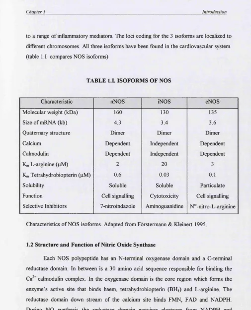
to a range of inflammatory mediators. The loci coding for the 3 isoforms are localized to different chromosomes. All three isoforms have been found in the cardiovascular system, (table 1.1 compares NOS isoforms)
TABLE 1.1. ISOFORMS OF NOS
Characteristic nNOS iNOS eNOS
Molecular weight (kDa) 160 130 135
Size of mRNA (kb) 4.3 3.4 3.6
Quaternary structure Dimer Dimer Dimer
Calcium Dependent Independent Dependent
Calmodulin Dependent Independent Dependent
Km L-arginine (jiM) 2 20 3
Km Tetrahydrobiopterin (|iM) 0.6 0.03 0.1
Solubility Soluble Soluble Particulate
Function Cell signalling Cytotoxicity Cell signalling Selective Inhibitors 7-nitroindazole Aminoguanidine N^-nitro-L-arginine
Characteristics of NOS isoforms. Adapted from Forstermann & Kleinert 1995.
1.2 Structure and Function of Nitric Oxide Synthase
Each NOS polypeptide has an N-terminal oxygenase domain and a C-terminal reductase domain. In between is a 30 amino acid sequence responsible for binding the Ca^^ calmodulin complex. In the oxygenase domain is the core region which forms the enzyme’s active site that binds haem, tetrahydrobiopterin (BH4) and L-arginine. The reductase domain down stream of the calcium site binds FMN, FAD and NADPH. During NO synthesis the reductase domain acquires electrons from NADPH and transfers them to the haem iron, which permits it to bind oxygen and catalyse NO synthesis. NO is liberated from organic nitrates and other nitrosovasodilators and has been used therapeutically for many years (Ahlner et al, 1991). Over the past decade
increasing evidence has appeared suggesting an important role for NO in mediating physiological and pathological changes within the heart.
1.3 Generation of Nitric Oxide
Sources of Endogenous Nitric oxide in the Myocardium
Each of the 3 isotypes of NOS has been identified in different regions and cell types within the heart.
i. Endothelial NOS: In animals and humans NO may be generated local to myocytes by eNOS present in the vascular endothelium of myocardial capillaries and venules and in the endocardial lining (Schulz et al., 1991; Smith et al., 1991; Andries et al., 1998). In addition eNOS expression bas been detected within rodent (Balligand et al., 1995; Seki et al., 1996), human atrial (Wei et al., 1996) and ventricular myocytes (Balligand and Cannon 1996; Gauthier et al., 1998). As in endothelial cells, myocyte eNOS is localised to caveolae glycosphingolipid rich microdomains present in the plasmalemma (Feron et al., 1996). Different caveolae subtypes have been identified with type 3 caveolin found in the T tubular system serving to localize eNOS to a site central in excitation contraction coupling. Evidence for a role for eNOS in modulating myocardial function is inconclusive. Early studies suggested a central role for eNOS in modulating muscurinic responses. However, in eNOS knockout mice the muscarinic and P-adrenergic regulation of heart rate, contractile force and Ca^^ current are preserved (Vandecasteele et al., 1999). Conversely over expression of eNOS has no effect on intrinsic heart rate or the response to acetylcholine (Brunner et al., 2001).
Chapter I____________________________________________________________________Introduction
independent NOS in the myocardium of patients with dilated cardiomyopathy, myocarditis and postpartum cardiomyopathy, but not in ischaemic or valvular heart disease. It was suggested that iNOS induction might have a central role in the aetiology of inflammatory cardiomyopathies.
Subsequent studies confirmed the presence of iNOS in the myocardium in dilated cardiomyopathy (DCM) (Winslaw et al., 1994; Haywood et al., 1996; Habib et al.,
1996; Satoh et al., 1997; De Belder et al., 1993; Vejlstrup et al., 1998; Drexler et al., 1998; Barbaro et al, 1999) but also found it in association with advance cardiac failure from a range o f other causes including ischaemic heart disease (Haywood et al, 1996; Winslaw et al, 1994; Fukuchi et a/., 1998; Drexler et al, 1998; Vejlstrup et al, 1998), septic shock (Thoenes et al, 1996) valvular heart disease (Haywood et al, 1996), transplant rejection (Lewis et al, 1996) and HTV related cardiomyopathy (Barbaro et al,
1999). It would appear that the induction of iNOS is related to heart failure per se, rather than being related to a particular underlying aetiology.
Though the bulk of studies have found iNOS expression in the failing human heart this has not been a universal finding (Stein et al, 1999). In contrast Haywood et al detected iNOS mRNA and protein in 57% of non-failing donor organs. (Results of studies looking at iNOS in the human myocardium are summarized in Table 1.11. NF = non failing, DCM = dilated cardiomyopathy, IHD - ischaemic heart disease and HCM = hypertrophic cardiomyopathy)
de Beider et al, IHD Citrulline assay
Lancet 1993 341 84 (+)
Br Heart J 1995 74 426 DCM +++
Winiaw et al, NF Plasma nitrate (+)
Lancet 1994 344 373 IHD
DCM
++ ++
Haywood et al. NF (post-mortem) 0 0
Circulation 1996 93 1087 NF (donor) 57% +ve +
IHD 59% +ve +
DCM 67% +ve +
Valve disease 100%+ve +
Thoenes et al. NF 0 CGMP+
J Mol Cell Cardiol IHD 0 CGMP+
1996 28165 DCM 0 CGMP+
Septic shock +++ CGMP+++
Habib et al. NF 0
Lancet 1996 3471151 IHD
DCM
+
+++
Lewis et al,
Circulation 1996 93 720
Allograft 48% +ve + cyclic GMP+
contractile dysfunction
Satoh et al. NF 0 0
JACC HCM 0 0
1997 29 716 DCM 54% +ve +
Fukuchi et al NF +/- Variable activity in
Circulation IHD (failing) + failing hearts (IHD and
DCM) correlated to
1998 98132 DCM + macrophage infiltration
Vejlstrup et al DCM + ++
JMCC 1998 30 1215 IHD + ++
Drexler et al NF
+/-JACC DCM ++ Citmlline assay
1998 32 955 IHD (failing) ++
Stein et al NF -
-JACC DCM & myocarditis +/-
-1998 321179 IHD (failing) +/-
-Chapter 1____________________________________________________________________Introduction
Demonstration of iNOS message and activity in myocardial homogenates cannot identify in which cell type NOS II is expressed. The exact localization of the iNOS protein is unclear. In animal models o f sepsis, iNOS appears within individual myocytes resulting in reduced contractile function. Inhibition of iNOS activity partially (Brady et a/., 1992) or completely (Stein et al., 1996; Balligand et al., 1993) reversed the depression of contractile amplitude. In the human myocardium iNOS activity has been localized to vascular endothelium and smooth muscle cells (Vejlstrup et al., 1998) as well as infiltrating macrophages (Fukuchi et al., 1998).
In the limited number of studies that have looked directly at tissue or cellular NOS enzyme activity, the in vitro assays were performed with non-limiting concentrations o f substrate and cofactors which may have given rise to overestimates of activity compared with in vivo.
Compared with humans, rodents readily express iNOS particularly during sepsis. In the mouse, a single gene for iNOS has been mapped to chromosome 11 (Mock et a/., 1994). However in primates, including man, multiple copies of the iNOS gene have been identified (Xu et al., 1994). The rapid evolutionary changes at the iNOS locus are evident when primates closely related to man are examined, suggesting that the iNOS duplication events have occurred recently in evolutionary terms (Xu et al., 1995). It is possible that expression of iNOS in human cells is under tighter regulation than in rodent cells and that much of the data relating to iNOS in rodent models are not relevant to human disease. In support of this, the levels of NO metabolites recorded in man during sepsis are far lower than those seen in rats or mice. Whether active iNOS is present within human myocytes in vivo remains contentious, however iNOS activity may be provided within the myocardium by infiltrating inflammatory, endothelial and vascular smooth muscle cells.
iii. Induction of iNOS in heart Failure
There have been a number of studies documenting the induction of iNOS in DCM, myocarditis and sepsis reviewed in table I I I Advanced cardiac failure and in
particular cardiac cachexia, are associated with systemic and cardiac cytokine activation (Torre-Amione et al., 1996). Cytokines are potent stimulators of iNOS expression in cardiomyocytes (Kan et al., 1999). It is attractive to attempt to tie together the experimental and clinical results by postulating that enhanced intracardiac synthesis of NO by iNOS is causally related to contractile dysfunction in heart failure.
There is no study in humans documenting the relation between iNOS expression, subsequent NO synthesis and reduced cardiac contractility. Most studies in table I II measured only iNOS expression by reverse transcription-polymerase chain reaction (RT- PCR) or immunological techniques. The presence of iNOS mRNA and protein can not be assumed to reflect functional iNOS activity (Luss et al., 1997). RT-PCR is a very sensitive method able to detect trace amounts of mRNA which may not be of biological significance. In dedifferentiated human myocytes stimulation with cytokines and LPS resulted in induction of iNOS mRNA but not protein (Luss et al., 1997). The same cells were capable of producing other functional proteins in response to the same stimulus, whilst transfection with hepatic iNOS cDNA resulted in message and protein, suggesting that cultured human myocytes lack the capacity to express endogenous iNOS protein.
Chapter 1____________________________________________________________________Introduction
iv. Neuronal NOS
Neuronal NOS has been identified in the guinea-pig atria, specialised conduction tissues such as the sinus and atrioventricular nodes (Tanaka et al, 1995), and in post ganglionic sympathetic neurons innervating the heart (Tanaka et al, 1993). It is not expressed in rat ventricular myocytes (Balligand, Kobzik et al, 1995; Belhasen et al
1996) and there are no reports of its identification in human cardiomyocytes. However, the recent finding of nNOS in the cardiac sarcoplasmic reticulum (Xu et al, 1999), has led to the suggestion that NO from nNOS may play a role in regulation of myocyte calcium fluxes. Contraction of ventricular myocytes isolated from mice with nNOS disruption (NOSl"^') exhibited greater contraction at all frequencies compared with wild type littermates. There was enhanced basal contraction and inotropic response to g- adrenoceptor stimulation (Ashley et al, 2002). Contrary to expectations the time to 50% relaxation was increased with nNOS inhibition or disruption. However, the response to isoprenaline reported by Ashley et al was not assessed across a full concentration range. At high concentrations of isoprenaline the Ca^^ transient appeared smaller in myocytes from NOSl^ mice (Barouch et al, 2002) suggesting the maximal inotropic response may in fact be decreased.
There is thus increasing evidence that the localization of nNOS to the SR membrane is consistent with a role modulating Ca^^ fluxes and hence inotropy in rodent models.
1.4 Paracrine Endothelial NO
Since endothelial cells have constitutive NOS expression it has been suggested that they may influence local myocardial function. Brutsaert et al (1988) reported that selective denudation of the endocardial endothelium altered contractile fimction in isolated muscle strips by reducing the force of contraction and twitch duration. These effects were shown to be mediated by release of difiusible factors by endocardial cells (Smith etal, 1991). The physiological relevance of these findings has been controversial. The endothelial monolayer of the endocardium is in close proximity to only a very small proportion of the total myocardium. Therefore its ability to exert a paracrine effect of relevance to the whole heart is in doubt.
The endothelial lining of the coronary microvasculature is a possible paracrine source, which is local to the majority of myocytes. It has been previously shown by microscopy that most myocytes are within a few microns of a capillary, a distance that could be bridged by a small readily diffusible molecule such as NO
1.5 The Role of NO in the Human Heart
In normal human hearts intracoronary L-NMMA had no effect on the response to systemic dobutamine (Hare et al., 1998). Inhibition of endogenous NO synthesis significantly reduced basal LV d p / d t m a x , suggesting a positive inotropic role for NO
whilst having no effect on heart rate, mean aortic pressure or right atrial pressure (Cotton et al, 2001). The administration of exogenous NO donor or an agonist of NO release (substance P) enhanced LV diastolic relaxation and resulted in a downward shift of the LV diastolic pressure-volume relationship (Paulus et al, 1994 and 1995).
The potential role of NO in diseased states is more complex. In DCM with moderate heart failure the non-specific NOS inhibitor L-NMMA has no effect on basal contractile function or the blunted force frequency relationship (Cotton et at., 2002). The inotropic response to P-adrenergic stimulation in severe DCM was potentiated by concurrent intracoronary L-NMMA suggesting that endogenous NO may depress the p- adrenergic response (Hare etal., 1995 and 1998). Given the non-specific NOS inhibition with L-NMMA the effect may be mediated through iNOS or nNOS.
1.6 Sources of Exogenous NO (NO Donors)
Chapter 1____________________________________________________________________Introduction
difiFerences between the newer classes of NO donor regarding the kinetics of NO release, the NO species generated and the breakdown products after NO liberation.
1.7 Pharmacology of Nitric Oxide Donors
NO donors as their name implies are able to provide NO or related species when applied to biological systems. They have been employed as tools to investigate the effect of NO where their addition has been used to mimic endogenous production. However, equating the effect of exogenous with endogenous NO may be inaccurate. Many classes of NO donor generate a range of redox forms of nitrogen monoxide (N 0 \ NO# or NO ) the proportions of which may vary depending on the local redox environment. As subsequently discussed the different NO species are capable of divergent interactions with other biomolecules. Thus the selection of NO donor may be crucial in the observed response.
R— 0 - N
\
O Organic nitrates
R—S—N = 0 ^'-Mtrosothiols
NONOates
R
O Sydnonimines
2Na+
N C ^ / C N
^%Fe *w
* i N ™
Sodium nitroprusside
Fig 1.2 Generic structures of NO donor groups
Organic Nitrates
Members of this group are the oldest known NO donors. They are nitric acid esters o f mono- and polyhydric alcohols. GTN, pentaerythritol, isosorbide mono- and dinitrate (ISMN, ISDN) all require enzymic or non-enzymic bioactivation and therefore are not considered spontaneous NO donors. Their exact metabolic cleavage pathway remains unclear but it is likely that multiple intra- and extra-cellular pathways are involved to generate NO in vivo. The relative importance of individual metabolic systems is not known (Ahlner et al., 1991; Feelisch 1993; Harrison & Bates 1993; Bennett et al,
Chapter 1____________________________________________________________________Introduction
proposed to account for the bioactivation of organic nitrates: an NADPH-dependent cytochrome P450 pathway (Schroder 1992; McDonald & Bennett 1993; McGuire et al.,
1994) and a system of enzymes related to the glutathione S-transferase family (Kenkare et al., 1994). In small coronary microvessels it has been demonstrated that administration of L-cysteine markedly enhances GTN-induced vasodilation. It has been postulated that the cysteine is converted to glutathione which participates in the intracellular enzymatic bioconversion of GTN to NO (Wheatley et al., 1994), but results have not been consistent. Interestingly, glutathione also improves the production and / or bioavailability of NO in the brachial artery of patients with documented coronary atherosclerosis (Vita et al., 1998). In the same study it did not affect the response to exogenous sources of NO suggesting the effect is not dependent on prolongation of NO t^^. The mechanism by which glutathione modulates NO production remains unclear. In isolated enzyme preparations of nNOS and iNOS optimal enzymatic activity was achieved in the presence of glutathione (Komori et al., 1995; Stuehr et al., 1990). Glutathione is able to prevent inactivation of NOS that is thought to result from peroxynitrite formation due to simultaneous generation o f NO and superoxide by the enzyme (Hobbs et al., 1994). More recently a mitochondrial aldehyde dehydrogenase has been found to catalyse the formation of 1,2-glyceryl dinitrate and nitrite from GTN (Chen et al., 2002). Data indicates that loss of this enzyme activity is associated with nitrate tolerance.
Non-enzymic liberation of NO appears to involve thiol groups (Fontecave and Pierre 1994) and their depletion may underlie nitrate tolerance. Although theoretically any thiol compound may decompose organic nitrates to yield inorganic nitrite only a few (cysteine, N-acetyl-cysteine and thiosalicylic acid) have been shown to promote simultaneous NO generation (Feelisch etal., 1988; Chong and Fung 1999).
Sodium Nitroprusside
Sodium nitroprusside (SNP) has been used for many years as an intravenous infusion to rapidly control severe hypertension. In vitro there is no spontaneous NO liberation. Generation of NO may occur after partial reduction (a one electron transfer)
which may be achieved by a variety of agents in vivo. Certainly thiols may perform this function (Fontecave and Pierre 1994).
2 RSH
2[Fe(CN)5(NO)]2- --- ► 2RSN0 +
2[Fe(CN)gH20]2-pH 7.4 L V /5 *2 J
t RSSR + 2N 0 SNP can also be decomposed directly to NO by light
2[Fe (CN)s(NO)f• NO + [Fe (CïOsHjO)]^
There is also a pathway of enzymatic cleavage (Kowaluk et al., 1992) which appears different from those involved in GTN. Breakdown o f SNP yields NO and 5 cyanide anions. The toxic cyanide byproduct inhibits the ferrocytochrome oxidase of the respiratory chain and therefore limits the duration of clinical exposure to SNP in man whilst in experimental systems complicates the interpretation of data. SNP is also a source of nitrosonium ions (NO^ which behave as a nitrosating electrophilic species.
Nitrosothiols
Nitrosothiols (R-SNO) are prepared by the reaction of a thiol with either acidified nitrous acid or an alkyl nitrite.
RSH + NO^ ^ RSNO +
RS" + RONO -> RSNO and RO
Chapter 1 Introduction
HOOC
S-Nitrosoglutathione
CH,
COOH
o
çy
OHS-N itroso-N -acetylpenidllam ine
Metal Ion; Cu^ ions play the central role in the metal catalysed breakdown of nitrosothiols in vivo. The exact nature of the chemical reaction and the identity of the intermediate structures formed have not been defined. Cu^ ions are generated fi’om the more abundant Cu^^. The Cu^ combines to the nitrogen atom of NO with an electron transfer. The NO is liberated with production of Cu^^ and RS'. The RS' is oxidised to RS# which then combines to form a homodimer (RS-RS) with reduction o f the copper to Cu^. The regeneration of Cu^ or the interaction o f Cu^ with the nitrosothiol may be the rate-limiting step depending on the chemical structure of the nitrosothiol donor.
2+
Cu + RS Cu + RSNO
^ Cu + RS"
2+
Cu + RS + NO
2 R S " ^ RSSR
(The generation o f C y from with subsequent cleavage o f nitrosothiol to yield NO and a homodimer RSSR)
Enzymic Decomposition: Gamma glutamyl transpeptidase may catalyse the decomposition o f GSNO (Askew et al., 1995) with formation of the less stable intermediate S-nitrosocysteinylglycine which then breaksdown to release NO in the presence of Cu^ ions.
Transnitrosation: This involves the spontaneous transfer of NO fi'om one thiol to another. If the donor thiol is relatively stable e.g. GSNO or SNAP and the recipient is an abundant low molecular weight thiol such as cysteine, the resultant S-nitrosocysteine is
relatively unstable and decomposes rapidly (Barnett et al.^ 1995), again in the presence of Cu^ ions.
Photochemical: Absorption of radiation at 365nm is capable of causing excitation sufficient to cause fission of the S-N bond and NO release. The resultant thiyl radical can then combine with another nitrosothiol molecule to liberate a further molecule of NO and to form a disulphide homodimer.
Thermal: The mechanism involved is similar to that for photochemical decomposition with the initial excitation energy coming fi'om a thermal source. The breakdown of the nitrosothiol with generation of an alkyl thiyl radical allows further decomposition with formation of disulphide homodimers and liberation of NO.
The relative importance of each of these routes in vivo remains a matter of debate. Thermal and photochemical breakdown is slow. The Cu^ mediated catalysis, either directly or indirectly via transnitrosation and enzymic metabolism appears to be the dominant factor. Thus the abundance of Cu^ either fi-ee or complexed to protein has a marked influence on NO liberation.
Interest in the S-nitrosothiols has increased with their detection in human plasma, airway secretions and other body fluids. S-nitrosoalbumin was found to be between 0.25 and 1 pM in plasma samples stored for 2-3 weeks (Stamler et al, 1992) with higher concentrations in fi-esh samples. GSNO has been detected in airway secretions (0.25 pM in healthy subjects and 4 pM in the presence of pneumonia ). Within red blood cells there is GSNO and S-nitrosohaemoglobin. Differences in nitrosohaemoglobin concentrations in arterial (0.3 pM) and venous blood (0.03 pM) has led to speculation that haemoglobin may serve as a reservoir of NO allowing this short lived locally active radical to be transported to more distant sites via the arterial circulation (Gow and Stamler 1998).
Chapter 1___________________________________________________________________ Introduction
et al, 1992; de Belder et al., 1994) and reduce platelet deposition and embolization following angioplasty (Langford etal., 1999), endarterectomy (Molloy et al., 1998) and saphenous vein grafting (Salas et al., 1998). The concentrations needed to inhibit platelet ftmction are low and cause only minimal change to systemic blood pressure and pulse rate. In a more traditional NO donor role the vascular efiects of nitrosothiols have a significant advantage over GTN and other organic nitrates in that they do not induce tolerance after a prolonged exposure. Tolerance to organic nitrates is thought to be the result of decreased availability of reduced thiol groups. In support of this the addition of L-cysteine restores the activation of soluble guanyl cyclase in tolerant isolated vascular strips (Isono et al, 1994) whilst co-administration of N-acetylcysteine and nitroglycerin partially reverses nitrate tolerance in human subjects with angina (Pizzuli et al., 1997).
Because the release of NO does not require an enzymatic degradation nitrosothiols have been used as a spontaneous source of NO in many biological experiments. However, it is wrong to assume that the addition of GSNO to a cell or tissue culture system results in immediate or quantitative NO release. Release kinetics are infiuenced by the presence of Cu^ and other low molecular weight thiols whilst the redox conditions influence the species of NO generated with nitrosothiols able to generate NO*,NO^ or NO
“NONOate” Nitric Oxide Donors
“Nonoates” or 1-substituted diazen-1 -ium-1,2-diolates are compound that contain a [N(0)N0]’ functional group and provide a consistent and reliable delivery of NO in vitro and in vivo. They are synthesized by exposing a suitable nucleophilic species to a few atmospheres of NO under anaerobic conditions.
X' + 2 N0 --- ^ X-[0)N0r
The resultant white powder is stable if stored under refrigerated dry conditions. When dissolved in solution the NONOates dissociate to liberate 2 mois of NO and 1 mol of the original nucleophilic starting material.
\ H \
N N 2 N 0 + N—H
O
Spontaneous NO release from all NONOates is governed exclusively by first order kinetics. The rate of this reaction is determined by the nature of the carrier nucleophile and the environmental temperature and pH. At 37®C decomposition is some 9 fold greater than at 22®C (Hrabie et al, 1993) whilst the dissociation reaction is catalyzed by an acidic environment and hence any drop of pH enhances NO liberation. The half-lives of the nonoate donors at 37®C and physiological pH have been previously determined and hence it is possible to select a particular donor to reliably deliver a known amount of NO over a specified time course.
° H,C, o ' CHjCHjCHj y O '
I
/ I - - - / N
^ N — o 'N a * Hjhren.CH.CH, N— O
DETANONOate 20 hrs DEA/NO 2-4mins PAPANOnoate 15mins
1.8 Nitric Oxide Species
Chapter 1____________________________________________________________________Introduction
The Nitrosonium Cation is the oxidised form o f nitric oxide. It is the key species in the process of nitrosation in which the NO^ is transferred from a carrier compound to a nucleophilic centre. The nitrosonium cation rapidly degrades in an aqueous environment to give nitrous acid. The lifetime of NO^ in aqueous solution at a neutral pH is 0.3 nS. Thus the NO^ cation is only found in solutions with a very high acidity. At physiological pH the nitrosation reaction occurs with the transfer of NO^ from carriers such as nitrosothiols. Nitric oxide can not in isolation act as a nitrosating agent. In the presence of oxidising agents it may be converted to N 0 \
Nitrosation is an important process in cell biology. Nitrosation of thiol residues (RS-) may produce nitrosothiols (RSNO) which may subsequently decompose to liberate NO with the formation of homodimers (RS-SR). These new disulphide linkages may bring about conformational changes in proteins such as ion channels with significant functional effects.
The Nitroxyl Ion may be produced by several routes within the cell and may exert direct and varied effects. NOS catalyses the oxidation of arginine to N-hydroxyarginine which may decompose to yield NO The binding of NO# to Fe (H) in a haem centre may also lead to the release of nitroxyl ions (Gow and Stamler 1998). Decomposition of S- nitrosothiols in the presence of thiols may lead to the formation of NO (Amelle and Stamler 1995). The suggestion that S-nitrosothiols can also donate NO to thiols further emphasises the importance of redox balance in determining the NO species and subsequent effect.
Once generated NO is short lived in aqueous solution lasting only milliseconds. It may act as EDRF though these effects may occur after oxidation to NO* At high pH NO may interact with an oxygen molecule to provide an alternative route for the generation of peroxynitrite (ONOO )
Peroxynitrite is produced mainly from the interaction of NO with superoxide O2 and
has a half life of 1 s at physiological pH. As discussed above there is evidence for a lesser
route of synthesis involving nitroxyl ions and oxygen (Sharpe and Cooper 1998). It is capable of nitration and hydroxylation of protein targets and is a more potent oxidising agent than either NO or superoxide alone. Macrophages are a known site of ONOO production (Ischiropoulos et al, 1992) where it may play a beneficial role in innate immunity (Zhu et al., 1992). Production of peroxynitrite elsewhere may cause detrimental local cellular and mitochondrial damage (Kayahara et al, 1998). Peroxynitrite reacts rapidly with carbon dioxide to give ONOOCO2 and this occurs
faster than the oxidation, nitration and hydroxylation reactions which account for its cytotoxicity. It is unclear what the fate of this CO2 derivative is. Thus peroxynitrite
formed in blood vessels is likely to react with CO2 whilst peroxynitrite formed in cells
will preferentially oxidise thiols.
1.9 Nitric Oxide targets in the Cardiovascular System
Soluble guanylate cyclase (White and Aurbach 1969) is the only proven receptor for NO. It catalyses the conversion of guanosine 5’-triphosphate (GXP) to cyclic guanosine 3 ’,5 ’-monophosphate (cGMP). Within the cardiovascular system the sGC signal transduction pathways play a central role in vascular tone, platelet adhesion and also to a lesser extent myocyte function. The binding of NO to the haem moiety of sGC results in enzyme activation by some 400 fold (Stone and Marietta 1994) elevation of cGMP levels and transmission of the signal to the downstream transduction elements such as cGMP dependent protein kinase (reviewed by Lehman et al., 1997), cGMP gated cation channels (reviewed by Seagate et al., 1996) and cGMP regulated phosphodiesterases (Degerman et al., 1997; Houslay and Milligan 1997). Activation of sGC results in elevation of cGMP but the diversity of targets for this second messenger may cause diverse and even opposing actions in different cell and tissue types.
Chapter 1___________________________________________________________________ Introduction
protease involved in apoptosis may also be inhibited by S-nitrosylation of its active site cysteine giving NO a role as an anti-apoptotic signal (Li et a/., 1997).
(Fig 1.3 summarizes possible targets for NO in the cardiovascular system)
Venous Tone
The Heart
Arterial tone
Heart Rate
Systolie / diastolic function
Sympathetic and
parasympathetic response
Coronary tone
Starling response
Cyclic Nucleotides
cGMP / cAMP levels
Cellular / Subcellular
Ion channels
Myofilament Ca++ sen sitiv ity
Chapter 1____________________________________________________________________Introduction
1.10 Calcium Homeostasis
Intracellular Ca^^ is the central regulator of cardiac contractility (reviewed by Bers 2000). A number of ion channels control the entry and sequestration of calcium within individual myocytes. During the action potential the sarcolemmal L-type calcium channels become activated and the Ca^^ enters via the /ca. A much smaller quantity of external Ca^^ enters via the Na-Ca^^ exchanger (NCX). This influx of external calcium triggers further release of Ca^^ from the internal stores within the sarcoplasmic reticulum (SR) through the SR Ca^^ channel (ryanodine receptor), a process termed calcium induced calcium release (CICR) (Fabiato 1978). The mechanism by which the trans- sarcolemmal Ca^^ activates the release of SR calcium is not clearly understood but appears to involve a conformational change in the SR Ca^^ release channel due to Ca^^ binding. The Ca^^ entry and that released by CICR raise the cytosolic free Ca^^ level. In turn this elevated level allows binding of Ca^^ to the thin myofilament protein troponin C, resulting in activation of the contraction process.
The autonomic nervous system may regulate the Ca^^ influx across the sarcolemma. Stimulation of P-adrenoceptor by agonists such as adrenaline and isoprenaline activates the enzyme adenyl cyclase via the receptor coupled G protein (Gs) (Bristow et al., 1982). The intracellular levels of cAMP rise with subsequent activation of protein kinase A (PKA). Several intracellular proteins can be phosphorylated by PKA, including phospholamban (PLB) and troponin I. PLB is a key regulator of SERCA in cardiac cells. Dephosphorylated PLB is closely associated with SERCA and acts as an inhibitor of the SR Ca^^ pump (James et al., 1989). On phosphorylation by PKA the inhibitory effect is relieved. This allows greater Ca^^ transport with faster contraction and relaxation. The inotropic and lusitropic effects of P-adrenergic activation were attenuated in PLB-KO mice. Phosphorylation of troponin I, which interacts with troponin C, decreases the affinity of troponin C for Ca^^ (Solaro et al., 1976).
For diastolic relaxation to occur there must be a decline in cytosolic Ca^^ levels allowing dissociation from troponin C and deactivation of the contraction process. There are four identified mechanisms to achieve this reduction of calcium level 1) reuptake in to
the SR by the SR - ATPase (SERCA) (Ikemoto et al., 1982; Hasselbach et al., 1983 2) extrusion of Ca^^ by the Na-Ca^^ exchanger 3) extrusion by the sarcolemmal Ca^^ - ATPase and 4) uptake in to mitochondria by the Ca^^ uniporter. The relative importance of each mechanism varies between species and has been examined by the use of specific inhibitors. In rabbit myocytes the SERCA removes 70% of the activated Ca^^ whilst the NCX removes 28% with a 1% contribution from each of the other 2 mechanisms. In contrast in the rat SERCA activity is greater with 92% of calcium taken back up in to the SR and only 7% extruded via the NCX.
In the failing human heart there is a change in the relative contributions of the 2 principle systems (reviewed by Ravens and Dobrev 2000). The mRNA levels for SERCA and phospholamban are consistently lower (Feldman et al., 1991) with reduced protein translation (Hasenfuss et al., 1994). With the reduction in SERCA expression, there is an increase in expression of the NCX. This shifts the balance of Ca^^ fluxes to favour extrusion of calcium from the myocytes and reduced reuptake in to the SR. The SR Ca^^ ATPase is a more energy efiBcient Ca^^ pump. It transports 2 Ca^^ ions for each molecule of ATP compared with one by the NCX. Thus in the failing heart the trans sarcolemmal Ca^^ cycling is less energy efficient.
In a steady state, the proportion of the activator Ca^^ extruded by the NCX during relaxation must be matched by the inward L-type channel Ica during the subsequent beat. Thus as predicted in the rabbit myocyte there is more Ca^^ entry via the Ica and less SR Ca^^ release compared with the rat. The cellular mechanisms of Ca^^
L-Type Ca^+ Channel
C a : +
CRC (Ryanodine Receptor)
Sarcoplasmic Reticulum
3Na' NCX
SERCA
Mitochondrion
C a : +
Contractile Apparatus
Sarcolemmal Ca^VATPase
Ca^+
Fig 1.4 A schematic representation of the regulatory mechanisms controlling the myocyte cytosolic Ca concentration. (Modified from Hague et al., 1998) NCX Sodium-Calcium exchanger
CRC Calcium release complex
1.11 Functional effects of NO on the h e a rt
Diastolic Function: There is a general consensus about the effect of NO on diastolic function. It selectively induces an earlier onset of isometric relaxation without affecting the rate of isometric tension development. This has been observed in various animal species using isolated myocytes (Shah et al., 1994; Ito et al., 1997), papillary muscles (Smith 1991), and whole heart studies (Grocott-Mason et al., 1994; Grocott-Mason et al, 1994) in response to endogenous NO release, stimulated by bradykinin or substance P and exogenous NO supplied by NO donors. In isolated guinea-pig hearts inhibition of endogenous NOS significantly attenuated preload induced increases in cardiac output (Prendergast et al., 1997). In man bicoronary injection of SNP (exogenous NO) or stimulation of endogenous NO release by substance P (Paulus et al., 1994) resulted in a downward displacement of individual diastolic left ventricular (LV) pressure - volume plots. It appears that NO is capable of enhancing LV distensibility. Using a porphyrinic electrode direct measurement of NO generation has provided further evidence that the intramyocardial NO concentration is associated with alteration of ventricular preload (Pinsky et al., 1997). Increased LV chamber stretch resulted in an increase of intracardiac NO release. During the usual cardiac cycle the peak NO concentrations are recorded around the time of early diastolic filling. This is in keeping with the proposed physiological role of NO to enhance myocardial relaxation, reduce diastolic tone and optimize coronary perfusion.
Chapter 1____________________________________________________________________Introduction
Balligand has proposed that myocyte eNOS represents an autocrine negative feedback system to modulate the myocyte contractile response to adrenergic stimulation. Activation of beta-adrenergic receptors produces an increase in intracellular Ca^^ via elevation of the Ca^^ current through voltage sensitive Ca^^ channels. This effect is mediated by a rise in cAMP and activation of protein kinase A. The resultant intracellular Ca^^ concentration is suflQcient to activate eNOS whose Ca^^ EC50 is 200-400nM. Inhibition of eNOS potentiates the positive inotropic effect to submaximal isoproterenol (2nM), by some 30%, in isolated rat ventricular myocytes (Balligand et al., 1993), with similar effects in rat atria (Sterin-Borda et al., 1998) and atrial strips of non-failing human hearts (Gauthier et al., 1998). In vivo endogenous NO blunts the contractile response to infused catecholamine (Kearney et al., 1996) or submaximal stimulation of the stellate ganglion in dogs (Takita et al., 1998).
The notion that eNOS attenuates the inotropic response to catecholamines was further supported by 2 independent reports of enhanced sensitivity to P-adrenoceptor stimulation in eNOS knockout mice (Godecke et al., 1998; Gyurko et al., 2000). However, subsequent studies have produced diametrically opposite results with the eNOS KO mice having a similar response to adrenergic stimulation as wild types (Han et al., 1998; Vandecasteele et al., 1999). Criticism o f these studies has centred on the high concentration of isoprenaline used (IpM) and in the use of older KO mice which have developed significant ventricular hypertrophy absent in the wild types. Thus further studies are awaited to clarify the modulatory effect of eNOS.
A range of cell types within the myocardium are capable of expressing iNOS following the appropriate inflammatory stimulus. In early studies, iNOS derived NO has been reported to depress isolated myocyte contractile amplitude in guinea-pig (Brady et al., 1992) and rat. (Balligand et al., 1993, Joe et al., 1998) Freshly isolated adult rat ventricular myocytes co-cultured with microvascular endothelial cells pretreated with IL- 1 exhibited decreased contractile responsiveness to P-adrenergic stimulation, indicating that NO fi'om iNOS in endothelial cells was sufficient to affect the systolic function of co-cultured myocytes (Ungureanu Longrois et al., 1995). In whole hearts IL-1 and TNF
reduced contractile amplitude in a time course consistent with iNOS induction (Schulz et al, 1995). Generally induction of iNOS appears to result in a negative effect on systolic amplitude. However, in the failing human myocyte exposure to a NOS inhibitor had no effect (Harding et al., 1998) despite the belief that the induction of functional iNOS may be a central event in the pathogenesis of cardiomyopathy. There are various possible explanations for this observation. Firstly there may be no functional iNOS induction or at least no significant depressant effect mediated by NO. In vitro studies with human cells have emphasised the relative difficulty of inducing iNOS which has required combinations of multiple cytokines (Luss et al., 1997). Alternatively, chronic NO exposure may cause permanent change in myocytes which is not reversed by brief exposure to a NOS inhibitor.
Pharmacological NO donors: Initial reports of the effect of exogenous NO donors on cardiac muscle or myocytes showed a modest depression of contractility, with SNP, SNAP or dissolved NO gas reducing basal contraction by 20-25% (Brady et a/., 1993; Vila-Petroff et al., 1999). There are also reports of exogenous NO reducing the response to adrenergic stimulation in adult rat ventricular myocytes (Balhgand et al., 1993).
Author Year Species Preparation NO donor Effect
Kojda 1995 rat Isolated myocytes, Langendorff
SPM 3672 (lOOpM)
Myocyte contraction amplitude increased by 75%. Heart LVP max and dp/dt max increased
18%
Kojda 1996 rat Isolated myocytes
SNAP(l^M) Myocyte contraction amplitude increased by 41%
Mohan 1997 cat Papillary muscle SlN-1, SNAP & SNP (0.01-
lOuM)
Positive effect on developed tension
Kojda 1997 rat Langendorff SNAP(lpM), GTN (1-100
pM)
Positive effect on peak LVP and dp/dt
Preckel 1997 dog Whole heart in vivo
DEANO (20|iM), SNAP
and GTN
DEANO produced a 6.3% increase in LVP. Other donors produced lesser positive effect
Prendergast 1998 Guinea-pig
Working heart SNP (1 and lOpM)
IpM SNP enhanced the response to dobutamine
Chesnais 1999 frog Atrial and ventricular fibres
SIN-1 (100 ^iM) Produced a 14% increase in developed tension
Vila-Petroff 1999 rat Isolated myocytes
SNAP(1 pM) Myocyte contraction amplitude increased by 38%
Muller-Strahl
2000 rat Langendorff and Woridng heart
GTN(lpM), DEANO and SNAP (0.1 pM)
Positive effect of 6-7.5% on LVP, cardiac output and dp/dtmax.
Table l.III. Summary of recent studies reporting positive inotropy in response to applied NO donors.
In fact the first report of a positive eflFect of NO on cardiac systolic function was made in 1971 using GTN on feline and human papillary muscle (Strauer 1971). Subsequent studies in patients undergoing cardiopulmonary bypass revealed a variable inotropic response to SNP, the direction of which was related to pre-operative P- adrenoceptor blockade (de Hert et al., 1997). Many of the more recent studies which observed positive inotropy demonstrated a biphasic response with high concentrations of NO donor inducing a negative effect (Kojda et al., 1996; Kojda et al., 1997; Mohan et al, 1996; Vila-Petroff et al., 1999). Direct measurement of cyclic nucleotides suggested that low concentrations of NO induce only modest rises in cGMP accompanied by elevation of cAMP, whilst high NO concentrations are associated with a greater increase in cGMP and a fall in cAMP. Figure 1.5 summarizes the interaction of cAMP and cGMP. Within cardiomyocytes 2 forms of the cAMP degrading enzyme phosphodiestrase (PDE) have been identified. PDE2 is activated by cGMP whilst PDE3 is inhibited. With low NO concentrations the modest rise of cGMP inhibit PDE3 resulting in a rise in cAMP and the subsequent activation of the L-type calcium channel and positive inotropy (Kirstein et al., 1995; Mery et al., 1993). Conversely high NO concentrations lead to a greater rise in cGMP and activation of PDE2. The fall in cAMP results in negative inotropy. This suggests that both the positive and negative inotropic effects are mediated by soluble guanyl cyclase.
F ig 1.5
NO Donors
eNOS
B-AR
Gs
NO
iNOS
sGC
cAM P
PDE
cGMP
1?KA
p k g
]
phosphorylation
L-type Channel
6-AR Beta Adrenoceptor
eNOS Endothelial nitric oxide synthase iNOS inducible nitric oxide synthase AC Adenyl cyclase
sGC soluble guanyl cyclase PDE phosphodiesterase PKA Protein kinase A PKG Protein kinase