^oHAru
^ î k T X " v t A x 4 t : 4%oo,
2 8 0 6 0 9 7 2 5 3
INSIGHTS INTO REGULATION OF LIPOPROTEIN
RECEPTORS IN HUMAN MESANGIAL CELLS: THE
EFFECTS OF INFLAMMATORY CYTOKINES AND
CALCIUM CHANNEL BLOCKERS
Thesis submitted for the degree of Doctor of Philosophy
To the Faculty of Medicitie
University of London
Xiong Zhong Ruan
Centre for Nephrology
Royal Free and University College Medical School
Rowland Hill Street
London NW3 2PF
August, 2000
ProQuest Number: U643416
All rights reserved
INFORMATION TO ALL USERS
The quality of this reproduction is dependent upon the quality of the copy submitted.
In the unlikely event that the author did not send a complete manuscript and there are missing pages, these will be noted. Also, if material had to be removed,
a note will indicate the deletion.
uest.
ProQuest U643416
Published by ProQuest LLC(2016). Copyright of the Dissertation is held by the Author.
All rights reserved.
This work is protected against unauthorized copying under Title 17, United States Code. Microform Edition © ProQuest LLC.
ProQuest LLC
789 East Eisenhower Parkway P.O. Box 1346
ABSTRACT
The involvement of abnormal lipid metabolism in the progression of renal disease
and the pathogenesis of chronic graft dysfunction is generally accepted. The
dysregulation of lipoprotein homeostasis is involved in the incidence of accelerated
cardiovascular disease in this population. Chronic renal dysfunction is also an
inflammatory condition. Therefore, the present study was undertaken to investigate
various mechanisms involved in the receptor-mediated regulation o f intracellular
lipoprotein transport and its interactions with inflammatory cytokines.
Using human mesangial cell line (HMCL) culture, we demonstrated that HMCL
express native LDL receptors . Phorbol 12-myristate 13-acetate (PMA), Angiotensin II
(Ang II), TNF-a, and IL-Ip induced acetylated-LDL internalisation, scavenger
receptor mRNA expression and promoter activity. Both AP-I and ets motifs were
specific response elements to PMA-induced scavenger receptor expression.
Conventionally, LDL receptor pathway is not thought to be involved in foam cells
formation due to the tight metabolic control through a feedback regulation.
However, our studies demonstrated that TNF-a, TGF-p, PDGF, or IL -ip increased
LDL binding, LDL receptor mRNA expression and LDL receptor promoter activity.
Both TNF-a, and IL-lp overrode the suppression of LDL receptor activity caused by
a high concentration of native LDL and caused foam cell formation in HMCL. TNF-
a , and IL-lp also increased the expression of a cleavage activating protein (SCAP)
for sterol regulatory element binding proteins (SREBP). Diltiazem and verapamil, not
nifedipine increased LDL binding, LDL receptor mRNA expression and LDL receptor
Our results suggest that inflammatory cytokines may contribute to lipid deposition
and foam cell formation in HMC through following pathways: 1) inducing scavenger
receptor expression; 2) disregulating LDL receptor gene expression by increasing
sterol-independent and mitogenesis-independent gene transcription. The implications
of these findings are that inflammatory cytokines are important risk factors for
glomerular atherosclerosis. Therefore, future strategies for controlling progression of
renal and cardiovascular diseases should include anti-oxidants, lipid-lowering, and
Acknowledgements
The experimental studies described in this thesis were carried out at the Renal
Research Laboratory, Royal Free and University College Medical School. The work
was conducted by the author and was supervised by Dr. Zac Varghese and Professor
John F. Moorhead. I am also grateful to Professor Stephen H. Powis for his kind help
and support during the execution of this work.
I thank Dr. Christopher K. Glass (University of California, San Diego) for his kind gift
of the human scavenger receptor promoter. Professor David W. Russell (Department
of Molecular Genetics, University of Texas) for his kind gift of the human LDLr
promoter, and Professor J. D. Sraer (Hôpital Tenon, Paris) for providing the
immortalised human mesangial cell line.
Finally I am indebted to Professor Moorhead and Dr. Varghese for the financial
support, advice and encouragement provided while I was conducting my
PUBLICATIONS
1. Ruan XZ, Varghese Z, Powis SH, Moorhead JF Human mesangial cell express
inducible macrophage scavenger receptor. Kidney Int 56:440-451, 1999
2. Ruan XZ, Varghese Z, Powis SH, Moorhead JF Human mesangial cell express
inducible macrophage scavenger receptor; an Ap-1 and ets mediated response.
Kidney Int 56. Suppl S163-S166, 1999.
3. Ruan XZ, Varghese Z, Fernando R, Moorhead JF. Cytokines regulate LDL receptor
gene transcription in human mesangial cells. Nephrol Dial Transplant 13:1391-
1397,1998
4. Ruan XZ, Varghese Z, Fernando R, Powis SH, Moorhead JF. LDL receptor
expression in human mesangial cell under the influence of calcium channel blockers.
Clin Nephrol 51: 263-271, 1999
5. Ruan XZ, Varghese Z, Powis SH, Moorhead JF. Functional transformation of B/E
LDL receptor under the influence of cytokines and its molecular mechanisms
(Abstract). J Am Soc Nephrol 10(9):470A, 1999 (oral presentation at the annual
meeting o f the American Society o f Nephrology in Miami, 1999)
6. Ruan XZ, Kang ZQ, Li XW, Zheng FL. Effects of LDL on mesangial cells
12(2):89-7. Ruan XZ, Varghese Z, Powis SH, Moorhead. A Constitutively Active Form of the
Retinoblastoma Gene Product inhibits Rat Mesangial Cell Proliferation (abstract).
{Presented at International Congress o f Nephrology, Sydney 1997).
8. Varghese Z, Ruan XZ, Fernando R, Moorhead JF. The effects of 1,25-
dihydroxyvitamin D3 on inhibition of growth of human mesangial cells (abstract).
{Presented in International Congress o f Nephrology, Sydney 1997).
9.Ruan XZ, Kang ZQ, Li XW, Zheng FL. Minimally oxidized low density lipoprotein
stimulates the secretion of tumor necrosis factor of mesangial cells (abstract). Kidney
CONTENTS
Page No
ABSTRACT 2
ACKNOW LEDGEMENTS 4
PUBLICATIONS 5
LIST OF FIGURES 16
LIST OF TABLES 20
LIST OF ABBREVIATIONS 21
CHAPTER 1. GENERAL INTRODUCTION 25
1.1. NORMAL LIPOPROTEIN METABOLISM 29
1.1.1 Lipoprotein, lipolytic enzymes and transfer proteins. 30
1.1.2.Lipoprotein receptors 33
1.1.2.1. LDL receptor 39
1.1.2.2. Scavenger receptors class A 42
1.1.2.3. Scavenger receptor class B 46
1.1.2.4. Lectin like receptor - a novel endothelial receptor 48
for Ox-LDL
1.1.2.5. VLDL receptor 48
1.1.2.6. LDL receptor related protein 49
1.1.2.7 Lipoprotein receptor in mesangial cells 49
1.2. LIPOPROTEIN ABNORMALITIES FOUND IN RENAL DISEASE 50
1.2.1. Nephrotic syndrome 50
1.2.2. Lipid abnormahties in chronic renal insufficiency 52
1.3. INVOLVEMENT OF LIPIDS IN PROGRESSIVE RENAL DISEASES 57
1.3.1. Hypothesis 57
1.3.2. Experimental evidences 59
1.3.3. Clinical evidences 62
1.3.4. Pathogenesis o f hpid mediated renal injury 63
1.3.4.1. Oxidation o f LDL 64
1.3.4.2. Recruitment o f monocyte-macrophages 68
1.3.4.3. Mesangial cell proliferation and matrix expansion 68
1.3.4.4. Tubulo-interstitial lesions 71
1.3.5. Effect o f lipid lowering drugs 74
1.4. AIM OF THE THESIS 77
CHAPTER 2. GENERAL METHODS 81
2.1. CELL CULTURE 82
2.2. CELL PROLIFERATION ASSAY 84
2.3. PREPARATION LIPOPROTEIN ^ 4
2.4. lODINATION OF LDL 86
2.5. BINDING OF ^^^I-LDL TO HMCL at 4 ®C 1^6
2.6. LDL MODIFICATION 87
2.7.MODIFIED LOWRY ASSAY FOR LIPOPROTEIN AND CELL 89
MEMBRANE PROTEIN ESTIMATION
2.8 CHARACTERISTICS OF LDL RECEPTOR AND 90
SCAVENGER RECEPTOR IN HMCL
2.9. HISTOLOGY 91
2.11. WESTERN BLOT ANALYSIS 93
2.12. RNA ISOLATION 97
2.13. DNA PROBE LABELLING 100
2.14. RNA ELECTROPHORESIS & NORTHERN BLOT ANALYSIS 102
2.15.RT-PCR 105
2.16. SOUTHERN BLOT ANALYSIS 106
2.17. CLONING IN PLASMID VECTORS 108
2.18. TRANSIENT EXPRESSION ASSAY 115
2.19. ESTABLISHMENT OF THE CELL LINE BY STABLE 118
TRANSFECTION
2.20. DATA ANALYSIS 118
CH APTER 3. HUMAN MESANGIAL CELLS EXPRESS AN 119
INDUCIBLE M ACROPHAGE SCAVENGER RECEPTO R
3.1. INTRODUCTION 120
3.2. METHODS 123
3.2.1. Cell culture 123
3.2.2. Preparation o f acetylated lipoprotein 123
3.2.3. lodination o f LDL 123
3.2.4.Binding o f '“ l-AcLDL to HMCL at 4°C 124
3.2.5. Cell labelling for flow cytometry 124
3.2.6. Flow cytometry analysis 125
3.2.7.Establishment o f stable cell line expressing a high level o f 125
scavenger receptor
3.2.9. RT-PCR 128
3.2.10. Southern blot analysis and quantitative evaluation 128
3.2.11. Construction o f wild type scavenger receptor promoter reporter 128
gene with V40 enhancer
3.2.11.1. Preparation o f scavenger receptor promoter gene 129
3.2.11.2. Preparation o f pGL3 enhancer vector 129
3.2.11.3. Ligation o f pGL3 enhancer and Scavenger 129
receptor promoter
3.2.12. Functional analysis o f scavenger receptor promoter: the role o f 132
transcription factor AP-1/ets
3.2.13. Transient Expression Assay 132
3.2.14. The effect o f various signal transduction inhibitors on PMA and 133
Ang II -mediated activity o f Scavenger receptor promoter
3.2.15. Data analysis 133
3.3. RESULTS 135
3.3.1 Estimation o f values for the dissociation constant (Kd) 135
for scavenger receptor in HMCL.
3.3.2. PMA and Ang II induced scavenger receptor expression 136
in HMCL
3.3.2.1. Analysis o f flow cytometry 136
3.3.2.2. Visualisation o f Ac-LDL uptake and lipid droplets 137
3.3.2.3. Expression o f scavenger receptor mRNA 138
3.3.2.4. Activity o f scavenger receptor promoter 138
3.3.2.5. The effect o f Angiotensin II on HMCL proliferation 139
inhibitors on scavenger receptor promoter activity
2)32.1. Functional analysis o f the scavenger receptor promoter 139
3.3.3. Inflammatory cytokines TNFa and IL -lp induced scavenger 140
receptor expression in HMCL
3.3.3.1. Analysis of flow cytometry 140
3.3.3.2. Expression o f scavenger receptor mRNA 150
3.3.3.3. Activity o f scavenger receptor promoter 150
3.3.3.4. Functional analysis o f the scavenger receptor promoter 150
3.4. DISCUSSION 155
CHAPTER 4. FUNCTIONAL TRANSFORMATION OF LDL 165
R ECEPTO R UNDER TH E INFLUENCE OF INFLAMMATORY
CYTOKINES IN CULTURED HUMAN MESANGIAL CELLS
4.1. INTRODUCTION 166
4.1.1. LDL receptor feedback regulation. 167
4.1.2. The effects o f hormones and cytokines on LDL receptor 168
regulation
4.1.3. The effects o f drug on LDL receptor regulation 170
4.1.4. Expression o f LDL receptor in mesangial cells 171
4.2. METHODS 172
4.2.1. Cell culture 172
4.2.2. Cell proliferation assay 172
4.2.3. Preparation o f Lipoprotein 173
4.2.5. lodination o f LDL 173
4.2.6. Binding o f '^’l-LDL to HMCL at 4 “C 174
4.2.7. Western blot analysis 174
4.2.8. Cell labelling and flow cytometry analysis 174
4.2.9. Northern Blot analysis 175
4.2.10. RT-PCR 175
4.2.11. Southern blot analysis and quantitative evaluation 176
4.2.12. LDL receptor promoter-report gene constructs 177
4.2.12.1. pGL3 enhancer Luciferase Reporter Vectors 177
4.2.12.2. Transformation for pGL3 enhancer Vectors 178
4.2.12.3. Mini-preparation and Identification o f pGL3 179
enhancer plasmid DNA
4.2.12.4. Maxi-preparation and purification o f pGL3 179
enhancer plasmid DNA
4.2.12.5. Protruding 5'-terminal dephosphorylation 179
4.2.12.6. Preparation o f Insert ( LDLR6500) for Cloning 179
4.2.12.7. Ligation 180
4.2.13. Transient Expression Assay 181
4.2.14. The effect o f various signal transduction inhibitors on 181
cytokines-mediated activity o f LDL promoter
4.3. RESULTS 182
4.3.1 Estimation o f values for the dissociation constant (Kd) 182
for LDL receptor in HMCL.
4.3.2. Inflammatory cytokines induced LDLr transcription and 184
4.3.2.1. The effect o f cytokines on LDL binding on HMCL 184
4.5.2.2. The effect o f cytokines on expression o f 184
LDL receptor mRNA
4.5.2.3. The effect o f cytokines on HMCL proliferation 184
4.3.2.4. The effect o f cytokines on activity o f LDL receptor 187
promoter
4.3.2.5. The effect o f various signal transduction pathway 189
inhibitors on scavenger receptor promoter activity
4.3.3. Inflammatory cytokines induce LDL receptor expression in 189
sterol-independent pathway
4.3.3.1. Visualisation o f Ac-LDL uptake and lipid droplets 189
4.3.3.2. Expression o f LDL receptor protein 192
4.3.3.3. Analysis of flow cytometry 192
4.3.3.4. Expression o f LDL receptor mRNA 196
4.3.3.5. Activity o f LDL receptor promoter 196
4.3.3.6. Expression o f SCAP mRNA 199
4.4. DISCUSSION 202
CHAPTER 5. THE EFFECTS OF CALCIUM CHANNEL BLOCKERS 210
(CCBs) ON THE REGULATION OF LDL RECEPTOR IN HUMAN
MESANGIAL CELLS
5.1. INTRODUCTION 211
5.2. METHODS 213
5.2.1. Cell culture 213
5.2.3. lodination o f LDL 213
5.2.4. Binding o f ‘^’l-LDL to HMCL at 4 "C 213
5.2.5. Northern Blot Analysis 213
5.2.6. Cell proliferation assay 214
5.2.7. LDL receptor promoter-report gene constructs 215
5.2.8. Transient E?q)ression Assay 215
5.2.9. The effect o f various signal transduction inhibitors on 216
CCBs-mediated activity o f LDL promoter
5. 3. RESULTS 216
5.3.1. The effect o f CCBs on LDL binding on HMCL 216
5.3.2. The effect o f CCBs on expression o f LDL receptor mRNA 216
5.3.3. The effect o f CCBs on HMCL proliferation 217
5.3.4. The effect o f CCBs on activity o f LDL receptor promoter 219
5.3.5. CCBs were not able to override the suppression o f LDL receptor 219
induced by high concentration o f LDL
5.3.6. The effect of various signal transduction pathway inhibitors 222
on CCBs-mediated activity o f LDL promoter
5.4. DISCUSSION 226
CHAPTER 6. GENERAL DISCUSSION AND CONCLUSION 232
6.1. INFLAMMATION IS A KEY FACTOR IN GLOMERULAR 233
ATHEROSCLEROSIS
6.2. INFECTION AND ATHEROSCLEROSIS 237
6.3. MARKERS OF INFLAMMATION 238
6.4. FUTURE WORKS 239
in different cell types
6.4.2. Identify the specific response elements in LDL receptor 240
promoter region
6.4.3. Further explore the signal transduction pathways 240
involved in scavenger receptor regulation
6.4.4. Identify the specific response elements to TNF-a and 241
IL -lp in scavenger receptor promoter region
6.4.5. Animal models 241
LIST OF FIGURES
Chapter 1 Page No.
Fig. 1.1. A unified concept o f progressive renal damage 28
Fig. 1.2. Structure o f plasma lipoprotein 31
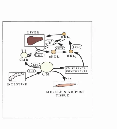
Figure 1.3. Chylomicron metabolism and HDL chylomicron interactions 37
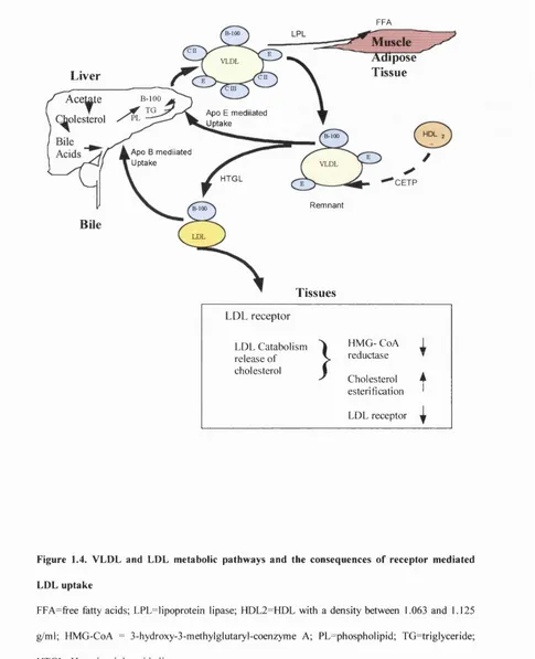
Figure 1.4. VLDL and LDL metabolic pathways and the consequences 38
o f receptor mediated LDL uptake
Fig. 1.5. LDL receptor pathway 40
Fig. 1.6. The structure o f the LDL receptor protein 41
Fig. 1.7 Models o f the predicted quaternary structure o f macrophage 44
scavenger receptors.
Fig 1.8. Mechanisms o f hyperlipidaemia in nephrotic syndrome 52
Figure. 1.9. Proposed mechanisms for the pathogenesis o f lipid-induced 58
glomerular atherosclerosis and tubulo-interstitial damage
in chronic progressive renal disease
Figure 1.10. A schematic o f the basic mechanisms involved in the 73
pathogenesis o f atherosclerosis
Chapter 3
Fig 3.1. Agarose gel electrophoretic mobility o f nLDL, Ac-LDL and Ox-LDL 125
Fig 3.2. Scavenger receptor mRNA expression in normal and transfected HMCL 127
Fig 3.3. Structure o f 5’stream o f scavenger receptor 130
Fig.3.5. Binding o f *^^I-AcLDL to HMCL at 4° C 135
Fig.3.6. Plot o f Scatchard transformed data o f ^^^I-AcLDL binding to HMCL 136
Fig.3.7 Analysis o f the mean fluorescence intensity (MFI) and the percentage 141
o f Dil-labelled cells in the PMA & Ang II treated-HMCL
Fig.3.8. Specificity o f the flow cytometric analysis o f uptake o f Dil-Ac-LDL 142
in HMCL
Fig.3.9. Visualisation o f Ac-LDL uptake and lipid droplets in HMCL after 143
PMA or Ang II treatment
Fig.3.10. Time dependent expression o f scavenger receptor mRNA in 144
response to PMA and Ang II
Fig.3.11. The effect o f different concentrations o f PMA and 145
Ang II on scavenger receptor mRNA expression
Fig.3.12. The response o f scavenger receptor promoter to PMA 146
Fig.3.13. The effect o f Angiotensin II on HMCL proliferation 147
Fig.3.14. Functional analysis o f the scavenger receptor promoter and AP-1/ets 149
motifs in the context of the minimal prolactin gene promoter
Fig3.15. Analysis o f the mean fluorescence intensity (MFI) o f Dil-labelled cells 151
in TNFa or IL-lp treated-HMCL
Fig.3.16. Time dependent expression o f scavenger receptor mRNA 152
in response to TNFa and IL-1 p
Fig.3.17. The response o f scavenger receptor promoter to TNFa and 153
IL -ip stimulation
Fig.3.18 Functional analysis o f the Scavenger receptor promoter and AP-1/ets 154
Chapter 4
Fig 4.1. Model for two-site proteolytic cleavage o f membrane-bound SREBPs 169
Fig 4.2. RNA electrophoresis 175
Fig 4.3. LDL receptor and GAPDH probe preperation 176
Fig 4.4. The map o f pGL3-Enhancer Vector 178
Fig 4.5. Preperation o f LDL receptor promoter 180
Fig 4.6. Identification o f positive clone 180
Fig.4.7. Binding o f ‘^’l-LDL to HMCL at 4°C 182
Fig.4.8. Plot o f Scatchard transformed data o f '^*I-LDL binding to HMCL 183
Fig.4.9. Effect of cytokines on LDL binding on HMCL 185
Fig.4.10. The effects o f cytokines on expression o f LDL receptor mRNA 186
Fig.4.11. Effects o f cytokines on HMCL proliferation 187
Fig.4.12. Cytokines stimulate LDL receptor promoter activity 188
Fig.4.13. Visualisation o f LDL uptake and lipid droplets in HMCL 191
after TNFa or IL-1 p treatment
Fig.4.14. The electrophoretic mobility o f LDL 193
Fig.4.15. Inflammatory cytokines overrode the LDL receptor protein 194
suppression induced by a high concentration o f native LDL.
Fig.4.16. Analysis o f the mean fluorescence intensity (MFI) in the TNFa or 195
IL -lp treated-HMCL
Fig.4.17. Inflammatory cytokines overrode the LDL receptor mRNA 197
suppression induced by a high concentration o f native LDL
Fig.4.18. The response o f LDL receptor promoter to TNFa or IL-1P 198
Fig.4.19. Intracellular concentrations o f cholesterol regulated SCAP mRNA 200
expression in HMCL
Fig.4.20. Inflammatory cytokines increased SCAP mRNA expression in HMCL 201
Chapter 5
Fig.5.1. Effect of CCBs on LDL binding on HMCL 218
Fig.5.2a.& 5.2b. The effects o f CCBs on expression o f LDL receptor mRNA 220
Fig.5.3a.& 5 .3b. Effects o f CCBs on HMCL proliferation 221
Fig.5.4. CCBs stimulate LDL receptor promoter activity 223
Fig.5.5. Effects o f CCBs on LDL receptor promoter activity 224
LIST OF TABLES
Page No. Chapter 1
Table 1.1. The five major density classes o f lipoproteins 34
Table 1.2. Composition of human plasma lipoproteins 35
Table 1.3. Apoprotein content o f human plasma lipoproteins 35
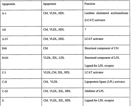
Table 1.4. Function o f Apoproteins 36
Table 1.5. Macrophage scavenger receptor ligands 45
Table 1.6. Plasma profile o f the major apolipoproteins in patients with CRF 53
Table 1.7. Factors that contribute to changes in triglyceride metabolism in 55
chronic renal insufficiency.
Chapter 2
Table 2.1. Comparison between parental human mesangial cells and the 82
immortalised human mesangial cell line
Chapter 3
Table 3.1. Effects o f PMA & Ang II on scavenger receptor promoter activity 148
in the presence of various signal transduction pathway inhibitors
Chapter 4
Tablet 4.1. Effect o f various signal transduction inhibitors on cytokines- 190
mediated luciferase activity driven by LDL receptor promoter
Chapter 5
Table 5.1. Effect o f various signal transduction inhibitors on CCBs- 225
LIST OF ABBREVIATIONS
ACAT Ac-LDL Ang n Apo AP-1 A-SAA bFGF BHT CBF CE CCBs CETP CHO CIAP CM CREBP CRF CyA Dil-Ac-LDL EDTA EGF ESRDAcyl Co A:Cholesterol acyl transferase
Acetylated LDL
Angiotensin n
Apolipoprotein
Activator protein-1
Acute-phase serum amyloid A proteins
Basic fibroblast growth factor
Butylated hydroxy toluene
CREBP binding protein
Cholesterol ester;
Calcium channel blocks
Cholesterol ester transfer protein
Chinese hamster ovary
Calf intestinal phosphatase
Chylomicrons
cAMP response element-binding protein
Chronic renal failure
Cyclosporin A
Ac-LDL labelled with l , l
’-dioctadecy-3,3,3’,3’,-tetramethylindocarbocyanine
Ethylene diamine tetra acetic acid
Epidermal growth factor
ET-1 FACS FSGS FSH GAPDH GM-CSF HDL Hep HMCL HMCL-Scr HMG CoA hsDNA EL-ip IGF LDL LCAT Lp(a), LPL LRP LOX-1 MFI M-CSF MCP-1 £ndothelin-l
Fluorescence-activated cell sorter analysis
Focal and segmental glomerulosclerosis
Follicle-Stimulating hormone
Glyceraldehyde phosphate dehydrogenase
Granulocyte monocyte colony stimulating
factor
High density lipoprotein
Heparin
Human mesangial cell line cells
Human mesangial cell line transfected by
human scavenger receptor full cDNA
3-Hydroxy-3-methyl glutaryl-Coenzyme A
Herring sperm DNA
Interleukin-ip
Insulin-like growth factor
Low density lipoprotein
Lecithin : cholesterol acyl transferase
Lipoprotein (a)
Lipoprotein lipase
LDL receptor-related protein
Lectin like Ox-LDL receptor
Mean fluorescence intensity
Monocyte colony stimulating factor
mOx-LDL m-CSF Ox-LDL P300: PBS PDGF PGL3SCR PGE pmp PMA Poly I PRD PPAR ROS RRT SCAP Scr SDS-PAGE ssDNA STAT SRE-1 SMC SRB-1
Mmimally oxidised LDL
Monocyte colony stimulating factor
Oxidised low-density lipoprotein
adenovirus E lA - associated protein
Phosphate buffered saline
Platelet derived growth factor
Scavenger receptor promoter reporter gene
Prostaglandin E
Per million population
Phorbol 12-myristate 13-acetate
Polyinosinic acid
Progression of renal disease
Peroxisome proUferator activated receptor
Reactive oxygen species
Renal replacement therapy
SREBP cleavage-activating protein
Scavenger receptors class A
Sodium dodecyl sulphate-polyacrylamide
Gel electrophoresis
Single strand DNA
Signal transducer and activator of
transcription
Sterol regulatoiy elem ent!
Smooth muscle cells
TG Triglyceride
TPA: 12-O-Tetradecanoyl-phorbol 13- acetate
TNF-a Tumour necrosis factor-a
TGF-P Transforming growth factor-g
TEARS Thiobarbituric acid reactive substances
VLDL Very low density lipoprotein
The incidence and prevalence of end-stage renal disease (ESRD) are increasing world
wide. Information available for the European Union from ERA-EDTA Registry for
1995 indicated that the stock of patients receiving renal replacement therapy (RRT)
was 644 per million population (pmp), with nearly 250,000 patients receiving
treatment in the European Union population of 373 million, with a mean expansion
rate of about 8.2 % per year (Berthoux et al. 1999). The RRT population of the
United States is approximately 300,000 against a background of 2 million patients
with chronic renal disease. The mean incidence of new patients starting RRT in
Europe is 120 pmp and the mean incidence of death among patients receiving RRT is
67 pmp (Berthoux et al. 1999). Considerable public health concern arises from the
high mortality rate and impaired quality of life of RRT patients. In the United
Kingdom RRT consumes nearly 2% of the NHS budget at a projected cost of £25,000
per patient per annum. Therefore, one of the major strategies of nephrology today is
to prevent the progression of renal disease (PRD) and extend the time to reach ESRD
and place patients on RRT.
In many patients decline in renal function occurs at a constant rate and may continue
despite the remission of the pathological processes which initiated kidney damage.
Studies over the last two decades have identified a large number of risk factors,
thought to be important in contributing to the rate of progression of chronic renal
disease and this has led to the formulation of several ideas which emphasise one
contributory factor or another. However, a unifying hypothesis is emerging which
indicates that ESRD progression occurs through a primary renal insult that
irreversibly damages a significant number of nephrons. In response to this decline in
glomerular hyperfiltration and hypertrophy (Hostetter, 1995). In the short term,
compensatory adaptations tend to normalise biochemical parameters of renal function.
However, other changes such as endotheUal injury, tubulo-interstitial damage,
hypertension, diabetes, hyperlipidaemia, platelet activation, oxidative stress and
proteinuria may cause long-term damage. In the long-term, factors contributing to
compensatory changes in the glomerulus and tubulo-interstitial damage may feed on
each other to create fiirther glomerular and tubulo-interstitial damage. El Nahas has
postulated the involvement of two pathways in the pathogenesis of
glomerulosclerosis: an intrinsic pathway involving infiltrating and resident glomerular
cells and a second extrinsic pathway involving the trans-differentiation of tubular
epithelial cells to myofibroblasts causing tubulo-interstitial damage (El Nahas, 1996).
From all these different ideas a consensus has emerged which suggests that over a
period of adaptive changes, glomerulosclerosis and tubular atrophy reduce nephron
number, fuelling a self-perpetuating cycle of nephron destruction culminating in
fibrosis, scarring and uraemia.
Abnormalities of lipid metabolism are seen in a variety of renal diseases, particularly in
those associated with nephrotic range of proteinuria. These lipid abnormalities persist
during all stages of renal disease and during RRT, including renal transplantation,
although the pattern of dyslipidaemia changes when patients move from one modality
of RRT to another (Chan et al. 1982). Cardiovascular disease is usually well
established by the time RRT starts, but is the most important cause of death at all
stages of PRD, accounting for approximately 50% of the mortality among patients on
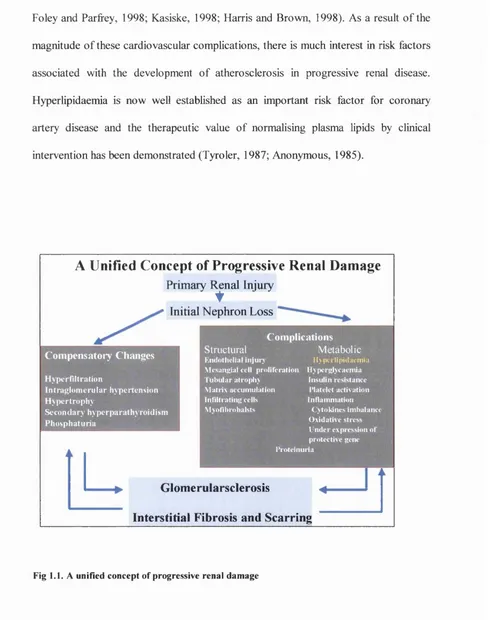
patients with ESRD is in far excess of that in control population (Spencer, 1980;
Foley and Parfrey, 1998; Kasiske, 1998; Harris and Brown, 1998). As a result of the
magnitude of these cardiovascular complications, there is much interest in risk factors
associated with the development of atherosclerosis in progressive renal disease.
Hyperlipidaemia is now well established as an important risk factor for coronary
artery disease and the therapeutic value of normalising plasma lipids by clinical
intervention has been demonstrated (Tyro 1er, 1987; Anonymous, 1985).
A Unified Concept of Progressive Renal Damage
Primary I^ n al InjuryInitial Nephron Loss
C o m p e n sa to iy C h an ges
Hyperfiltration
Intraglom erular hypertension H ypertrophy
Secondary hyperparathyroidism Phosphaturia
C o m p lica tio n s M etabolic
Ily p crlip id a cm iu
Structural
K n d oth elial I n ju n
Mcsanjpal cell proliferation liypcrglvcaem ia
tub u lar atrophy Matrix accum ulation Infiltrating cells M vofibrobalsts
Insulin resistance Platelet activation Inflammation
( yloldnes Imbalance O xidative stress I nder expression o f protective gene Proteinuria
Glomerularsclerosis ^
Interstitial Fibrosis and Scarring
Atherosclerotic renal artery stenosis is a contributory factor for ESRD and is
particularly significant in the elderly group of patients starting RRT (Scoble et al.
1989). However, only limited studies directed at the treatment of the lipid
abnormalities of chronic renal failure patients are available, and major efforts are
needed in controlling hyperlipidaemia in this vulnerable group o f patients. It is logical
to pose the question: is hyperlipidaemia the cause or the consequence of renal
diseases? Before the publication of the lipid nephrotoxicity hypothesis of Moorhead
and colleagues in 1982 (Moorhead et al. 1982), it was generally considered that
abnormal lipid metabolism was a consequence of renal disease. They pointed out that
in some circumstances, circulating lipoproteins may directly damage glomerular
structures and suggested that hyperlipidaemia is an aggravating factor in the
progression of initial glomerular injury to glomerulosclerosis. A large number of
animal experiments, cell culture and clinical studies have suggested an association
between abnormal lipid metabolism and PRD. In both the remnant kidney model and
in obese Zucker rats, lowering lipids with drugs lessens injury.
In the rest of this introductory chapter 1 will briefly discuss normal lipoprotein
metabolism, lipoprotein abnormalities in renal diseases, involvement of lipids in PRD,
and the pathogenesis of lipoprotein-induced glomerulosclerosis. Finally the specific
purpose of the project will be discussed.
1.1. NORMAL LIPOPROTEIN METABOLISM
macromolecular complexes called lipoproteins. Five major classes of lipoproteins are
recognised; chylomicrons (CM), very low density lipoprotein (VLDL), low density
lipoprotein (LDL), Lipoprotein (a) (Lp(a)), and high density lipoprotein (HDL).
These five classes of lipoproteins are heterogeneous in terms of their size, lipid and
apoprotein content and can be fractionated by using techniques such as
electrophoresis, gradient ultracentrifugation and affinity chromatography (Table 1.1).
Typical values for the major composition of the five classes of lipoproteins are shown
in Table 1.2 and the apo content is described in Table 1.3. (Illingworth, 1993). The
main triglyceride-carrying lipoproteins are chylomicrons and VLDL. Chylomicrons
are not normally present in blood after a 12 h fasting. In the fasting state VLDL
account for approximately 60% of the total plasma triglyceride (Powell et al. 1987;
Young, 1990; Illingworth, 1993). Lp(a) is an LDL-like particle in which apo-BlOO is
linked to another apoprotein-apo(a) (Kronenberg et al. 1996).
1.1.1. Lipoproteins, lipolytic enzymes and transfer proteins.
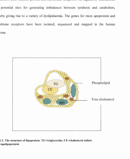
Lipids, being insoluble in aqueous solution, are transported in plasma in association
with specialised proteins. Lipoproteins are composed of an inner core of non-polar
neutral lipids (esterihed cholesterol and triglyceride) surrounded by an outer coating
of polar molecules (phospholipids, free cholesterol and apoproteins) (Fig. 1.2). They
are synthesised in the liver and intestine and transport dietary and endogenously
synthesised lipids in the circulation.
Lipid synthesis and assembly into lipoproteins, and the transport, storage and
cholesterol ester transfer protein and membrane receptors. All regulatory mechanisms
are potential sites for generating imbalances between synthesis and catabolism,
thereby giving rise to a variety of dyslipidaemia. The genes for most apoprotein and
membrane receptors have been isolated, sequenced and mapped in the human
genome.
Phospholipid TG
CE
Free cholesterol apo
Fig. 1.2. The structure of lipoprotein. TG=trig!yceride; CE=cholesterol ester ;
apo=apolipoprotein
Chylomicrons (CM) are rich in triglycerides derived from food. They are synthesised
in small intestine, then secreted into the lymphatics and subsequently enter the
circulation. They become emulsified through the addition of apo-B48, apo-AI, apo-
AII qpd apo-AIV, then acquired apo-CI, -CII, -CIII and apo-E fi*om HDL. They are
chylomicrons are removed by the action of lipoprotein lipase (LPL). Apo-CII
activates LPL and apo-CIII inhibits this enzyme. Some apoproteins namely apo-AI
and apo-AII are transferred to HDLs during this delipidation process and the partially
delipidated chylomicrons are known as chylomicron remnants. The chylomicron-
mediated pathway represents exogenous lipoprotein metabolism (Windier and Havel,
1985).
VLDL also is triglyceride-rich lipoprotein which synthesised in the liver. The
delipidation pathway of VLDL is similar to that of chylomicrons in that LPL on the
endothelial surface concerts VLDL to IDL. About 50% of IDL are removed directly
by the liver and remaining fraction is further delipidated by hepatic lipase and
converted to LDL (Young, 1990). During the delipidation process of VLDL, surface
phospholipids and apoproteins are transferred to HDL as they are from chylomicrons.
Cholesteryl ester, which is formed in the plasma as a result of the action of
lecithin;cholesterol acyl transferase (LCAT) on the HDL particle, is transferred from
HDL to VLDL through the mediation of cholesterol ester transfer protein (CETP).
LDL is the major lipoprotein fraction of fasting plasma and it carries approximately
75% of the total cholesterol in plasma. A single molecule of apo-BlOO is the only
apoprotein present in LDL. In normal situation, there is a substrate product
relationship between VLDL and LDL and almost all LDL apo-B can be accounted for
as a product of VLDL metabolism.
HDL particles are synthesised and secreted by the liver and the small intestine. Newly
apo-AI and apo-E. This is an ideal substrate for LCAT which converts free
cholesterol, acquired from extrahepatic tissue and other lipoproteins, into cholesteryl
esters. During this process HDL becomes a spherical particle having cholesteryl ester
as its core lipid. HDL particles undergo a series of remodelling processes in the
plasma. In normal human plasma, the major species are the smaller HDL3 and the
larger HDL2. The continued LCAT activity in HDL3 converts these particles to
cholesteryl ester rich HDL2. This mature HDL2 is necessary for the transport of the
lipoprotein lipase activator apo-CII to nascent chylomicrons and VLDL. CETP can
exchange cholesteryl ester from HDL for triglycerides in VLDL and IDL. Hepatic
lipase can act on HDL triglycerides and convert HDL2 to HDL3. Thus during the
hydrolysis of chylomicrons and VLDL, excess surface components are transferred to
HDL2 and this increases the size of these particles. In addition, VLDL triglycerides
can exchange with cholesteryl esters in the presence of CETP and the hydrolysis of
resulting HDL2 triglycerides by hepatic lipase can cause a decrease in HDL2 size and
convert HDL2 to HDL3. Therefore, HDL is a key player in modulating the
delipidation cascade of chylomicrons, VLDL and DDL, and also provides a medium
for the reverse transport of cholesterol from extrahepatic tissue to the liver.
1.1.2. Lipoprotein receptors
The removal of lipoprotein from the circulation occurs largely by a receptor-mediated
process in the liver and extrahepatic tissues. Evidence for the existence of cell surface
receptors for apo-B was first provided by Goldstein and Brown in studies of human
skin fibroblasts (Brown and Goldstein, 1975). Over last two decades, a number of
protein/alpha2-macroglobulin receptor (LRP). As the present study is designed to
investigate involvement of lipoprotein receptors in lipid accumulation in the
progression of renal dysfunction using HMC in culture, a brief description of current
knowledge of lipoprotein receptors is therefore apposite.
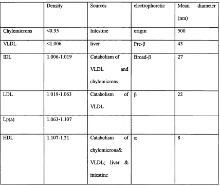
Table 1.1. The five m ajor density classes of lipoproteins
Density Sources electrophoretic Mean diameter
(nm)
Chylomicrons <0.95 Intestine origin 500
VLDL <1.006 liver Pre-p 43
IDL 1.006-1.019 Catabolism o f
VLDL and
chylomicrons
Broad-P 27
LDL 1.019-1.063 Catabolism of
VLDL
P 22
Lp(a) 1.063-1.107
HDL 1.107-1.21 Catabolism of
chylomicrons&
VLDL; liver &
intestine
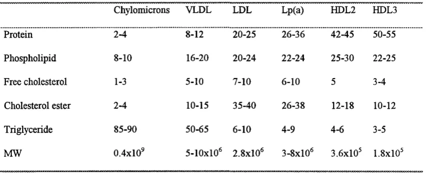
Table 1.2. Composition of human plasma lipoproteins.
(% of total dry w eight of the lipoprotein)
Chylomicrons VLDL LDL Lp(a) HDL2 HDL3
Protein 2-4 8-12 20-25 26-36 42-45 50-55
Phospholipid 8-10 16-20 20-24 22-24 25-30 22-25
Free cholesterol 1-3 5-10 7-10 6-10 5 3-4
Cholesterol ester 2-4 10-15 35-40 26-38 12-18 10-12
Triglyceride 85-90 50-65 6-10 4-9 4-6 3-5
MW 0.4x10^ 5-10x10^ 2.8x10® 3-8x10® 3.6x10® 1.8x10®
Table 1.3. Apoprotein content of human plasm a lipoproteins
Chylomicrons VLDL LDL L p(a) HDL
M ajor apoproteins
Apo B-48 Apo B-lOO Apo B-100 Apo B-100
Apo C-I Apo C-I Apo (a)
Apo C-II Apo C-II
Apo C-III Apo C-III
Apo E Apo E
M inor apoproteins
Apo A-I Apo D Apo C-I
Apo A-II Apo C-II
Apo A-IV Apo C-III
Table 1.4. Function of Apoproteins
Apoprotein lipoprotein Function
A-1 CM, VLDL, HDL Lecithin cholesterol acyltransferase
(LCAT) activator
A ll CM, VLDL, HDL ?
A-IV CM, VLDL, HDL LCAT activator
B48 CM Structural component of CM
BlOO VLDL, IDL, LDL Structural component of LDL
Ligand for LDL receptor
C-I VLDL,CM, IDL, HDL LCAT activator
C-II CM, VLDL Lipoprotein lipase (LPL) activator
C-III CM, VLDL, IDL, HDL Inhibitor of LPL
L I V E R
H D L
n H D L
C M R
C M S U R F A C E C O M P O N E N T S
F F A
I N T E S T I N E
M U S C L E & A D I P O S E T I S S U E
Figure 1.3. Chylomicron metabolism and HDL chylomicron interactions
HDL2 = HDL with a density between 1.063 g/ml and 1.125 g/m l; HDL3 = HDL with a density
between 1.125 and 1.21 g/m l; FFA = free fatty acids, LPL= lipoprotein lipase; C ETP = cholesterol
FFA
Liver
B-lO O
ipose Tissue VLDL
A cetate B -100
Apo E mediiated Uptake olesterol
B -lO O
Apo B mediiated
Uptake VLDL
H TGL
Remnant
B-lO O
Tissues
L D L re c e p to r
LDL C atabolism ^ HM G - CoA 1 release o f J^ reductase ▼ cholesterol 1
C holesterol A estérification '
LDL receptor ^
Figure 1.4. VLDL and LDL metabolic pathways and the consequences of receptor mediated LDL uptake
FFA=free fatty acids; LPL=lipoprotein lipase; HDL2=F1DL with a density between 1.063 and 1.125
g/m l; HM G-CoA = 3-hydroxy-3-m ethylglutaryl-coenzym e A; PL=phospholipid; TG =triglyceride;
1.1.2.1 The LDL receptor
The LDL receptor is the primary receptor for binding and internalising plasma-derived
LDL-cholesterol and regulates plasma LDL (Goldstein and Brown, 1985; Brown and
Goldstein, 1986a). In normal lipidaemic subjects 60-80% of LDL is removed through
the LDL receptor pathway. Defect of LDL receptor in familial hypercholesterolaemia
(FH) patients leads to raise level of plasma cholesterol and atherosclerosis. The
transport of macromolecules into cells by receptor-mediated endocytosis first
emerged as a distinct mechanism following studies carried out on human fibroblasts
by Brown and Goldstein (Brown and Goldstein, 1975). On exposure of human
fibroblasts to high concentrations of LDL, cellular endogenous cholesterol synthesis
was decreased while the intracellular content of cholesterol remained largely
unchanged. Following further biochemical studies it emerged that in mammals, the
delivery of LDL-derived cholesterol into hepatic and extra-hepatic cells was mediated
by a specific cell surface receptor known as the apo B/E receptor. The sequential
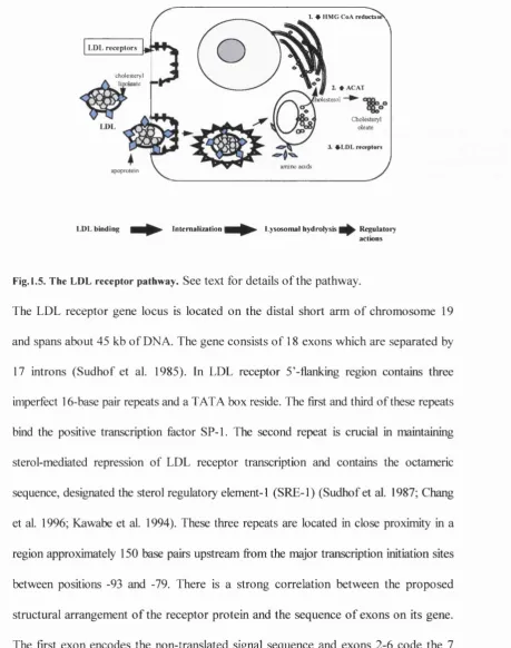
process of receptor-mediated endocytosis of LDL and the subsequent regulation of
cellular synthesis of cholesterol has been termed the LDL receptor pathway. This
important receptor pathway is represented schematically in fig. 1.5. It depicts the
events that are involved in the receptor-mediated endocytosis of LDL, which allows
cells to control their intracellular cholesterol content and establish intracellular
1. * H M G CoA reductases
LDL receptors
cholesteryl leate
2. * ACAT olesterol
C holesteiyl d e a te
3. * L D L receptors
am ino acids apoprotein
LDL binding Internalization I Lysosomal hydrolysis Regulatory
actions
Fig. 1.5. The LDL receptor pathway. See text for details of the pathway.
The LDL receptor gene locus is located on the distal short arm of chromosome 19
and spans about 45 kb of DNA. The gene consists of 18 exons which are separated by
17 introns (Sudhof et al. 1985). In LDL receptor 5'-hanking region contains three
imperfect 16-base pair repeats and a TATA box reside. The first and third of these repeats
bind the positive transcription factor SP-1. The second repeat is crucial in maintaining
sterol-mediated repression of LDL receptor transcription and contains the octameric
sequence, designated the sterol regulatory element-1 (SRE-1) (Sudhof et al. 1987; Chang
et al. 1996; Kawabe et al. 1994). These three repeats are located in close proximity in a
region approximately 150 base pairs upstream from the major transcription initiation sites
between positions -93 and -79. There is a strong correlation between the proposed
structural arrangement of the receptor protein and the sequence of exons on its gene.
The first exon encodes the non-translated signal sequence and exons 2-6 code the 7
cysteine-rich repeats of the LDL binding domain (Sudhof et al. 1985; Schneider,
1989). The next 8 exons code for the EOF precursor homology domain and the third
between introns 14 and 15. The membrane spanning and cytoplasmic domains are
encoded by 2 exons, and the 18*^ exon is translated into the carboxyl end amino acids
of the receptor protein and also contains a 2.5 kb non-translated stretch of mRNA
(Yamamoto et al. 1984; Schmid and Jelinek, 1982).
The structure of the LDL receptor from four species (human, rabbit, bovine and
hamster) has been well characterised (Schneider et al. 1982) (Fig. 1.6.). It is a highly
conserved integral membrane glycoprotein with five main domains. These domains
listed in order of the appearance from the amino terminus of the protein are: 1) The
LDL binding domain, 2) a domain which has a strong homology to the epidermal
growth factor (EGF) precursor, 3) a domain in which there is a cluster of o-linked
carbohydrate chains, 4) a transmembrane domain and 5) a short region that extends
into the cytoplasm (Schneider, 1989).
A detailed description of LDL receptor regulation is given in Chapter 4.
NH2 I I n I M i- T ^ A A
3 4 5
Homology with EGF- O -linked M em brane Cytoplasm ic Cysteine-rich precursor sugars spanning tail repeats
Extracellular Intracellular
1.1.2.2 Scavenger receptors class A
Brown and Goldstein found that cholesterol uptake by the LDL receptor pathway did
not lead to massive accumulation of cholesterol in cells because the uptake was tightly
coupled to the concentration of intracellular cholesterol. They noticed however, when
LDL was chemically modified, macrophages in culture were able to accumulate large
amounts of lipid and convert into cholesterol ester droplet-filled cells. These cells
show a striking morphological similarity to foam cells found in atherosclerotic plaques
(Goldstein et al. 1983; Brown and Goldstein, 1983; Brown and Goldstein, 1986a;
Goldstein and Brown, 1977). These receptors were first termed acetyl LDL receptors
but are now known as macrophage scavenger receptors because of their multi-ligand
binding capacity.
Scavenger receptor class A cDNA has been cloned in bovine, mouse, rabbit and
human. Human scavenger receptor genomic DNA is located on chromosome 8. Two
mRNA, 4.0 and 3.2 kb, have been detected in human liver, placenta, and brain
(Matsumoto et al. 1990). The two isoforms of human scavenger receptor class A, SR-
AI and SR-AII are produced by alternative splicing of a message encoded by a single
gene located on chromosome 8 in humans (Kodama et al. 1990; Naito et al. 1992;
Matsumoto et al. 1990).
Analysis of the ligand-binding properties of scavenger receptor activities on
macrophages and endothelial cells lead to the suggestion that there are multiple
classes of scavenger receptors. Scavenger receptor class A was identified as the first
family in 1990 (Kodama et al. 1990; Rohrer et al. 1990). Two types of scavenger
1991; Matsumoto et al. 1990). Although their normal physiological role remains
uncertain, biochemical studies have demonstrated that both isoforms of scavenger
receptor class A are capable of binding and internalising acetylated LDL (Ac-LDL)
and oxidised LDL (Ox-LDL) (Kodama et al. 1990; Goldstein et al. 1979; Freeman et
al. 1991; Steinbrecher et al. 1989; Parthasarathy et al. 1986). Unlike LDL receptor,
the activity of scavenger receptor is not suppressed by rising intracellular cholesterol
concentrations, thus providing a mechanism for unregulated cholesterol uptake. The
massive accumulation of cholesterol in foam cells present in atherosclerotic plaques is
thought to involve scavenger receptors class A. Several lines o f evidence support this
view: firstly, macrophage-like cells, and CHO cells transfected with scavenger class A
receptor accumulate modified LDL and become lipid-laden foam cells in vitro
(Freeman et al. 1991); secondly, scavenger receptor class A mRNA is expressed and
the ligand for scavenger receptor class A (Ox-LDL) is present in atherosclerotic
plaques (Hiltunen et al. 1998; Hiltunen and Yla-Herttuala, 1998); finally, the
antioxidant drug probucol inhibits formation of atherosclerotic plaques in animal
models of atherosclerosis (Donetti et al. 1998; Braesen et al. 1995).
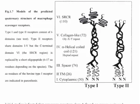
Type I receptors made up of 451-454 amino-acids with an elongated homotrimeric
integral membrane protein structure. This protein is organised as 6 distinct domains
(Fig 1.7.).
I: The N-terminal cytoplasmic domain [amino acid (aa) residues 1-50].
II: A single transmembrane domain per chain (aa 51-76).
Ill: A spacer region (aa 77-150).
V: A second coiled-coil domain composed of a right handed, collagenous triple helix
containing 23 or 24 uninterrupted Gly-X-Y triplet repeats (aa 272-343).
VI: A C-terminal cysteine-rich domain (SRCR) which is thought to fold into a
globular structure (aa 344-453).
Type II receptors have domains I -V found in type I receptors but lack the cysteine-
rich domain VI which is replaced by a truncated C-terminus consisting of 6-17 aa.
Although type II receptors lack SRCR they still have a broad ligand-specificity
suggesting that this region is not essential to ensure binding of multiple ligands.
Fig. 1.7 Models of the predicted
quaternary structure of macrophage
scavenger receptors.
Type I and type II receptors consist of 6
dom ains (see text). Type II receptors
share dom ains I-V but the C -term inal
dom ain VI (the SRCR region) is
replaced by a short oligopeptide (6-17 aa
residues depending on the species). The
aa residues o f the bovine type I receptor
are indicated in parenthesis.
VI. SRCR
(110)
V. Collagen-like (72)
Gly-X-Y repeat
IV. a-Helical coiled coil (121)
Heptad repeat
III. Spacer (74)
II.TM (26)
I. Cytoplasmic (50)
N N N
N N N
Type I
Type II
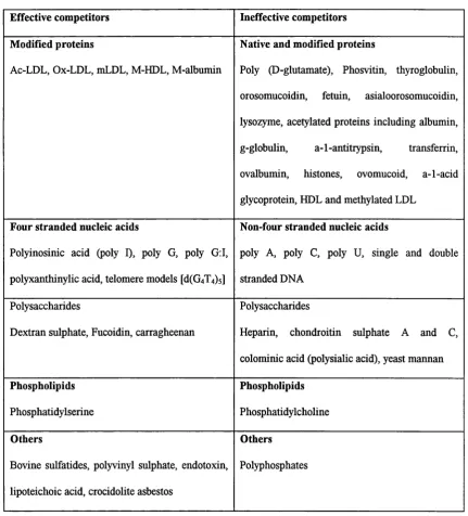
Initial studies indicated that scavenger receptors class A found on mouse peritoneal
macrophages were capable of high affinity binding, internalisation and degradation of
competitively inhibit this binding. Using direct binding assay and competitive
inhibition studies a variety of compounds that can bind scavenger receptor class A
with a high affinity have been identified (Table. 1.5.). Ligands that bind macrophage
scavenger receptor class A identified so far are either polyanionic molecules or
macromolecular complexes. The regulation of scavenger receptor class A is discussed
in chapter 3.
Table 1.5. M acrophage scavenger receptor ligands
Effective com petitors Ineffective competitors
M odified proteins
Ac-LDL, Ox-LDL, mLDL, M-HDL, M-albumin
Native and m odified proteins
Poly (D-glutamate), Phosvitin, thyroglobulin,
orosomucoidin, fetuin, asialoorosomucoidin,
lysozyme, acetylated proteins including albumin,
g-globulin, a-1-antitrypsin, transferrin,
ovalbumin, histones, ovomucoid, a-1-acid
glycoprotein, HDL and methylated LDL
Four stranded nucleic acids
Polyinosinic acid (poly I), poly G, poly 0:1,
polyxanthinylic acid, telomere models [d(G4U )5]
Non-four stranded nucleic acids
poly A, poly C, poly U, single and double
stranded DNA
Polysaccharides
Dextran sulphate, Fucoidin, carragheenan
Polysaccharides
Heparin, chondroitin sulphate A and C,
colominic acid (polysialic acid), yeast maiman
Phospholipids
Phosphatidylserine
Phospholipids
Phosphatidylcholine
Others
Bovine sulfatides, polyvinyl sulphate, endotoxin,
Others
1.1.2.3 Scavenger receptors class B
The class B scavenger receptor family is divided into two: CD36 and scavenger
receptor class B-1 (SRB-1). Endemann and colleagues reported the identification of
the first class B scavenger receptor, CD36 (Endemann et al. 1993). The cell surface
protein of CD36 family was shown to bind modified lipoprotein proteins (acetylated
LDL, oxidised LDL), but not the broad array of other polyanions which are ligands of
the class A receptors (Acton et al. 1994; Endemann et al. 1993). CD36 may play a
quantitatively significant role in modified LDL binding to macrophages (Endemann et
al. 1993). In addition to binding modified LDL, CD36 binds thrombospondin (Asch et
al. 1987), anionic phospholipids (Rigotti et al. 1995), long-chain fatty acids (Abumrad
et al. 1993), collagen (Tandon et al. 1989), and plasmodium falciparum-infected
erythrocytes. CD36 is expressed in a variety of tissues, including adipocytes,
macrophages, epithelial cells, monocytes, endothelial cells, platelets, and a wide
variety of cultured lines (Abumrad et al. 1993). CD36 has been reported to be
clustered in specialised domains of the plasma membrane (Lisanti et al. 1994).
Although the physiological functions of CD36 have not been fully described, it may
serve as an adhesion molecule owing to its collagen-binding properties. CD36 may
also serve as a receptor on macrophages for damaged or senescent neutrophils (Savili
et al. 1992).
Another member of the class B scavenger receptor SR-BI was isolated by expression
cloning from a Chinese hamster ovary cell variant Var-261, which expresses a
scavenger receptor activity distinct from that scavenger receptor class A (Acton et al.
1994). The protein sequence of SR-BI (509 amino acids) is approximately 30%
affinity binding for acetylated LDL, oxidised LDL, maleylated bovine serum albumin,
but not the broad array of other polyanions (e.g. fucoidin, polyguanosinic acid,
carragheenan) which are ligands of the class A receptors. It also binds anionic
phospholipid suggesting that SR-BI might be involved in recognising senescent or
apoptotic cells (Acton et al. 1994; Rigotti et al. 1997).
Recently SR-BI has been identified as HDL receptor (Acton et al. 1996; Stangl et al.
1998). Cholesteryl ester delivery from HDL to cells through SR-BI receptor pathway
is fundamentally different from that of the LDL receptor pathway, because it does not
involve endocytosis and degradation of the entire lipoprotein particle (Krieger, 1999).
Instead, HDL binds to the cell surface and transfer cholesteryl esters to the cell and
then the lipid-depleted HDL dissociates from the cell surface and re-enters the
circulation. This novel receptor exchange mechanism for HDL cholesterol uptake is
called selective lipid uptake (Krieger, 1999). Human SR-BI has been mapped to
human chromosome 12 (12q24.2-qter) (Acton et al. 1994). The partial and complete
genomic structures for the murine and human SR-BI homologues have been reported
(Cao et al. 1997). Northern blot analysis of murine tissues showed that SR-BI was
most abundantly expressed in adipose tissue and was present at moderate levels in
lung and liver. Furthermore, SR-BI mRNA expression was induced upon
differentiation of 3T3-L1 cells into adipocytes. Thus, the tissue distribution of
expression and ligand binding properties of SR-BI raise the possibility that this cell
1.1.2.4 Lectin like receptor - a novel endothelial receptor for Ox-LDL
Vascular endothelial cells in culture and in vivo internalise and degrade Ox-LDL
through a putative receptor-mediated pathway that does not involve macrophage
scavenger receptors. Sawamura reported a molecular cloning of an Ox-LDL receptor
from vascular endothelial cells. The cloned receptor is a membrane protein that
belongs structurally to the C-type lectin family, and is expressed in vivo in vascular
endothelium and vascular-rich organs (Sawamura et al. 1997). Lectin like Ox-LDL
receptor (LOX-1) is a receptor for Ox-LDL but not for Ac-LDL. LOX-1 recognizes
protein moiety of Ox-LDL, and its ligand specificity is distinct from other receptors
for Ox-LDL, including class A and B scavenger receptors (Moriwaki et al. 1998).
1.1.2.5 VLDL receptor
The VLDL receptor has been described as a new member of the LDL receptor
supergene family. The predicted human VLDL receptor protein shows approximately
75% sequence homology to LDL receptor proteins (Webb et al. 1994). Isolation and
characterisation of cDNAs encoding human very low density lipoprotein (VLDL)
receptor revealed the presence of two forms of the receptor: one consists of five
domains that resemble the LDL receptor, and a variant form which lacks an 0-linked
sugar domain (Sakai et al. 1994). The ligands for VLDL receptor are apolipoprotein
E-rich lipoproteins such as p-migrating VLDL, IDL, chylomicron remnants, LRP
receptor-associated protein (RAP). In human tissues in vivo, the mRNA was
expressed predominantly in heart and skeletal muscle, and also in ovary and kidney,
but not in the liver (Takahashi et al. 1992). Based on the structural features, ligand
of apolipoprotein E-containing lipoproteins enriched with triglyceride in non-hepatic
tissues that are active in fatty acid metabolism (Takahashi et al. 1992).
1.1.2.6 The LDL receptor-related protein.
LDL receptor-related protein (LRP) is primarily expressed in the liver, brain and
placenta. The cDNA for this receptor was cloned in 1988 using a homology screening
approach. The predicted protein structure of this receptor includes many structural
motifs found in the LDL receptor. These include clusters of ligand binding
(complement-type) domains which contain repeats of cysteine-rich regions, cysteine-
rich EGF repeats, EGF-precursor homologous domains (Herz et al. 1988). The main
ligands for LRP are thought to be apo E containing lipoprotein “remnants” that are
formed from VLDL and chylomicrons by modifications caused by removal or addition
of apoproteins by the action of LPL. There is strong evidence however that LRP is
also a multiligand receptor for other important ligands such as P-VLDL, lactoferrin,
az-macroglobulin and complexes of plasminogen activator/inhibitor (Krieger and
Herz, 1994).
1.1.2.7. Lipoprotein receptor in mesangial cells
Lipoprotein receptors have variable degree of expression in different tissues and cell
types. Its expression, centrally, in the hepatic tissue is important in the modulation of
the concentration of plasma lipids. However, the expression of various lipoprotein
completely elucidated. Our group have previously showed that human and rat
mesangial cells have LDL receptors (Wheeler et al. 1991a; Wheeler et al. 1990b).
Furthermore we showed that rat mesangial cells have the ability to oxidise native LDL
and they have receptors for modified LDL (Fernando et al. 1993). Wanner and Anami
demonstrated that human mesangial cells (HMC) express VLDL receptors (Anami et
al. 1997; Quaschning et al. 1997). The evaluation of the expression and regulation of
these receptors on peripheral tissues may help in the understanding of the influence of
lipid-mediated injury.
1.2. LIPOPROTEIN ABNORMALITIES FOUND IN RENAL DISEASE
1.2.1 Nephrotic syndrome
The nephrotic syndrome is characterised by proteinuria of greater than 3.5 g/day,
hypoproteinaemia, oedema, and hyperlipidaemia and is a result of an alteration in
glomerular filtration barrier selectivity and permeability (Cameron, 1987). The
nephrotic plasma lipid profile is characterised by an increase in plasma cholesterol
concentration and an elevated plasma triglyceride levels, particularly in patients with
heavy proteinuria (>10 g/day). This characteristic profile is a result of an increase in
LDL, VLDL and/or IDL particles (Joven et al. 1990) and a decreased or unchanged
level o f the HDL fi*action (Joven et al. 1990; Kaysen, 1991). There is also evidence
that the relative levels of HDL subtypes also undergo change in the nephrotic patient
(Muls et al. 1985). The HDL3:HDL2 ratio increases due to a small elevation in HDLg
levels and a more dramatic decrease in HDL2 levels (Muls et al. 1985; Short et al.
factor together with the increase in VLDL, DDL and LDL cholesterol puts these
patients in to a group which has a high risk of developing premature cardiovascular
disease (Miller et al. 1981).
Qualitative and quantitative changes in the composition of the plasma lipoprotein
fractions in nephrotic syndrome increased ratio o f cholesterol to TG and of free
cholesterol, cholesterol esters and phospholipid to protein in these fractions (Gherardi
et al. 1977). There is also an accumulation in the plasma of lipoprotein particles that
are similar to VLDL, DDL and CM remnants which are rich in esterified and non-
esterified cholesterol and phospholipids. ApoB and C-III levels increase in nephrotic
syndrome while apo A-I, A-II and C-D remain unchanged (Joven et al. 1990). Apo C-
II is an essential co-factor involved in LPL activity and apo C-III is a competitive
inhibitor of the action of apo C-II. The increase in the apo Clll/apo C-II ratio may
reduce LPL activity resulting in a delayed clearance of triglyceride rich lipoprotein
particles (Brown and Baginsky, 1972).
There is general agreement that the hyperlipidaemia of the nephrotic syndrome is a
result of both increased hepatic synthesis of lipids and apolipoproteins (Makar et al.
1998) and decreased lipoprotein catabolism (Garber et al. 1984). Low plasma oncotic
pressure and viscosity are thought to be the reason for increasing the synthesis of
albumin and apoprotein B (Yedgar et al. 1982; Conwill et al. 1977). Delayed
clearance of lipoproteins due to defective catabolism may also cause hyperlipidaemia
(Garber et al. 1984; Staprans et al. 1987; Davies et al. 1990; Mene et al.