OncoTargets and Therapy 2018:11 4367–4375
OncoTargets and Therapy
Dove
press
submit your manuscript | www.dovepress.com 4367
S T u dy P r O T O c O l
open access to scientific and medical research
Open Access Full Text Article
Elevated expression of Tiam1 is associated with
poor prognosis and promotes tumor progression
in pancreatic cancer
Mina ding1
yue li1
yang yang2
Kun Zhu1
Shuanlong che2
Zhenhua lin2,3
liyan chen1,3
1department of Biochemistry and
Molecular Biology, yanbian university Medical college, yanji 133002, china;
2department of Pathology and cancer
research center, yanbian university Medical college, yanji 133002, china;
3department of Jilin Province, Key
laboratory of the Science and Technology, yanji 133002, china
Objective: T-cell lymphoma invasion and metastasis inducing factor 1 (Tiam1) is known to be involved in tumor progression. However, its molecular roles and mechanism in pancreatic ductal adenocarcinoma (PDAC) remain unclear. The purpose of this study is to determine Tiam1 expression levels and investigate its underlying molecular mechanism in PDAC.
Materials and methods: Tiam1 protein expression levels in PDAC tissues were examined using immunohistochemistry. Tiam1 expression was confirmed in pancreatic cancer (PC) cells by Western blot and immunofluorescence staining. Tiam1-silenced PC cells were created using short interfering RNA. Subsequently, colony formation, scratch, and migration and invasion assays were carried out to explore the molecular mechanisms of Tiam1 in PC cells.
Results: The results indicated that Tiam1 expression was significantly higher in PDAC tissues than in paired non-tumor tissues, and overexpression of Tiam1 was significantly correlated with histological grade (P=0.040) and lymph node metastasis (P=0.031) in PDAC. The PDAC patients with high Tiam1 expression had significantly lower 5-year overall survival than patients with low Tiam1 expression. More importantly, univariate and multivariate analysis suggested that Tiam1 expression, along with lymph node metastasis, is a significant independent prognostic factor for patients with PDAC. Furthermore, we also demonstrated that the downregulation of Tiam1 was associated with decreased cell proliferation and reduced migratory and invasive capability.
Conclusion: High expression of Tiam1 plays a significant role in the progression of PDAC and may be a potential biomarker of poor prognosis as well as a therapeutic target.
Keywords: Tiam1, pancreatic cancer, prognosis, survival analysis, carcinogenesis
Introduction
Pancreatic cancer (PC) is a highly lethal malignancy in which mortality closely parallels incidence. Pancreatic ductal adenocarcinoma (PDAC) is the most universal type of
PC, and it has a 5-year survival rate of 5%.1 Currently, although great improvements
in the diagnosis of PDAC have been made, the prognosis of PDAC patients is still generally poor. Tumor progression and metastasis are the major causes of mortality in
PDAC patients.2,3 The high propensity of this cancer is included in the major causes
due to the lack of progress in improving the prognosis for chemoresistance and early
distant metastasis.4 Furthermore, the molecular mechanisms that underlie these events
continue to be unclear.
T-cell lymphoma invasion and metastasis inducing factor 1 (Tiam1), a member of the guanine nucleotide exchange factors family that mediates the specific activation of Rac1, was first discovered by Habets et al as an invasion and metastasis-related gene
in mice with aggressive T-cell lymphoma.5–8 The Tiam1 gene is located in the q22
correspondence: liyan chen department of Biochemistry and Molecular Biology, yanbian university Medical college, No. 977 Gongyuan road, yanji 133002, china
Email lychen@ybu.edu.cn
Journal name: OncoTargets and Therapy Article Designation: Study Protocol Year: 2018
Volume: 11
Running head verso: Ding et al
Running head recto: High Tiam1 plays a significant role in the progression of PDAC DOI: 171425
OncoTargets and Therapy downloaded from https://www.dovepress.com/ by 118.70.13.36 on 25-Aug-2020
For personal use only.
Dovepress
ding et al
band of chromosome 21 and the centromeric end of the Aml 21 gene, and it contains 2 exons separated by 1 intron; the 2
exons were ~7.3 kb, and the intron was 14 kb. Previous
stud-ies have showed that Tiam1 is mainly expressed in normal brain and testis tissues and is minimally or not expressed in
other normal tissues.9 In recent years, it has been confirmed
that upregulation of Tiam1 expression is implicated in the
formation of solid tumors, such as esophageal carcinoma,10,11
lung adenocarcinoma, and breast cancer.12,13 The potential
prognostic relevance of Tiam1 expression in PDAC, how-ever, has not been adequately explored.
In the present study, we concentrated on the analysis of alterations in Tiam1 expression in PDAC to clarify its associations with clinicopathological features and prognosis and identify the mechanism by which Tiam1 contributes to proliferation and epithelial–mesenchymal transition (EMT) events. These findings may provide a potential therapeutic target for the treatment of PDAC.
Materials and methods
Patients and specimens
Tissue samples from patients with PDAC tissues and paired adjacent paracancerous tissue samples were acquired from 81 patients. These patients were diagnosed at the Yanbian Hospital, China, and recruited in the present study. This study was approved by the Yanbian University’s ethics committee. Among the 81 PDAC patients, 33 were females and 48 males with age ranging from 38 to 85 years (median, 59 years). The average time of survival was 28.2 months for these patients, and it ranged from 1 to 81 months. The histological types were assigned according to the criteria of the World Health Organization Classification. No patients in this study received any adjuvant systemic treatment ahead of surgery. The subjects or their caregivers provided written informed consent.
cell lines
The human PC cell BxPC-3, SW1990, Mia-paca-2, AsPC-1, and Capan-1 were supplied by the Cancer Research Center of Yanbian University, which were approved by Yanbian University’s ethics committee. The cells were seeded in high glucose DMEM (Gibco, Grand Island, NY, USA), contain-ing 10% fetal bovine serum (FBS) (HyClone Laboratories Inc., Novato, CA, USA). And then, the cells were cultured
at 37°C in 5% CO2 incubator.
Immunohistochemistry and scoring
Immunohistochemistry staining was confirmed to study altered protein expression in PC and paracancerous tissues. Briefly, all paraffin sections were dewaxed, dehydrated,
and rehydrated. Anti-Tiam1 antibody (1:100; Santa Cruz Biotechnology Inc., Santa Cruz, CA, USA) was
incu-bated with the sections and incuincu-bated at 4°C overnight.
Following incubation with biotinylated secondary antisera, the streptavidin-biotin complex/horseradish peroxidase was employed. Finally, the sections were treated with diamin-obenzidine and counterstained with hematoxylin. As depicted previously, staining for Tiam1 protein in tumor tissues was scored through a semiquantitative analysis. Staining intensity was graded as 0, negative; 1,weak; 2, medium; and 3, strong. Scores for the staining extent were as fol-lows: 0 (0%), 1 (1%–25%), 2 (26%–50%), 3 (51%–75%), or 4 (76%–100%). Based on the semiquantitative grade estimated by multiplying these 2 values, we finally defined
3 intensity of the staining: negative (score=0); low
expres-sion (score=1–3), or high expression (4). Each section was
captured with a microscope.
Immunofluorescence staining assay
Cells were cultured on coverslips overnight and fixed in 4% paraformaldehyde for 15 minutes. After fixation, the cells were permeabilized with 0.5% Triton X-100 for 10 minutes. And then incubated with rabbit anti-Tiam1 (1:200; Santa
Cruz Biotechnology Inc.) at 4°C overnight followed by a
fluorescein isothiocyanate-conjugated secondary anti-rabbit
antibodies and a nuclear counterstain 4′
,6-diamidino-2-phenylindole. Finally, the images were taken using immu-nofluorescence microscopy.
Western blot
The cells were collected in cell lysis buffer. Protein samples were separated on 6%–8% sodium dodecyl sulfate polyacryl-amide gel electrophoresis and transferred to polyvinylidene fluoride membranes (Immobilon P; EMD Millipore, Bedford, MA, USA). The membranes were incubated with primary
antibodies at 4°C overnight, including Tiam1 (1:1,000; Santa
Cruz Biotechnology Inc.), E-cadherin (1:1,000; CST, Bos-ton, MA, USA), Snail (1:1,000; CST), Slug (1:1,000; CST),
Vimentin (1:1,000; CST), β-actin (1:1,000; Santa Cruz
Bio-technology Inc.), followed by incubation with the correspond-ing secondary antibodies. Finally, the signals were visualized with the enhanced chemiluminescence substrate.
cell transfection
The PC cells were plated in 6-well plates for transfection.
Then special Tiam1 siRNA (5 µL) was transfected using
Lipofectamine 3,000 (Life Technologies, Carlsbad, CA, USA) according to the manufacturer’s instructions. For Tiam1 knockdown, a control siRNA and 3 different siRNAs were
OncoTargets and Therapy downloaded from https://www.dovepress.com/ by 118.70.13.36 on 25-Aug-2020
Dovepress High Tiam1 plays a significant role in the progression of PDAC
purchased from RiboBio (Shanghai, China). The sequence was
as follows: si-Tiam1-1:5′-CCTCCGTACAGTAATTATA-3′;
si-Tiam1-2:5′-GGAGCTGATTTGCAAGACA-3′; and
si-Tiam1-3:5′-GCAGTTTGCATG AGATGAA-3′. The
effects were confirmed by Western blot analysis.
colony formation assay
Cells were seeded in 6-well plates at a concentration of
5×10³ cells/well and treated with siRNA. Cells were fixed in
4% paraformaldehyde (PFA) and stained with 0.1% crystal violet after culturing for 10 days. The images were observed by a camera.
Wound healing assay
For the wound healing assays, BxPC-3 and SW1990 cells were cultured in 6-well plates. The cells were scratched
by scraping with a 10 µL tip after overnight incubation.
The cells were incubated in low-serum (2%) medium with treatments, and images were obtained at 0, 24, and 48 hours using a microscope.
Migration and invasion assay
Cell migration and invasion ability were tested using a transwell assay. For migration assays, cells were plated and
transfected with siRNA. The treated cells in 100 µL
serum-free medium were placed in the top chamber and 500 µL
of medium containing 20% FBS was added to the lower chamber. After incubation for 48 hours, the cells on the upper surface were removed and cells migrated to the lower chamber were fixed with 4% PFA and stained. The invasion assay was similar to that of the cell migration assay, except that the top chamber was pre-coated with Matrigel. The image of migrat-ing and invadmigrat-ing cells was captured under a microscope.
Statistical analysis
All data were expressed as mean ± SD and analyzed for
statistical significance using GraphPad Prism 6 (Graph-Pad Software, Inc., San Diego, CA, USA) and the JMP software program (SAS Institute Inc, Cary, NC, USA).
The χ2 test was used to calculate the correlations between
the expression of Tiam1 protein and clinicopathological characteristics. The probability of survival was undertaken using the Kaplan–Meier method. The significance of various variables in the prognosis of the disease was assessed by the Cox proportional hazards regression model for univariate and multivariate analyses. Statistical analysis approaches mainly included Student’s t-test, log-rank test, and one-way
analysis of variance test. A P-value 0.05 was considered
to be significant in all cases.
Results
Elevated expression of Tiam1 in PdAc
To analyze the clinical relevance of Tiam1 expression in human PDAC, first we obtained information about the alteration of Tiam1 protein expression in clinical specimens from the humanprotein atlas (www.proteinatlas.org). The results showed that
expression of Tiam1 was higher in PC tissues than in normal tissues (Figure 1A). In addition, according to Oncomine data (www.oncomine.org), the Tiam1 mRNA levels were higher
in PDAC tissues than in normal tissues (P0.001; Figure 1B).
These data indicated that Tiam1 is overexpressed in PDAC. Next, we examined the expression of Tiam1 in a cohort of 81 archived paraffin-embedded PDAC specimens and non-cancerous specimens by immunohistochemistry (Figure 1C). Seventy-five of these tumor tissue samples showed high expression of Tiam1 (92.59%) and 30 had low expression (37.04%). The strongly positive expression rate in tumor tissues was higher than that of the matched non-cancerous
tissues (41.98% vs 17.28%, P0.001). There was a
sig-nificant difference in the overexpression of Tiam1 between tumor and paracancerous tissue samples (Table 1).
Subsequently, Tiam1 expression and subcellular localiza-tion in PC cell lines were explored using immunofluorescence staining. It was found that Tiam1 was also highly expressed in BxPC-3 and SW1990 cells (Figure 1D). Tiam1 protein was localized to the nucleus and the cytoplasm, mainly to the former (Figure 1E).
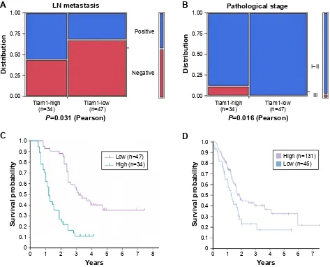
Tiam1 expression and clinicopathologic
features
To determine the connection between PC progression and Tiam1 protein expression, we analyzed the correlations between Tiam1 expression and the clinicopathological attributes of PC. These data showed that the expression of Tiam1 was positively correlated with lymph node
metasta-sis (P=0.031; Figure 2A) and histological grade (P=0.040;
Figure 2B). However, Tiam1 overexpression was not associ-ated with the sex, age, tumor location, or T classification of the patients with PDAC (Table 2).
Tiam1 expression is inversely related to
prognosis in PdAc patients
To further substantiate the significance of Tiam1 expression in PC progression, the association between the survival period and Tiam1 expression was evaluated by applying Kaplan– Meier analysis using the log-rank test. Total survival analysis using the Kaplan–Meier approach indicated that patients with high Tiam1 expression had lower 5-year survival rates than patients without Tiam1 overexpression (log-rank
OncoTargets and Therapy downloaded from https://www.dovepress.com/ by 118.70.13.36 on 25-Aug-2020
Dovepress
ding et al
(
'$3, 7LDP%[3&
6:
0HUJH
&
D E F%
'
7LDP
%[3& 0LDSDFD&DSDQ6:$V3&
$FWLQ
±
1RUPDO 7XPRU
P51$
OHYHORI7
LDP
ORJPHGLDQFHQWHUHG
LQWHQVLW\
%DGHDSDQFUHDVVWDWLVWLFV
Q
$
'
1RUPDO
7LDP
/RZ +LJK
Figure 1 High expression of Tiam1 in pancreatic cancer tissues samples and pancreatic cancer cells.
Notes: (A) Immunohistochemistry of Tiam1 proteins in clinical specimens from the human protein atlas (www.proteinatlas.org). (B) comparison of Tiam1 mrNA expression levels between the normal tissues and primary tumor or metastatic tumor tissues according to Oncomine data (www.oncomine.org) ***P0.001. (C) The expression of Tiam1 in pancreatic tissue samples was detected by immunohistochemistry (dAB, ×200). a: Negative expression of Tiam1; b: weakly positive expression of Tiam1; c: strongly positive expression of Tiam1. (D) Western blot assays performed on pancreatic cancer cell lines as indicated. (E) Immunofluorescence staining of the Tiam1 protein in BxPc-3 and SW1990 pancreatic cancer cells; a nuclear staining pattern was also observed.
Abbreviations: Tiam1, T-cell lymphoma invasion and metastasis inducing factor 1; dAPI, 4′,6-diamidino-2-phenylindole; dAB, diaminobezidin.
Table 1 Tiam1 protein expression in different groups
Group Case Negative Positive Positive
rate (%)
Strongly positive rate (%)
+ ++ +++
Pancreatic cancer tissues 81 6 41 29 5 92.59a 41.98a
Paracancerous tissues 81 51 16 14 0 37.04 17.28
Notes: Positive rate: percentage of positive cases with +, ++, and +++ staining scores. Strong positive rate: percentage of positive cases with ++ and +++ staining scores.
aP0.01.
Abbreviation: Tiam1, T-cell lymphoma invasion and metastasis inducing factor 1.
OncoTargets and Therapy downloaded from https://www.dovepress.com/ by 118.70.13.36 on 25-Aug-2020
Dovepress High Tiam1 plays a significant role in the progression of PDAC
Figure 2 cancer patients with high Tiam1 expression are associated with poor prognosis.
Notes: (A and B) correlations between Tiam1 expression and lymph node metastasis (A), pathological stage (B) of PdAc (P0.05, P0.05, respectively). (C) Kaplan–Meier survival curves in PdAc patients with high and low Tiam1 protein expression levels. (D) Patients with high or low Tiam1 mrNA levels obtained from the human protein atlas (www.proteinatlas.org).
Abbreviations: Tiam1, T-cell lymphoma invasion and metastasis inducing factor 1; PdAc, pancreatic ductal adenocarcinoma.
3RVLWLYH
1HJDWLYH
3 3HDUVRQ
/1PHWDVWDVLV
'LVWULEXWLRQ
7LDPKLJK
Q 7LDPORZQ
,±,,
,,,
3DWKRORJLFDOVWDJH
3 3HDUVRQ
'LVWULEXWLRQ
7LDPKLJK
Q 7LDPORZQ
$
%
<HDUV
6XUYLYDOSUREDELOLW\
+LJKQ /RZQ
6XUYLYDOSUREDELOLW\
<HDUV
+LJKQ /RZQ
&
'
value=25.93, P=0.000; Figure 2C). However, the data from
the human protein atlas (www.proteinatlas.org) until now
showed that PC patients with low Tiam1 mRNA had lower 5-year survival rates (Figure 2D). Further investigation is required to determine the relationship between expression of Tiam1 mRNA and protein and prognosis of PC.
Univariate analysis using the Cox proportional hazards
model revealed that lymph node metastasis (P=0.023),
patho-logical grade (P=0.006), and Tiam1 expression (P=0.000)
were associated with overall survival. Moreover, multivariate analysis showed that the high expression level of Tiam1 was a strong predictor of a negative prognosis (hazard ratio, 2.753;
95% CI: 1.670–4.536; P=0.000; Table 3). Consequently,
we demonstrated that Tiam1 expression was significantly associated with prognosis and was also an independent risk factor for PDAC patients.
Alteration of Tiam1 expression affects
proliferation, migration, and invasion of
Pc cells
To further elucidate the functional significance of Tiam1 in the growth and progression of PC, loss-of-function experi-ments were first performed to knockdown the expression of Tiam1 in SW1990 cells and BxPC-3 using a specific plasmid infection. The efficiency of the knockdown of Tiam1 expres-sion was confirmed by Western blot (Figure 3A). A colony formation assay showed that suppressing Tiam1 expression significantly inhibited the proliferation of siTiam1-BxPC-3 and siTiam1-SW1990 cells compared with the proliferation of the corresponding group of mock cells (Figure 3B).
Additionally, we examined the influential role of Tiam1 in the migration rates of PC cells using wound healing assays. The results showed that BxPC-3 and SW1990 cells
OncoTargets and Therapy downloaded from https://www.dovepress.com/ by 118.70.13.36 on 25-Aug-2020
Dovepress
ding et al
(Figure 3D). Altogether, these findings suggest that decreased expression of Tiam1 could significantly suppress PC cell proliferation, migration, and invasion.
Tiam1 promoted Pc cell metastasis via
activating EMT
An increasing number of reports have revealed that cell invasion and migration are associated with altered levels of
EMT biomarkers.14 To ascertain Tiam1’s underlying
mecha-nism in EMT, Western blot analysis was used to observe the expression of epithelial and mesenchymal protein markers. The results indicated that the silencing of Tiam1 expression in BxPC-3 and SW1990 cells increased the expression of E-cadherin while reducing the expression of mesenchymal markers, such as vimentin, slug, and snail (Figure 4). These results indicate that Tiam1 is a necessary mediator for promotion of PC EMT processes.
Discussion
PC is one of the most lethal malignancies in the world, and
each year ~40,000 patients die from this cancer. In contrast
to the steady decrease in mortality for most cancers, the incidence and death rates of PC are increasing. Therefore, there is an urgent need to examine the molecular mechanisms that are relevant to the progression of PDAC and search for novel therapeutic strategies.
A number of roles in the regulation of cellular func-tions are exhibited by Tiam1, and these roles depend on
the substratum, the specific cell type, and other factors.15
Li et al and Ding et al confirmed that the overexpression of Tiam1 predicts poor prognosis in ovarian carcinoma and
hepatocellular carcinomas,16,17 and Yang et al indicated that
upregulation of Tiam1 correlates with poor prognosis in
head and neck squamous cell carcinoma.18 In line with these
results, in this study, we assessed the expression level of Tiam1 in PDAC and found for the first time that Tiam1 was upregulated significantly in PDAC compared with that in non-cancerous tissues. Furthermore, elevated Tiam1 expres-sion was positively correlated with lymph node metastasis and pathological status in PDAC patients, which indicated that Tiam1 potentially played an important role in the pro-gression of PDAC.
Few studies to date have reported an association between Tiam1 expression and prognosis in PDAC. To determine whether the levels of Tiam1 expression might function as a prognostic factor for human PDAC, we analyzed Tiam1 expression and patient survival rates. We discovered that
Table 2 The relationship between high expression of Tiam1 protein and the clinicopathological parameters of patients with PdAc
Characteristic Case Tiam1
expression
Chi-squared P-value
Low High
Sex 0.005 0.946
Male 48 28 20
Female 33 19 14
Age (years) 0.650 0.420
60 40 25 15
60 41 22 19
Tumor location 0.406 0.524
Head 54 30 24
Body/tail 27 17 10
Tumor size 0.101 0.750
4 cm 54 32 22
4 cm 27 15 12
lymph node status 4.653 0.031a
yes 34 15 19
No 47 32 15
Pathological status 6.448 0.040a
I 1 1 0
II 76 46 30
III 4 0 4
T stage 5.011 0.082
T1 3 2 1
T2 68 36 32
T3 10 9 1
Note:aP0.05.
Abbreviations: Tiam1, T-cell lymphoma invasion and metastasis inducing factor 1; PdAc, pancreatic ductal adenocarcinoma.
Table 3 univariate and multivariate analyses of prognosis in patients with pancreatic cancer
Variable HR (95% CI) P-value
univariate
Gender 1.260 (0.800–1.984) 0.319
Age, years 0.918 (0.591–1.426) 0.703
Tumor location 0.950 (0.594–1.518) 0.829
Tumor size, d/cm 0.926 (0.581–1.473) 0.744
lymph node status 1.697 (1.077–2.674) 0.023a
Pathological status 4.478 (1.540–13.018) 0.006a
T classification 0.689 (0.415–1.144) 0.150
Tiam1 expression 2.861 (1.765–4.636) 0.000b
Multivariate
lymph node status 1.672 (1.051–2.660) 0.030a
Pathological status 2.336 (0.802–6.802) 0.120
Tiam1 expression 2.753 (1.670–4.536) 0.000b
Notes:aP0.05. bP0.01.
Abbreviations: Hr, hazard ratio; Tiam1, T-cell lymphoma invasion and metastasis inducing factor 1.
with decreased Tiam1 had delayed wound closure compared with that in control cells (Figure 3C). Matrigel transwell assays showed a similar phenomenon, in which decreased Tiam1 levels inhibited BxPC-3 and SW1990 cell invasion
OncoTargets and Therapy downloaded from https://www.dovepress.com/ by 118.70.13.36 on 25-Aug-2020
Dovepress High Tiam1 plays a significant role in the progression of PDAC
Figure 3 Tiam1 knockdown inhibited the proliferation, migration, and invasion of pancreatic cancer cells.
Notes: (A) Western blot showing Tiam1 expression in BxPc-3 and SW1990 cells upon Tiam1 knockdown. (B) Proliferating capability of transfected cells was evaluated using colony formation assays. (C) A scratch wound healing assay was used to determine the effects of si-Tiam1 on pancreatic cancer cells motility. (D) Migration and invasion of Tiam1 knockdown cells was measured by transwell assay. **P0.01.
Abbreviations: con, control; Tiam1, T-cell lymphoma invasion and metastasis inducing factor 1.
0RFN
%[3F
6:
VLFRQ VL7LDP VL7LDP
1XPEH
U
RIFRORQLH
V
%[3& 6:
0RFNVLFR 0RFN
Q
VLFR Q VL7LDP
VL7LDP
VL7LDP
VL7LDP
VL7LDP
VL7LDP 6: %[3&
7LDP
β$FWLQ
$
%
0RFN
VLFRQ VL7LDPVL7LDP
0RFN
K
K
K
VLFRQ VL7LDP VL7LDP
%[3&
K
K
K
0RFN VLFRQ VL7LDP VL7LDP
6:
&
%[3&
$UHDRIZRXQG JDSV
K K K
6:
$UHDRIZRXQG JDSV
K K K
0RFN VLFRQ VL7LDP VL7LDP
0RFN VLFRQ VL7LDP VL7LDP
6:
0LJUDWLRQ
%[3&
0RFN VLFRQ VL7LDP VL7LDP
6:
,QYDVLRQ
%[3&
'
0RFN VLFRQ VL7LDP VL7LDP
0LJUDWLRQFHOOV
%[3& 6:
,QYDVLRQFHOOV
%[3& 6:
OncoTargets and Therapy downloaded from https://www.dovepress.com/ by 118.70.13.36 on 25-Aug-2020
Dovepress
ding et al
7LDP
%[3&
0RFN VLFRQ VL7LDPVL7LDP
(FDGKHULQ
9LPHQWLQ
6QDLO
6OXJ
β$FWLQ
6:
0RFN VLFRQ VL7LDPVL7LDP
Figure 4 Tiam1 promoted pancreatic cancer cell migration and invasion by promoting EMT.
Note: The changes in the protein expression levels of the epithelial marker E-cadherin and the mesenchymal markers vimentin, snail, and slug in si-Tiam1-transfected BxPc-3 and SW1990 cells as detected by Western blot.
Abbreviations: con, control; Tiam1, T-cell lymphoma invasion and metastasis inducing factor 1; EMT, epithelial–mesenchymal transition.
compared with patients with high Tiam1 expression, those with low Tiam1 expression had a longer 5-year overall survival. Consistent with our findings, the data from the human protein atlas showed that high levels of Tiam1 mRNA in PC were negatively correlated with overall patient survival. Most importantly, both the univariate and multivariate analyses confirmed that Tiam1 expression was an independent risk factor for poor prognosis, along with lymph node status.
Uncontrolled growth and metastasis, which are impor-tant features of malignant tumors, are the main causes of cancer-related death. Michiels et al considered that Tiam1
overexpression may contribute to oncogenic transformation.7
Moreover, Tiam1 was found to play an important role in
regulating the tumor microenvironment,19 as well as
modi-fication of cell polarity and the actin cytoskeleton,20 all of
which are thought to be associated with tumor progression. Here, we knocked down the expression of Tiam1 in BxPC-3 and SW1990 cells via siRNA transfection and found that cell growth was significantly inhibited. Downregulation of Tiam1 also significantly inhibited cell invasion and migration. These findings demonstrated that Tiam1 may function as an oncogene in PC cells and that its expression may facilitate PC development and progression.
EMT is a process whereby cancer cells develop migration capability and invasiveness, which leads to poor prognosis
and cancer metastasis.21,22 Consistent with the studies by Liu
et al, in which Tiam1 overexpression was associated with
thyroid carcinoma metastasis,23 in this study, we discovered
that Tiam1 overexpression promoted EMT by decreasing the expression of the mesenchymal markers vimentin and snail and by elevating the expression of the epithelial marker E-cadherin. These results confirmed that Tiam1 promoted cell migration and invasion by promoting the process of EMT in PC. However, the role of Tiam1 in regulating PC progression and metastasis requires further in-depth studies in the future.
This study demonstrated that Tiam1 overexpression is strongly related to PDAC progression and is an independent prognostic factor. Moreover, the suppression of Tiam1 in PC cells significantly inhibited cell proliferation, migration, and invasion. Our findings indicated that Tiam1 may also repre-sent a new molecular biomarker for human PDAC.
Acknowledgment
This study was supported by grants from National Natural Science Funds of China (Nos 81660436, 81460399).
Disclosure
The authors report no conflicts of interest in this work.
References
1. Lin QJ, Yang F, Jin C, Fu DL. Current status and progress of pancreatic cancer in China. World J Gastroenterol. 2015;21(26):7988–8003. 2. Siegel RL, Miller KD, Jemal A. Cancer statistics, 2015. CA Cancer J
Clin. 2015;65(1):5–29.
3. Polireddy K, Chen Q. Cancer of the Pancreas: Molecular pathways and current advancement in treatment. J Cancer. 2016;7(11):1497–1514.
OncoTargets and Therapy downloaded from https://www.dovepress.com/ by 118.70.13.36 on 25-Aug-2020
OncoTargets and Therapy
Publish your work in this journal
Submit your manuscript here: http://www.dovepress.com/oncotargets-and-therapy-journal OncoTargets and Therapy is an international, peer-reviewed, open access journal focusing on the pathological basis of all cancers, potential targets for therapy and treatment protocols employed to improve the management of cancer patients. The journal also focuses on the impact of management programs and new therapeutic agents and protocols on
patient perspectives such as quality of life, adherence and satisfaction. The manuscript management system is completely online and includes a very quick and fair peer-review system, which is all easy to use. Visit http://www.dovepress.com/testimonials.php to read real quotes from published authors.
Dovepress
Dove
press
High Tiam1 plays a significant role in the progression of PDAC
4. Hidalgo M. Pancreatic cancer. N Engl J Med. 2010;362(17): 1605–1617.
5. Habets GG, Scholtes EH, Zuydgeest D, et al. Identification of an invasion-inducing gene, Tiam-1, that encodes a protein with homol-ogy to GDP-GTP exchangers for Rho-like proteins. Cell. 1994;77(4): 537–549.
6. Hordijk PL, Ten Klooster JP, van der Kammen RA, et al. Inhibition of invasion of epithelial cells by Tiam1-Rac signaling. Science. 1997; 278(5342):1464–1466.
7. Michiels F, Habets GG, Stam JC, van der Kammen RA, Collard JG. A role for Rac in Tiam1-induced membrane ruffling and invasion.
Nature. 1995;375(6529):338–340.
8. Mertens AE, Roovers RC, Collard JG. Regulation of Tiam1-Rac signal-ling. FEBS Lett. 2003;546(1):11–16.
9. Habets GG, van der Kammen RA, Jenkins NA, et al. The invasion-inducing TIAM1 gene maps to human chromosome band 21q22 and mouse chromosome 16. Cytogenet Cell Genet. 1995;70(1–2):48–51. 10. Liu H, Shi G, Liu X, Wu H, Fan Q, Wang X. Overexpression of Tiam1
predicts poor prognosis in patients with esophageal squamous cell carcinoma. Oncol Rep. 2011;25(3):841–848.
11. Wu QY, Wang Y, Tong JC, Zhang M, Zhang K. Expression and clinical significance of Tiam1 gene in esophageal carcinoma. Int J Clin Exp
Med. 2015;8(11):21229–21234.
12. Liu S, Li Y, Qi W, et al. Expression of Tiam1 predicts lymph node metastasis and poor survival of lung adenocarcinoma patients. Diagn
Pathol. 2014;9:69.
13. Xu K, Tian X, Oh SY, et al. The fibroblast Tiam1-osteopontin pathway modulates breast cancer invasion and metastasis. Breast Cancer Res. 2016;18(1):14.
14. Voulgari A, Pintzas A. Epithelial-mesenchymal transition in cancer metastasis: mechanisms, markers and strategies to overcome drug resistance in the clinic. Biochim Biophys Acta. 2009;1796(2):75–90. 15. Huang J, Ye X, Guan J, et al. Tiam1 is associated with hepatocellular
carcinoma metastasis. Int J Cancer. 2013;132(1):90–100.
16. Li H, Cui X, Chen D, et al. Clinical implication of Tiam1 overex-pression in the prognosis of patients with serous ovarian carcinoma.
Oncol Lett. 2016;12(5):3492–3498.
17. Yang W, Lv S, Liu X, et al. Up-regulation of Tiam1 and Rac1 correlates with poor prognosis in hepatocellular carcinoma. Jpn J Clin Oncol. 2010;40(11):1053–1059.
18. Yang H, Cai YC, Cao Y, et al. The prognostic value of Tiam1 protein expression in head and neck squamous cell carcinoma: a retrospective study. Chin J Cancer. 2015;34(12):614–621.
19. Xu K, Rajagopal S, Klebba I, et al. The role of fibroblast Tiam1 in tumor cell invasion and metastasis. Oncogene. 2010;29(50):6533–6542. 20. Mertens AE, Pegtel DM, Collard JG. Tiam1 takes PARt in cell polarity.
Trends Cell Biol. 2006;16(6):308–316.
21. Ombrato L, Malanchi I. The EMT universe: space between cancer cell dissemination and metastasis initiation. Crit Rev Oncog. 2014;19(5): 349–361.
22. Son H, Moon A. Epithelial-mesenchymal transition and cell invasion.
Toxicol Res. 2010;26(4):245–252.
23. Liu L, Wu B, Cai H, et al. Tiam1 promotes thyroid carcinoma metastasis
by modulating EMT via Wnt/β-catenin signaling. Exp Cell Res. 2018;
362(2):532–540.
OncoTargets and Therapy downloaded from https://www.dovepress.com/ by 118.70.13.36 on 25-Aug-2020