Isolation and expression analysis of
foxj1 and foxj1.2 in zebrafish embryos
EMIL AAMAR and IGOR B. DAWID*
Laboratory of Molecular Genetics, National Institute of Child Health and Human Development, National Institutes of Health, Bethesda MD, USA
ABSTRACT In this report, we present the isolation and identification of a zebrafish homolog of the winged helix\forkhead transcription factor Foxj1. Foxj1 was identified in other species but not in zebrafish. Foxj1 was shown in mice to be required in ciliogenesis and left-right asymmetry establishment. Here we present a spatio-temporal expression pattern of zebrafish foxj1, showing that this gene is expressed in dorsal forerunner cells, Kupffer’s vesicle, the floor plate, pronephric ducts and kidney. This expression pattern is overlapping but different from that of the foxj1.2, the closest related gene in zebrafish. Foxj1 in zebrafish appears to have similar functions as those reported in other species connected to ciliogenesis and left-right asymmetry.
KEY WORDS:
Foxj1, Foxj1.2, dorsal forerunner cell, Kupffer’s vesicle, floor plate, pronephric duct
Hepatocyte nuclear factor-3 (HNF-3)/forkhead homologue 4 (HFH-4)/Foxj1 is a winged helix/forkhead transcription factor. A 100-amino-acid DNA-binding motif, known as the winged helix, was first identified in mammalian hepatocyte nuclear factor-3 (HNF-3) and Drosophila Forkhead transcription factors (Avraham et al., 1995; Lai et al., 1993). Subsequently, many additional proteins containing the winged helix motif have been identified (Avraham et al., 1995). In humans (Pelletier et al., 1998) rats (Hackett et al., 1995) and mice, Foxj1 is expressed in ciliated cells of various tissues including the respiratory tract, brain, and ependyma in late development through adulthood, as well as in oviduct, testis, embryonic kidney (Blatt et al., 1999; Brody et al., 2000; Tichelaar et al., 1999a; Tichelaar et al., 1999b) and the choroid plexus (Blatt et al., 1999; Brody et al., 1997; Lim et al., 1997; Swetloff and Ferretti, 2005).
Foxj1 is involved in the regulation of ciliogenesis and axonemal structural proteins. Foxj1 regulates basal body anchoring to the cytoskeleton of ciliated pulmonary epithelial cells, and is required for apical localization of ezrin and the formation of axonemal structures (Gomperts et al., 2004; Gomperts et al., 2007). Further, Foxj1 promotes RhoA-mediated apical actin enrichment required for ciliogenesis (Pan et al., 2007). In Foxj1 null mice, classic motile type cilia with a 9+2 microtubule ultrastructure were absent in epithelial cells, resulting in defective ciliogenesis in the airways. In other organs, sensory cilia with a 9+0 microtubule pattern, such as those on olfactory neuroepithelial cells, were still present. Foxj1 is expressed in the ciliated cells of Hensen’s node in the
BIOLOGY
www.intjdevbiol.com*Address correspondence to: Igor B. Dawid. Laboratory of Molecular Genetics, National Institute of Child Health and Human Development, National Institutes of Health, Building 6B, Room 413, Bethesda, MD, 20892 USA. e-mail: [email protected]
Published online: 8th May 2008.
0214-6282/2008/$35.00
© UBC Press Printed in Spain
Abbreviations used in this paper: HNF, hepatocyte nuclear factor;
chick, and is required for left/right asymmetry determination (Blatt et al., 1999; Chen et al., 1998; Feistel and Blum, 2006; Maiti et al., 2000; Tamakoshi et al., 2006; Zhang et al., 2004). However analysis of the node in Foxj1 null mice revealed that, in contrast to the absence of airway cilia, node cilia were present. These observations indicate that there are independent regulatory path-ways for node ciliogenesis compared with 9+2 type ciliogenesis in airways, and support a central role for Foxj1 in ciliogenesis and left-right axis formation (Brody et al., 2000; Chen et al., 1998). At high levels of expression, Foxj1/HFH-4 altered epithelial cell differentiation and inhibited branching morphogenesis in the developing mouse lung, restricting the expression of markers typical of nonciliated cells of the distal lung parenchyma (Blatt et al., 1999; Maiti et al., 2000).
et al., 2005). Foxj1 expression in lung bud morphogenesis is further regulated by BMP4 signaling(Hyatt et al., 2004).
Here we report the cloning of foxj1 in zebrafish and the protein encoded by it, and we compare the predicted zebrafish protein sequence to Foxj1 sequences of other species. We further present the temporal and spatial expression of foxj1 in zebrafish embryos. Zebrafish foxj1 is expressed in the dorsal forerunner cells, floor plate, Kupffer’s vesicle, and in the pro-nephric duct and developing kidney. These sites are similar but not identical to the sites of expression of foxj1 in other verte-brate species. We compare this expression pattern with that of foxj1.2 and find that they have distinct and partially overlapping patterns. Foxj1.2 is the closest to Foxj1 in Danio rerio. Even though a sequence for Foxj1.2 is available, no work has been done to describe its expression pattern or function in zebrafish or other species, except for Xenopus tropicalis, where a very brief expression pattern is described. In X. tropicalis FoxJ1.2 is expressed in the otic vesicle during late neurula stages and is
then also expressed in the presumptive nephrostomes of the pronephros during tailbud stages (Choi et al., 2006).
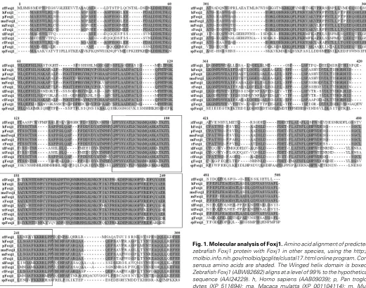
Isolation of zebrafish foxj1 and comparison of
verte-brate Foxj1 proteins
proteins is illustrated in Figure 2.
Expression of
foxj1
during zebrafish
embryogen-esis
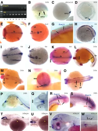
Expression of foxj1 in zebrafish embryos was examined by RT-PCR and whole-mount in situ hybridization. Foxj1 mRNA is not detected maternally and begins to be ex-pressed weakly at high-dome stages as shown by RT-PCR (Fig. 3A lane 3). The earliest expression that could be detected by in situ hybridization occurs at about 30% epiboly (Fig. 3B) in dorsal forerunner cells. This expression continues through shield stage (Fig. 3C) and is maintained until late epiboly stages (Fig. 3D). Consistently with the fact that dorsal forerunner cells are the precursors of Kupffer’s vesicle we see that foxj1 is strongly expressed in Kupffer’s vesicle starting at bud stage (Fig. 3F, H and J). Foxj1 transcripts are also detected in the presump-tive floor plate at the dorsal midline, with increasing extent and intensity as development proceeds (Fig. 3E-K). This expression pattern might correspond to that described in rabbits where foxj1 is expressed in the notochordal plate as the cells migrate from Hensen’s node anteriorly, and eventually is expressed in the notochord (Feistel and Blum, 2006). However, in zebrafish, foxj1 expression was not detected in the notochord but in the floor plate as visualized by DIC optics (not shown) and by double in situ staining with ntl as a notochord marker (Fig. 3G, R and S). This expression in the floor plate is maintained through somitogenesis stages up to about 2 days where it becomes weaker in the floor plate and shows weakening expression at the posterior neural tube at the tail tip. Both expression domains weaken or disappear by three and four days of age (not shown). Although at 16 somites (16s) to 24hpf stages the expression in the floor plate is relatively
weaker in the trunk and most of the tail, it is strongly maintained in the tail tip in the floor plate and neural tube, and at the ventral edge of the mid-hindbrain boundary (MHB) (Fig. 3L-S). The MHB is the anterior limit of foxj1 expression (Fig. 3P, Q), as also seen by double in situ staining with pax2.1 which marks the MHB (Fig. 3E, L-N). Expression in Kupffer’s vesicle is no longer detected at about 16s stages since the vesicle is much smaller (Fig. 3L).
By bud stage, foxj1 expression is also detected in the presump-tive pronephric ducts (Fig. 3E-F). This expression continues through the 24hpf stage where it is detected also in the forming kidney (Fig. 3H-L, H-P). Double in situ hybridization using foxj1 and pax2.1 confirmed expression in the pronephric duct and its precursors, as both markers align in this region (Fig. 3E-F, K-M and O). Foxj1 expression in the pronephros at the 24hpf stage is usually weaker as compared to the 16s stage.
At 2-4 days after fertilization, foxj1 expression fades or has disappeared, except in the kidney (Fig. 3T-W) and around the mid-hindbrain boundary, which strengthens in intensity and ex-tends to the tectal ventricle (Fig. 3T-V). Expression in the tectal ventricle decreases by 3 days (not shown) and 4days of develop-ment (Fig. 3V vs. T), while expression in the olfactory pits starts at about 3days and increases by 4days (Fig. 3V). By 4days a weak expression is also seen in the otoliths of the otic vesicles (Fig. 3W top).
Expression of
foxj1.2
is overlapping but different from
foxj1
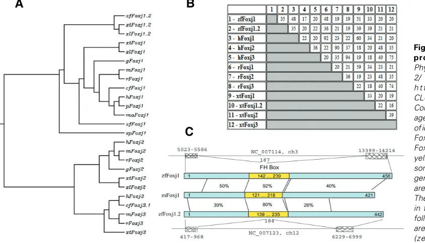
The closest sequence in Danio rerio to Foxj1 is listed under accession number NP_001008648.1 as a hypothetical protein. The predicted protein is most closely related to Foxj1.2 in X. tropicalis, and we therefore identify this sequence as zebrafish Foxj1.2. Zebrafish foxj1 and foxj1.2 genes are located on
chromo-B
C
A
somes 3 (ch3, NC_007114 REGION: 63826357.63840654) and 12 (ch12, NC_007123 REGION: 19967418.19975131), respec-tively, and their ORFs are similarly encoded by two exons (Fig. 2C; the exon start and end positions are given). Foxj1 and foxj1.2 share very limited homology at the level of cDNA sequence which is mostly restricted to a part of the forkhead box (FH box) domain (82% identity in 293nt). The proteins share 35% identity mainly in the FH box (Fig. 2B). Zebrafish Foxj1 has higher similarity to Foxj1 proteins from other species than Foxj1.2 does (Fig. 2).
Foxj1.2 mRNA is not detected maternally and begins to be expressed at about 40% epiboly, as shown by RT-PCR (Fig. 4Q lane 4). In situ hybridization shows strong expression in the epiblast by shield stage (Fig. 4A), and at 80% epiboly (Fig. 4B), different from the pattern of foxj1 which is restricted to the dorsal forerunner cells (compare Fig. 4A and B to Fig. 3B-D). During the bud through early somite stages, the expression of foxj1.2 over-laps with foxj1 in the floor plate and pronephric duct. Unlike foxj1
anterior tectal ventricle. This expression pattern is partially over-lapping with that described for foxj1.2 in Xenopus tropicalis (Choi et al., 2006).
As conclusion, the expression pattern of Foxj1.2 is different from that of Foxj1, which supports the conclusion that our se-quence is the one which is more likely to be called Foxj1.
Concluding remarks
Here we present the isolation of the zebrafish foxj1 gene; we illustrate its dynamic expression during development and com-pare it to foxj1.2. Zebrafish foxj1 is expressed in three major domains, dorsal forerunner cells and Kupffer’s vesicle, the floor plate, and the pronephros. This pattern is similar to that of Foxj1 genes in other vertebrates. Foxj1 function has been connected to
Fig. 4. Zebrafish foxj1.2 expression. (A-P)Spatio-temporal expression pattern of foxj1.2. Whole-mount in situ hybridization with foxj1.2 probe alone (A-D, F-G, L, M4), or combined with pax2.1 and charon (both red) (E, H-K, M-P). Shield-60% epiboly (A), 80% epiboly (B), bud (C-E), 4 somites (F-G), 7 somites (H-J), 15 somites (K,P), 16 somites (L), 1 day (M), 2 days (N), 3 and 4 days (P left and right respectively). Views are as follows: dorsal (A, B); dorsal with anterior to the left (C bottom, E, F bottom, H and H bottom), dorsal with anterior up (P right), lateral with dorsal to the right, (A left bottom, B left bottom), lateral with anterior to left (C top, F, I, K-O except for M4, P left), posterior with dorsal up (D, G, J), anterior ventral with dorsal up (M4), animal view (A left top, B left top), ventral with posterior to the right (J right bottom). (Q)RT-PCR expression analysis of foxj1.2 and β-actin as control was performed for different stages (1-8: unfertilized eggs, 100-200 cells, high-dome, 40-50% epiboly, 80-90%, bud, 13-somites, 24hpf and 3days old embryos respectively, and –RT in lane 10). d, diencephalon; dt, dorsal tectum; e, eye; fp, floor plate; k, kidney; KV, Kupffer’s vesicle; MHB, mid-hindbrain boundary; nt, notochord; op, olfactory pit; ot; otolith; ov, otic vesicle; p, pineal gland; pd, pronephric duct; pnt, posterior neural tube; t, tectum; tg, trigeminal ganglion; tv, tectal ventricle.
ciliogenesis and the regulation of left/right asymmetry, which fits the major expression sites in zebrafish embryos, Kupffer’s vesicle and the pronephric ducts, both of which are known to contain highly ciliated cells. Therefore it is likely that zebrafish Foxj1 functions similarly to Foxj1 of other vertebrate.
Experimental Procedures
Embryos
Zebrafish (Danio rerio) were raised and maintained accordingto standard procedure (Westerfield, 2000). Embryos were raised at 28.5°C and staged as described (Kimmel et al., 1995).
RT-PCR
RNA isolation was performed using the RNeasy Mini Kit (Qiagen, http:/ /www1.qiagen.com). Reverse transcription and PCR were performed as
G
O
B
C
D
E
F
H
I
J
K
L
P
Q
A
M
M1
N
M2
M3
M4
L1
described in the SuperScript™ II Reverse Transcriptase manual (Invitrogen, http://www.invitrogen.com). The expression levels of zebrafish
foxj1 were compared to histone 4 (H4, AM422106). The primers used for H4 were:
forward 5'- GAAGAGGCAAAGGAAGCAAA -3' and
reverse 5'- TGGCGTGCTCTGTGTAGGTA -3' (58ºC, 25 cycles). The primers used for foxj1 were:
forward 5'- GATTCCAGCCAGGATTTTCA -3'and
reverse 5'- AATGCAAATGTGCCAACAAA -3' (58ºC, 30 cycles). The expression levels of zebrafish foxj1.2 were compared to β-actin
(BC154531).
The primers used for β-actin were:
forward 5'- GAGGAGCACCCCGTCCTGC -3' and
reverse 5'- GATGGCTGGAACAGGGCC -3' (58°C, 25 cycles). The primers used for foxj1.2 were:
forward 5'- CGTGAAGCCACCCTATTCAT-3' and
reverse 5’- GGATTGAGTTCTGCCAGCTC -3' (58ºC, 35 cycles).
Cloning and construction of expression plasmid
A partial zebrafish foxj1 clone (#5134) was identified in an expression patternscreen (Kudoh et al., 2001). The 5' end of foxj1 was subsequently generatedby 5'-RACE using the SMART RACE Kit (Clontech) with a gene-specificprimer (5'- CGTGTTCGTGTGGGCGATTTTAAGAG -3'), and NestedGSP primer 5’ AGCTCGAATGTTAGCGGGAATTGGAC -3’, according to the manufacturer’s instructions.
Whole-mount in situ hybridization
In situ hybridizations were performed as described by Thisse and Thisse (http://zfin.org/zf_info/zfbook/chapt9/9.82.html) (Westerfield, 2000). Antisense digoxigenin or fluorescein-labeled probes were synthesized for the following zebrafish markers: Foxj, using the original EST clone (5134), which is a 3’UTR of the gene; Foxj1.2, using the clone IM-AGE:7228406 (http://www.openbiosystems.com/), ntl (Schulte-Merker
et al., 1994), pax2a/2.1 (Krauss et al., 1991; Pfeffer et al., 1998), charon
(a gift from Dr. Rebagliati Michael) (Gourronc et al., 2007), aanat2 (Falcon
et al., 2003; Ziv et al., 2005), and otx5 (Gamse et al., 2002; Ziv et al.,
2005). The labeling kit from Roche Molecular Biochemicals was used as described (Westerfield, 2000).
Acknowledgments
This research was supported by the Intramural Research Program of the National Institute of Child Health and Human Development, National Institutes of Health.
References
AVRAHAM, K. B., FLETCHER, C., OVERDIER, D. G., CLEVIDENCE, D. E., LAI, E., COSTA, R. H., JENKINS, N. A., and COPELAND, N. G. (1995). Murine chromosomal location of eight members of the hepatocyte nuclear factor 3/fork
head winged helix family of transcription factors. Genomics 25: 388-93.
BLATT, E. N., YAN, X. H., WUERFFEL, M. K., HAMILOS, D. L., and BRODY, S. L. (1999). Forkhead transcription factor HFH-4 expression is temporally related to
ciliogenesis. Am J Respir Cell Mol Biol 21: 168-76.
BRODY, S. L., HACKETT, B. P., and WHITE, R. A. (1997). Structural characteriza-tion of the mouse Hfh4 gene, a developmentally regulated forkhead family
member. Genomics 45: 509-18.
BRODY, S. L., YAN, X. H., WUERFFEL, M. K., SONG, S. K., and SHAPIRO, S. D. (2000). Ciliogenesis and left-right axis defects in forkhead factor HFH-4-null
mice. Am J Respir Cell Mol Biol 23: 45-51.
CHEN, J., KNOWLES, H. J., HEBERT, J. L., and HACKETT, B. P. (1998). Mutation of the mouse hepatocyte nuclear factor/forkhead homologue 4 gene results in
an absence of cilia and random left-right asymmetry. J Clin Invest 102: 1077-82.
CHOI, V. M., HARLAND, R. M., and KHOKHA, M. K. (2006). Developmental
expression of FoxJ1.2, FoxJ2, and FoxQ1 in Xenopus tropicalis. Gene Expr
Patterns 6: 443-7.
FALCON, J., GOTHILF, Y., COON, S. L., BOEUF, G., and KLEIN, D. C. (2003). Genetic, temporal and developmental differences between melatonin rhythm
generating systems in the teleost fish pineal organ and retina. J Neuroendocrinol
15: 378-82.
FEISTEL, K., and BLUM, M. (2006). Three types of cilia including a novel 9+4
axoneme on the notochordal plate of the rabbit embryo. Dev Dyn 235: 3348-58.
GAMSE, J. T., SHEN, Y. C., THISSE, C., THISSE, B., RAYMOND, P. A., HALPERN, M. E., AND LIANG, J. O. (2002). Otx5 regulates genes that show circadian
expression in the zebrafish pineal complex. Nat Genet 30: 117-21.
GOMPERTS, B. N., GONG-COOPER, X., and HACKETT, B. P. (2004). Foxj1 regulates basal body anchoring to the cytoskeleton of ciliated pulmonary
epithelial cells. J Cell Sci 117: 1329-37.
GOMPERTS, B. N., KIM, L. S., FLAHERTY, S. A., and HACKETT, B. P. (2007).
IL-13 Regulates Cilia Loss and foxj1 Expression in Human Airway Epithelium. Am
J Respir Cell Mol Biol.
GOURRONC, F., AHMAD, N., NEDZA, N., EGGLESTON, T., and REBAGLIATI, M. (2007). Nodal activity around Kupffer’s vesicle depends on the T-box
transcrip-tion factors Notail and Spadetail and on Notch signaling. Dev Dyn 236: 2131-46.
HACKETT, B. P., BRODY, S. L., LIANG, M., ZEITZ, I. D., BRUNS, L. A., and GITLIN, J. D. (1995). Primary structure of hepatocyte nuclear factor/forkhead homologue 4 and characterization of gene expression in the developing
respi-ratory and reproductive epithelium. Proc Natl Acad Sci USA 92: 4249-53.
HYATT, B. A., SHANGGUAN, X., and SHANNON, J. M. (2004). FGF-10 induces SP-C and Bmp4 and regulates proximal-distal patterning in embryonic tracheal
epithelium. Am J Physiol Lung Cell Mol Physiol 287: L1116-26.
JIN, R., ZHANG, J., and CHEN, W. (2006). Thymic output: influence factors and
molecular mechanism. Cell Mol Immunol 3: 341-50.
KIMMEL, C. B., BALLARD, W. W., KIMMEL, S. R., ULLMANN, B., and SCHILLING,
T. F. (1995). Stages of embryonic development of the zebrafish. Dev Dyn 203:
253-310.
KRAUSS, S., JOHANSEN, T., KORZH, V., and FJOSE, A. (1991). Expression of the
zebrafish paired box gene pax[zf-b] during early neurogenesis. Development
113: 1193-206.
KUDOH, T., TSANG, M., HUKRIEDE, N. A., CHEN, X., DEDEKIAN, M., CLARKE, C. J., KIANG, A., SCHULTZ, S., EPSTEIN, J. A., TOYAMA, R., and DAWID, I.
B. (2001). A gene expression screen in zebrafish embryogenesis. Genome Res
11: 1979-87.
LAI, E., CLARK, K. L., BURLEY, S. K., and DARNELL, J. E., JR. (1993). Hepatocyte nuclear factor 3/fork head or «winged helix» proteins: a family of transcription
factors of diverse biologic function. Proc Natl Acad Sci USA 90: 10421-3.
LIM, L., ZHOU, H., and COSTA, R. H. (1997). The winged helix transcription factor HFH-4 is expressed during choroid plexus epithelial development in the mouse
embryo. Proc Natl Acad Sci USA 94: 3094-9.
LIN, L., BRODY, S. L., and PENG, S. L. (2005). Restraint of B cell activation by
Foxj1-mediated antagonism of NF-kappa B and IL-6. J Immunol 175: 951-8.
LIN, L., and PENG, S. L. (2006). Coordination of NF-kappaB and NFAT antagonism
by the forkhead transcription factor Foxd1. J Immunol 176: 4793-803.
MAITI, A. K., BARTOLONI, L., MITCHISON, H. M., MEEKS, M., CHUNG, E., SPIDEN, S., GEHRIG, C., ROSSIER, C., DELOZIER-BLANCHET, C. D., BLOUIN, J., GARDINER, R. M., and ANTONARAKIS, S. E. (2000). No delete-rious mutations in the FOXJ1 (alias HFH-4) gene in patients with primary ciliary
dyskinesia (PCD). Cytogenet Cell Genet 90: 119-22.
PAN, J., YOU, Y., HUANG, T., and BRODY, S. L. (2007). RhoA-mediated apical
actin enrichment is required for ciliogenesis and promoted by Foxj1. J Cell Sci
120: 1868-76.
PARK, K. S., WELLS, J. M., ZORN, A. M., WERT, S. E., and WHITSETT, J. A.
(2006). Sox17 influences the differentiation of respiratory epithelial cells. Dev
Biol 294: 192-202.
PELLETIER, G. J., BRODY, S. L., LIAPIS, H., WHITE, R. A., and HACKETT, B. P. (1998). A human forkhead/winged-helix transcription factor expressed in
devel-oping pulmonary and renal epithelium. Am J Physiol 274: L351-9.
PFEFFER, P. L., GERSTER, T., LUN, K., BRAND, M., and BUSSLINGER, M. (1998). Characterization of three novel members of the zebrafish Pax2/5/8 family: dependency of Pax5 and Pax8 expression on the Pax2.1 (noi) function.
Development 125: 3063-74.
NUSSLEIN-VOLHARD, C. (1994). no tail (ntl) is the zebrafish homologue of the
mouse T (Brachyury) gene. Development 120: 1009-15.
SELA, U., DAYAN, M., HERSHKOVIZ, R., CAHALON, L., LIDER, O., and MOZES, E. (2006). The negative regulators Foxj1 and Foxo3a are up-regulated by a peptide that inhibits systemic lupus erythematosus-associated T cell responses.
Eur J Immunol 36: 2971-80.
SRIVATSAN, S., and PENG, S. L. (2005). Cutting edge: Foxj1 protects against
autoimmunity and inhibits thymocyte egress. J Immunol 175: 7805-9.
SWETLOFF, A., and FERRETTI, P. (2005). Changes in E2F5 intracellular
localiza-tion in mouse and human choroid plexus epithelium with development. Int J Dev
Biol 49: 859-65.
TAMAKOSHI, T., ITAKURA, T., CHANDRA, A., UEZATO, T., YANG, Z., XUE, X. D., WANG, B., HACKETT, B. P., YOKOYAMA, T., and MIURA, N. (2006). Roles of the Foxj1 and Inv genes in the left-right determination of internal organs in mice.
Biochem Biophys Res Commun 339: 932-8.
TICHELAAR, J. W., LIM, L., COSTA, R. H., and WHITSETT, J. A. (1999a). HNF-3/forkhead homologue-4 influences lung morphogenesis and respiratory
epi-thelial cell differentiation in vivo. Dev Biol 213: 405-17.
TICHELAAR, J. W., WERT, S. E., COSTA, R. H., KIMURA, S., and WHITSETT, J. A. (1999b). HNF-3/forkhead homologue-4 (HFH-4) is expressed in ciliated
epithelial cells in the developing mouse lung. J Histochem Cytochem 47:
823-32.
WAN, H., DINGLE, S., XU, Y., BESNARD, V., KAESTNER, K. H., ANG, S. L., WERT, S., STAHLMAN, M. T., and WHITSETT, J. A. (2005). Compensatory
roles of Foxa1 and Foxa2 during lung morphogenesis. J Biol Chem 280:
13809-16.
WESTERFIELD, M. (2000). « THE ZEBRAFISH BOOK; A guide for the laboratory use of zebrafish (Danio rerio).» University of Oregon Press, Eugene, OR.
ZHANG, M., BOLFING, M. F., KNOWLES, H. J., KARNES, H., and HACKETT, B. P. (2004). Foxj1 regulates asymmetric gene expression during left-right axis
patterning in mice. Biochem Biophys Res Commun 324: 1413-20.
ZIV, L., LEVKOVITZ, S., TOYAMA, R., FALCON, J., and GOTHILF, Y. (2005). Functional development of the zebrafish pineal gland: light-induced expression
of period2 is required for onset of the circadian clock. J Neuroendocrinol 17:
314-20.
Further Related Reading, published previously in the
Int. J. Dev. Biol.
See our recent Special Issue Ear Development edited by Fernando Giraldez and Bernd Fritzsch at: http://www.ijdb.ehu.es/web/contents.php?vol=51&issue=6-7
Expression and comparative genomics of two serum response factor genes in zebrafish
Jody L. Davis, Xiaochun Long, Mary A. Georger, Ian C. Scott, Adam Rich and Joseph M. Miano Int. J. Dev. Biol. (2008) 52: 2514-2514
Expression of protocadherin 18 in the CNS and pharyngeal arches of zebrafish embryos
Fumitaka Kubota, Tohru Murakami, Yuki Tajika and Hiroshi Yorifuji Int. J. Dev. Biol. (2008) 52: 2424-2424
Embryonic heat shock reveals latent hsp90 translation in zebrafish (Danio rerio)
Michelle H. Connolly and Brian K. Hall Int. J. Dev. Biol. (2008) 52: 71-79
The retinoic acid metabolising gene, CYP26B1, patterns the cartilaginous cranial neural crest in zebrafish
Susan Reijntjes, Adam Rodaway and Malcolm Maden Int. J. Dev. Biol. (2007) 51: 351-360
Pax7 identifies neural crest, chromatophore lineages and pigment stem cells during zebrafish development
Ana M Lacosta, Jesús Canudas, Cristina González, Pedro Muniesa, Manuel Sarasa and Luis Domínguez
Int. J. Dev. Biol. (2007) 51: 327-331
Spatiotemporal expression of the creatine metabolism related genes agat, gamt and ct1 during zebrafish embryogenesis
Lifeng Wang, Ying Zhang, Ming Shao and Hongwei Zhang Int. J. Dev. Biol. (2007) 51: 247-253
Zebrafish spata2 is expressed at early developmental stages
Enrico Moro, Claudio Maran, M. Liliana Slongo, Francesco Argenton, Stefano Toppo and Maurizio Onisto
Int. J. Dev. Biol. (2007) 51: 241-246
Cadherin-6 is required for zebrafish nephrogenesis during early development
Fumitaka Kubota, Tohru Murakami, Kenji Mogi and Hiroshi Yorifuji Int. J. Dev. Biol. (2007) 51: 123-129
Changes in E2F5 intracellular localization in mouse and human choroid plexus epithelium with development
Adam Swetloff and Patrizia Ferretti Int. J. Dev. Biol. (2005) 49: 859-865