Ahmed Saadi, MD; Abderrazak Bouzouita, MD; Mohamed Cherif, MD; Mohamed Hedi Rebai, MD;
Walid Kerkeni, MD; Haroun Ayed, MD; Amine Derouiche, MD; Riadh Ben Slama, MD; Mohamed Chebil, MD
Department of Urology, Charles Nicolle Hospital, Tunis, Tunisia
Cite as: Can Urol Assoc J 2015;9(5-6):E374-8. http://dx.doi.org/10.5489/cuaj.2782 Published online June 18, 2015.
Abstract
Introduction: The hydatid cyst is a real public health problem in
Tunisia. The retrovesical localization is rare. It is considered an aberrant or ectopic location defined by the development of the parasite in the subvesical and retrovesical fat.
Methods: From 2004 to 2013, 4 patients with retrovesical hydatid
cyst were hospitalized and operated in the Department of Urology at the Charles Nicolle hospital of Tunis in Tunisia. The average patient age was 40.75 years (range: 23 –76). Signs of bladder irri-tation were the most frequent presenting complaint. No cases of hydaturia were noted. The diagnosis was made on the ultrasound and the computed tomographic urography. Hydatid serology was positive for 3 patients. In 3 cases, a hydatid cyst of the liver was associated. A total cysto-pericystectomy was performed for 1 patient, for others it was partial.
Results: The postoperative course was uneventful. No urinary
fis-tula or infection of the residual cavity was observed. One patient had a retroperitoneal cyst recurrence requiring reoperation.
Conclusion: Retrovesical location of hydatid cyst is rare and
treat-ment is primarily surgical.
Introduction
Hydatidosis is a parasitic disease that is endemic in Tunisia. The hydatid cyst or hydatid disease is due to the develop-ment in human body of the larval form of the dog tapeworm: Echinococcus granulosus.1
The retrovesical location of hydatid cyst is very rare.2,3 It represents 1% to 2% of Tunisian series and less than 1% of the European series.2 It is considered as aberrant and ectopic defined by the development of a parasite in sub-vesical and retrosub-vesical fat.4,5 It can occur either by
hema-togenous dissemination or by cracking an intraperitoneal hydatid cyst.2,5
Methods
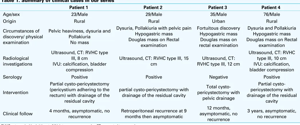
This is a retrospective study of 4 men hospitalized and operated in the department of urology at the Charles Nicolle Hospital of Tunis in Tunisia. Patients were treated for retrovesical hydatid cyst over 9 years (January 2004– December 2013). The average patient age was 40.75 years (range: 23–76). The geographical origin was specified for all patients, 3 patients came from the rural northwest part of Tunisia.
Signs of bladder irritation and pelvic pain were the most frequent presenting complaint. In 1 patient the discovery was fortuitous. No case of hydaturia was noted. At physical examination, 3 patients had an hypogastric mass and a mass in the pelvic pouch of Douglas on rectal examination (Table 1). Complete blood count has shown a hyper-eosinophilia in 2 patients. Hydatid serology was performed for all patients and was positive in 3 patients (ELISA test).
All patients have benefited from an abdominal ultrasound which demonstrated hydatid cyst retrovesical in all cases. The cyst was multivesicular type III according to the clas-sification of Gharbi (Fig. 1) in 3 patients and monovesicular in 1 case. This same ultrasound revealed a second liver localization in 3 cases.
A plain film of urinary tract was done for all patients and revealed a calcified cyst in 1 case (Fig. 2). A complement by intravenous urography was done for 2 patients, revealing compression of the bladder (Fig. 3).
A computed tomography (CT) scan was performed in all patients preoperatively to clarify the relationship with adja-cent organs. The CT scan revealed typical scannographic images of a hydatid cyst, multi-loculated, thick-walled and calcified in some places, displacing the bladder forward, without contrast (Fig. 4). The cyst was type I in 1 case (Fig. 5).
Fig. 1. Ultrasound: Retrovesical cystic mass multi-loculated, type III Classification Gharbi.
All patients were operated with a midline subumbilical laparotomy. After protecting the surgical fields imbibed with hypertonic saline, we performed an aspiration of cystic contents; we then injected a scolicide solution (hypertonic saline) into the cyst cavity and held for 10 minutes. After release of the cyst, we performed a partial cystopericystec-tomy with drainage of the residual cavity in 3 patients. Total cystopericystectomy was performed in a patient. The other 3 patients with hydatid cyst of the liver underwent surgery at a later time.
Results
The postoperative course was favourable for all patients. No urinary fistula or suppuration of the residual cavity was
noted. The drain was removed on average on postoperative day 5. The average length of postoperative hospital stay was 9 days. The mean follow-up time was 16 months (range: from 4 months to 3 years). Monitoring was clinical and by ultrasound. One patient had a retroperitoneal recurrence of the cyst at 9 months and a further operation was performed.
Discussion
Hydatidosis is a common parasitic disease in North Africa, especially in Tunisia where it is endemic.3 The surgical inci-dence is 15/100000 inhabitants.2,5 The most affected organs by this parasite are the liver and lungs. Hydatid location at the urogenital tract, dominated by the kidney comes in third place, and 2% to 5% from other locations. Retrovesical
Fig. 4. Computed tomographic appearance of a retrovesical hydatid cyst Type III with bladder compression.
Table 1. Summary of clinical cases in our series
Patient 1 Patient 2 Patient 3 Patient 4
Age/sex 23/Male 29/Male 35/Male 76/Male
Origin Rural Rural Urban Rural
Circumstances of discovery/ physical examination
Pelvic heaviness, dysuria and Pollakiuria
No mass
Dysuria, Pollakiuria with pelvic pain Hypogastric mass Douglas mass on Rectal
examination Douglas mass on Rectal
examination
Radiological investigations
Ultrasound, CT: RVHC type III, 8 cm
IVU: calcification, bladder compression
Ultrasound, CT: RVHC type III, 15 cm
Ultrasound, CT: RVHC type III, 12 cm
Ultrasound, CT: RVHC type III, 10 cm IVU: calcification, bladder compression
Serology Positive Positive Negative Positive
Intervention
Partial cysto-pericystectomy (pericystium adhering to the rectum) with drainage of the
residual cavity
partial cysto-pericystectomy with drainage of the residual cavity
Total cysto-pericystectomy with
pelvic drainage
Partial cysto-pericystectomy with drainage of the residual
cavity
Clinical follow 4 months, asymptomatic, no recurrence
Retroperitoneal recurrence at 9 months then asymptomatic
RVHC: retrovesical hydatid cyst , IVU: intravenous urography; CT: computed tomography.
localization is rare, accounting for only 0.1% to 0.5%,2,3,5 and 1 to 2% of Tunisian series.2,6
A double etiopathogenic mechanism is possible, with the primitive blood-graft of oncosphere or hexcanth and a graft in the pelvic pouch of Douglas of protoscolices coming from the cracking of abdominal hydatid cysts.2,5 Moreover, in our case there were three concurrent liver locations. Another possible location is the lymphatic pathway that joins the venous system of Retzius and the anastomoses of Schmiedel.2,5
Hydatidosis affects mainly young adults, with no gender preference.2,5 In our series, all patients were male. This male predomination should not however lead us to epidemiologi-cal extrapolations given the small sample size. Three patients were all from the northwest part of Tunisia. This hyperen-demic region for hydatidosis has several factors favouring the parasitic cycle: temperate climate allowing the longevity of embryophores and patient location at a large sheep farm, the main hosts of the parasite in Tunisia.7
The clinical manifestations occur at a late stage of opment of the cyst explained by a slow and insidious devel-opment of this affection.2,5 The discovery of the cyst was fortuitous in 1/4 patients. Clinical signs are dominated by palpation of retropubic mass and compressive manifesta-tions, such as the signs of bladder irritation or transit dis-orders.2,5 Three of our patients had a hypogastric mass on palpation and of pelvic pouch of Douglas at rectal exami-nation. The appearance of a hydaturia is pathognomonic, but rare; it signals a cracking cyst in the bladder.2,5 None of our patients had a hydaturia. Sometimes the cyst is dis-covered during a complication: suppuration of the cyst, an anaphylactic shock after his break or a renal insufficiency by ureteral compression.2
The diagnosis of hydatid cyst is mainly based on the ultrasound to specify the location of the cyst, its vascular relations, and the existence of another locations.2,5 There are 5 types of hydatid cyst.8 Type 1 is the monovesicular simple cyst. Types 2, 3, and 5 do not pose a diagnostic problem, but Type 4, called pseudotumoral, poses a problem of dif-ferential diagnosis.
In view of its rarity, a hydatid cyst may not be the first differential diagnosis in a patient presenting with an iso-lated pelvic cyst. On imaging, a retrovesical hydatid cyst may mimic the following conditions: cyst of the seminal vesicle, posterior bladder diverticulum, rectal duplication cyst, hydronephrosis in a pelvic kidney, and large ectopic ureterocoele.
All 4 patients in our series were male. In female patients, however, a retrovesical hydatid cyst may mimic any one of the gynecological conditions, such as ovarian neoplasm or a hydrosalpinx.
When the diagnosis is not clear, a scanner can be used to confirm the pseudo fluid nature of the mass and show
mural calcifications.2 Moreover, the scanner can help us locate other abdominal locations. It can show a cystic mass, although limited, containing daughter vesicles or membrane debonded and which remains unchanged after injection of the contrast agent.2 The presence of daughter cysts on CT is pathognomonic. It also allows us to study the reports of the cyst with adjacent organs, to appreciate the repercussion on the upper urinary tract, and to diagnose atypical forms.2 An intravenous urography may be useful in cases of dilated renal cavities on ultrasound and allows us to assess the impact of this mass on the upper urinary tract.2
The magnetic resonance imaging is not a technique of choice in hydatid disease. It is justified only when other imaging does not establish a diagnosis.2,9 The cyst appears as a circumscribed mass, hyposignal intensity on T1 sequence, hypersignal intensity on T2 and that changes little or not at all after contrast injection. The identification of daughter cysts with septa hyposignal intensity on T1 and T2 sequences is pathognomonic of hydatic cystic.2
Biologically, hydatid serology is moderately sensitive in extrahepatic locations in 30% to 70% of the time,5 but can help the diagnosis if there are doubts or if the ultrasound images are not typical. Indeed this simple and inexpensive test is useful in the positive diagnosis and in the postopera-tive follow-up. All patients had a hydatid serology, which was positive in 3 patients. The hyper eosinophilia suggests hydatid disease in 33% to 53% of cases.5 Two of our patients had hyper eosinophilia.
The treatment of hydatid cyst is surgical. The goal of sur-gery is total cyst excision without spillage and contamination of the field. Location within the narrow confines of the pelvis along with dense adhesions to surrounding structures may render dissection a formidable task.
For some authors, the approach must be extraperitone-al,10,11 to minimize the risk of hydatid dissemination, second-ary suppurations, and postoperative occlusion.10,12 However, a middle laparotomy is recommended whenever the diag-nosis remains hesitant or when there is doubt about the existence of an associated intraperitoneal localization. This approach allows us to simultaneously treat the intra- and extraperitoneal cysts.10 It consists of an injection of a scoli-cide solution (hypertonic saline or hydrogen peroxide) for 10 minutes and draining the cyst.5,10 The technique of choice is total cystopericystectomy.5 The cystopericystectomy can be partial and includes resecting most of the pericystium and sparing the plates in contact with hazardous areas, such as the ureters, vessels, or gastrointestinal tract.5,10 Three of our patients had a partial cystopericystectomy and only 1 patient had a total cystopericystectomy.
irrigation-drainage system to prevent secondary collections and suppu-rations of the residual cavity and detect for urinary fistula.10 Traditional surgery is associated with a wide range of complications motivating the development of minimally invasive managements with minimal morbidity. Kumar and colleagues described 2 cases of retrovesical hydatid cysts which were managed laparoscopically with laparoscopic cyst aspiration, instillation and suction.13
The Palanivelu hydatid system (PHS) is a recently described device to remove the internal contents and the endocyst without excising the entire cyst.It allows minimally invasive management with low chances of spillage of fluid or injury to adjacent organs.14 The PHS has primarily been described in the management of hepatobilliary hydatid cysts where complete excision was extremely difficult.14 The first reported case of laparoscopically treating a pelvic hydatid cyst with the PHS was reported by Subramaniam and col-leagues.15 The use of this technique in the pelvis, particularly for lesions with dense adhesions to surrounding structures, allows a safe minimally invasive option to the existing surgi-cal techniques.15
Postoperative monitoring is needed, but also the abdom-inal-pelvic ultrasound and immunology for many years for early detection of any recurrence.2,5,10
Conclusion
The retrovesical location of the hydatic cyst is rare. The diagnosis is often delayed and clinical symptomatology is dominated by signs of bladder irritation. Ultrasound and hydatid serology allow the diagnosis in most cases. The scanner allows precise preoperative topographical evalua-tion. Surgical treatment consists of a total cystopericystec-tomy in the best case. Retrovesical hydatid disease can be managed currently laparoscopically with the PHS, a mini-mally invasive approach with minimal morbidity. We must also emphasize the significance of primary prevention of this disease to reduce its incidence, morbidity and cost.
Competing interests: The authors declare no competing financial or personal interests.
This paper has been peer-reviewed.
References
1. Ameur A, Lezrek M, Boumdin H, et al. Hydatid cyst of the kidney based on a series of 34 cases. Prog Urol 2002;12:409-14.
2. Ben Ahmed Y, Khemekhem R, Nouira F, et al. Retrovesical hydatic cyst in children: About four cases.
Jpp 2012;25:131-5.
3. Hafsa C, Golli M, Kriaa S, et al. Retrovesical hydatid cyst in children: Report of 3 cases. J Radiol
2007;88:7-8.
4. Hommadi A, Ziadi T, Kasmaoui H, et al. Retrovesical location of hydatid cyst. J Maroc Urol 2008;12:31-3. 5. Khouaja MK, Ben Sorba N, Haddad N, et al. Retrovesical hydatid cyst: Diagnosis and treatment in 8
cases. Prog Urol 2004;14:489-92.
6. Ameur A, Boumadian H, Aqira A, et al. Retrovesical hydatid cyst. A propos of 6 cases. Prog Urol
1998;8:557-60.
7. Hsairi M, Chahed MK, Bchir A, et al. Epidémiologie de l’hydatidose en Tunisie. Tunis Chir 1997; Spécial 18ème Congrès de Chirurgie.
8. Hassine W, Dupuch K, Gharbi HA. Value of ultrasonography in hydatid liver disease in children: A report on 42 cases. J Radiol 1980;61:323-27.
9. Cherkaoui MM, Nassar I, Jroundi L, et al. Hydatid disease of the urinary bladder: A case report. J Radiol
2002;83:45-6.
10. Ben Adbullah R, Hajri M, Aoun K, et al. Retrovesical and retroperitoneal extrarenal hydatid cyst: Descriptive study of 9 cases. Prog Urol 2000;10: 424-31.
11. Bennani S, El Mrini M, Raji A, et al. Isolated retrovesical and retroperitoneal hydatid cysts. 5 case reports.
Ann Urol 1992;26:344-9.
12. Amar J, J Garnier, Faraj A, et al. Isolated retroperitoneal hydatid cyst. A propos of 2 new cases. J Urol
1983;89:147-52.
13. Kumar S, Pandya S, Agrawal S, et al. Laparoscopic management of genitourinary hydatid cyst disease.
J Endourol 2008;22:1709-14. http://dx.doi.org/10.1089/end.2008.0128
14. Palanivelu C, Senthilkumar R, Jani K, et al. Palanivelu hydatid system for safe and efficacious laparoscopic management of hepatic hydatid disease. Surg Endosc 2006;20:1909-13. http://dx.doi.org/10.1007/ s00464-005-0274-7
15. Subramaniam B, Abrol N, Kumar R. Laparoscopic Palanivelu-hydatid-system aided management of retrovesi-cal hydatid cyst. Indian J Urol 2013; 29:59-60. http://dx.doi.org/10.4103/0970-1591.109987