m onoam ine efflux following adm inistration o f antidepressant
drugs: A m icrodialysis study
in vivo
Faddy Sadideen, BSc (Hons)
A thesis submitted in part fulfilment of the
requirements of the University of London, Faculty of Medicine
for the Degree of Doctor of Philosophy
Department of Pharmacology,
School of Pharmacy, University of London,
Brunswick Square, London WCIN 1 AX
All rights reserved
INFORMATION TO ALL USERS
The quality of this reproduction is dependent upon the quality of the copy submitted.
In the unlikely event that the author did not send a complete manuscript and there are missing pages, these will be noted. Also, if material had to be removed,
a note will indicate the deletion.
uest.
ProQuest 10104831
Published by ProQuest LLC(2016). Copyright of the Dissertation is held by the Author.
All rights reserved.
This work is protected against unauthorized copying under Title 17, United States Code. Microform Edition © ProQuest LLC.
ProQuest LLC
789 East Eisenhower Parkway P.O. Box 1346
It is widely accepted that the symptoms o f depression are due, in part, to abnormal
monoaminergic tone in the brain, primarily serotonin, noradrenaline and to a lesser
extent dopamine. This constitutes the monoamine theory o f depression. Antidepressants
(ADs) work by increasing the extracellular concentration o f monoamines at the synapse.
Though, their mechanism is not fully understood, it has been suggested that chronic AD
treatments can affect NMDA receptor function in the brain.
Using in vivo microdialysis in freely moving rats, the effects o f acute, 7-day subchronic and chronic doses o f the ADs paroxetine and clomipramine treatment on the NMDA-
evoked efflux of extracellular DA, 5-HT and their metabolites, DOPAC and 5-HIAA
respectively in the frontal cortex were investigated. The duration of these effects after
48 hours and 14 days o f drug cessation, and the effect of the co-administration of
NMDA antagonists with paroxetine on monoamine levels and their metabolites was also
investigated.
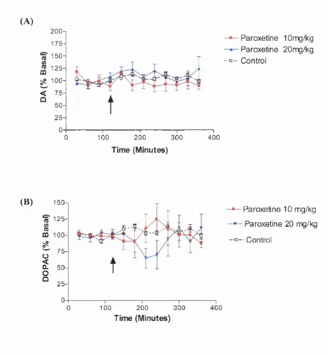
Acute injection of paroxetine (10 and 20 mg/kg i.p.) did not affect dialysate DA or
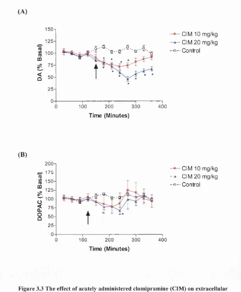
5-HT content in the frontal cortex. Clomipramine at 10 and 20 mg/kg caused a decrease
in extracellular DA without exerting any influence on dialysate 5-HT levels. Local
infusion of lOOpM NMDA into the frontal cortex decreased both extracellular DA and
5-HT levels in this region. 21 day treatment of rats with paroxetine and clomipramine
increased 5-HT levels to 150% and 147% above basal levels respectively. The same
treatment increased DA levels to 200% and 186% above basal levels. When NMDA
infusion was preceded by a single injection of paroxetine/clomipramine no marked
differences between NMDA and NMDA+paroxetine/clomipramine treated groups were
observed. Subchronic (7-days) and chronic (21-days) treatment with
paroxetine/clomipramine were able to abolish the NMDA-evoked decrease in dialysate
DA and 5-HT levels. This effect lasted for a period o f 48 hours but was abolished
following a 14-day ‘drug holiday’. This suggests that adaptive functional changes occur
in NMDA receptor function during treatment with AD drugs. These results suggest that
the NMDA receptor is subject to adaptive changes following chronic AD treatment.
Interestingly, the co-administration o f acute paroxetine with NMDA antagonists
(amantadine, budipine, CGP 40116 and ifenprodil) causes an increase in extracellular
For my family
disease.... Psychodynamic theories are based on stories not facts... nevertheless every person is a story. For the patient, this is the most crucial fact of all.
Donald Goodwin
Professor o f Psychiatry,
I’d like to acknowledge a number o f people who have contributed to the production of
this thesis. I thank Drs Andy Fisher and Philip Thomas who really helped me with their
continual advice throughout my PhD. A big, special ‘thank you’ goes to Dr Clare
Stanford for her invaluable time and effort. I’d like to extend my gratitude to Prof.
Trevor Smart who saved me in times of desperation. Time after time, he was there to
give me the ‘final push’. I also thank Dr Peter Whitton for using his protocols and his
laboratory.
I will not forget others in the Department o f Pharmacology who gave me advice and
assistance. I am grateful to Dave Khan, Helena De Silva, Joanna Watts, Chris Courtice,
the floor crowd (Donna, Steve and Dave), Drs Tracy Assari, Ian Diguid, Owei
Eradiri, Les Fowler, Alistair Hosie, Martin Mortenson and Brian Pearce.
Finally and most importantly, I would like to thank my family one by one. I cannot
forget the support of my dearest mother, Abla, my father, Munir, and sister Massa, who
have always been there for me. My uncle, Mohedin A1 Shatta, has been someone very
special in my life and it has been a great tragedy for us o f the passing away of my
grandparents during the course o f my PhD. Finally and most importantly, I will never
forget the help of my brother, the medic, Hazim for his continuing support. I couldn’t
Ballotta M, Segieth J, Sadideen F, Whitton PS (2001). Repeated but not acute
clomipramine decreases the effect o f Wmethyl-D-aspartate receptor activation on
serotonergic transmission between the raphe nuclei and frontal cortex.
Neuropharmacology. 41, 294-300
Sadideen F. Karbor A, Whitton PS (2001) The effects o f acute and chronic paroxetine
treatment on basal and NMDA-stimulated dopamine release in the frontal cortex of
Title 1
Abstract 2
Dedication 3
Quotation 4
Acknowledgements 5
Publications 6
Table of Contents 7
List of Figures 15
List of Tables 21
Abbreviations 22
Chapter 1 Introduction 25
1.1 Introduction: Depression 26
1.2 Epidemiology 27
1.2.1 Social epidemiology of depression 27
1.3 Causes of depression 28
1.4 Clinical diagnosis of depression 28
1.5 Treatment of depression 31
1.5.1 The pharmacological treatment of depression 31
1.5.2 Clinically used antidepressants: 33
1.5.2.1 Monoamine oxidase inhibitors (MAOIs) 33
1.5.2.2 Tricyclic antidepressants (TCAs) 33
1.5.2.3 Selective serotonin reuptake inhibitors (SSRIs) 34
1.5.2.4 Selective serotonin and noradrenaline reuptake
inhibitors (SNRIs) 36
1.5.2.5 Selective noradrenaline reuptake inhibitors (SNARIs) 36
1.6 Beyond pharmacological treatment 38
1.6.1 Electroconvulsive shock therapy (ECT) 38
1.7 The monoamine theory o f depression 39
1.8 Monoamine theory: Revised 39
1.9 Role of 5-HT in depression 40
1.12 Monoaminergic neurotransmitter interactions 47
1.12.1 DA-5-HT interactions 47
1.13 Depression and the glutamatergic system 49
1.13.1 The glutamatergic System 49
1.13.2 NMDA receptors 50
1.13.2.1 Pharmacology 50
1.13.3 Molecular biology 51
1.13.4 NMDA receptor antagonists 54
1.14 NMDA and depression: 54
1.14.1 Molecular evidence for NMDAR involvement in depression 55
1.14.2 NMDA, monoamines and intracellular second messengers 56
1.15 Principle drugs used in this study: Mechanisms of action 58
1.15.1 Paroxetine 58
1.15.2 Clomipramine 59
1.16 Aims o f this study 61
Chapter 2 Materials and Methods 62
2.1 Materials and Methods: Determining levels/concentrations
of brain extracellular fluids: In Vivo mehods 63
2.2 In Vivo sampling techniques 63
2.3 Microdialysis: What is being measured? 66
2.4 Animals used for dialysis experiments 68
2.5 Stereotaxic surgery and probe implantation 68
2.6 Microdialysis probe construction 72
2.7 The in vitro recovery o f perfusate from constructed microdialysis probes 73 2.8 HPLC-ElectroChemical Detection (BCD) parameters and hardware 77
2.9 Chromatograph calibration 77
2.10 Experimental drugs 7 8
2.11 Experimental protocol 78
treatment on NMDA- evoked extracellular DA and DOPAC
changes in the frontal cortex. 84
3.1 Introduction 85
3.2 Results 88
3.2.1 Basal levels o f DA and DOPAC measured in the frontal cortex. 88
3.2.2 The effect of acute paroxetine on extracellular levels of DA
and DOPAC in the frontal cortex. 88
3.2.3 The effect o f acute clomipramine on extracellular levels of
DA and DOPAC levels in the frontal cortex. 88
3.2.4 The effect o f NMDA infusion into the frontal cortex on
extracellular DA and DOPAC levels in the frontal cortex 92
3.2.5 Effect o f acute paroxetine on 100 pM NMDA-evoked changes
in extracellular DA and DOPAC in the frontal cortex 92
3.2.6 Effect o f acute clomipramine on 100 pM NMDA-evoked
changes in extracellular DA and DOPAC in the frontal cortex 95
3.2.7 Effect o f 7-day (sub-chronic) dosing o f paroxetine on
100 pM NMDA-evoked changes in extracellular DA and
DOPAC levels in the frontal cortex 95
3.2.8 Effect of 21 -day (chronic) dosing o f paroxetine on
100 pM NMDA-evoked changes in extracellular DA and
DOPAC levels in the frontal cortex 98
3.2.9 Effect o f 21 -day (chronic) dosing o f paroxetine with 48 hours
‘drug holiday’ on 100 pM NMDA-evoked changes in
extracellular DA and DOPAC levels in the frontal cortex 98
3.2.10 Effect o f 21-day (chronic) dosing o f paroxetine with 14 days
‘drug holiday’ on 100 pM NMDA-evoked changes in DA and
DOPAC levels in the frontal cortex 102
3.2.11 Effect o f 7-day (sub-chronic) dosing of clomipramine on
100 pM NMDA-evoked changes in extracellular DA and
100 pM NMDA-evoked changes in extracellular DA and
DOPAC extracellular levels in the frontal cortex 104
3.2.13 Effect o f 14-day (chronic) dosing o f clomipramine on
100 pM NMDA-evoked changes in extracellular DA and
DOPAC in the frontal cortex 105
3.2.14 Effect o f 14-day (chronic) dosing o f clomipramine with
48 hours ‘drug holiday’ on 100 pM NMDA-evoked changes
in extracellular DA and DOPAC in the frontal cortex 109
3.2.15 Effect o f 14-day (chronic) dosing of clomipramine with
14 days ‘drug holiday’ on 100 pM NMDA-evoked changes
in DA and DOPAC levels in the frontal cortex 109
3.3 Discussion 114
3.3.1 The effect o f acute, subchronic (7-day) and chronic
(21-day) paroxetine treatment on extracellular DA and
DOPAC changes in the frontal cortex 114
3.3.1.1 Acute paroxetine and clomipramine treatment 114
3.3.1.2 Local infusion o f paroxetine 117
3.3.1.3 Subchronic and chronic paroxetine/clomipramine
treatment 118
3.3.1.4 Adaptation following AD treatment? 121
3.3.2 Effects of NMDA on basal DA and DOPAC efflux in the
frontal cortex 123
3.3.3 Effects o f NMDA on basal and
paroxetine/clomipramine-induced DA and DOPAC efflux in the frontal cortex 125
3.3.4 Drug holiday o f paroxetine/clomipramine treatment. Any
difference in NMDA-induced DA and DOPAC efflux in the
frontal cortex 126
3.3.5 Change in the NMDA receptor following chronic AD treatment? 129
treatment on NMDA-evoked extracellular 5-HT and 5-HIAA
release in the frontal cortex. 132
4.1 Introduction 133
4.2 Results 135
4.2.1 Basal levels o f 5-HT and 5-HIAA measured in the frontal cortex. 135
4.2.2 The effect of acute paroxetine on extracellular levels of
5-HT and 5-HIAA in the frontal cortex 135
4.2.3 The effect o f acute clomipramine on extracellular levels of
5-HT and 5-HIAA in the frontal cortex 138
4.2.4 The effect o f NMDA infusion into the frontal cortex on
extracellular 5 -HT and 5 -HIAA 138
4.2.5 Effect of acute paroxetine on 100 pM NMDA-evoked changes
in extracellular 5-HT and 5-HIAA in the frontal cortex 141
4.2.6 Effect o f acute clomipramine on 100 pM NMDA-evoked
changes in extracellular 5-HT and 5-HIAA in the frontal cortex 141
4.2.7 Effect of 7-day (sub-chronic) dosing of paroxetine on
100 pM NMDA-evoked changes in extracellular 5-HT and
5-HIAA levels in the frontal cortex 144
4.2.8 Effect o f 21 -day (chronic) dosing o f paroxetine on
100 pM NMDA-evoked changes in extracellular 5-HT and
5-HIAA levels in the frontal cortex 144
4.2.9 Effect o f 21 -day (chronic) dosing o f paroxetine with 48 hours
‘drug holiday’ on 100 pM NMDA-evoked changes in
extracellular 5-HT and 5-HIAA in the frontal cortex 147
4.2.10 Effect o f 21-day (chronic) dosing o f paroxetine with 14 days
‘drug holiday’ on 100 pM NMDA-evoked changes in 5-HT
and 5-HIAA levels in the frontal cortex 149
4.2.11 Effect of 7-day (sub-chronic) dosing of clomipramine on
100 pM NMDA-evoked changes in extracellular 5-HT and
5-HIAA levels in the frontal cortex 149
100 pM NMDA-evoked changes in extracellular 5-HT and
5-HIAA levels in the frontal cortex 152
4.2.13 Effect o f 14-day (chronic) dosing o f clomipramine on
100 pM NMDA-evoked changes in extracellular 5-HT and
5-HIAA in the frontal cortex. 152
4.2.14 Effect o f 14-day (chronic) dosing o f clomipramine with
48 hours ‘drug holiday’ on 100 pM NMDA-evoked
changes in extracellular 5-HT and 5-HIAA in the frontal cortex. 153
4.2.15 Effect o f 14-day (chronic) dosing o f clomipramine with 14 days
‘drug holiday’ on 100 pM NMDA-evoked changes in 5-HT
and 5-HIAA levels in the frontal cortex 157
4.3 Discussion 160
4.3.1 The effect of acute, subchronic (7-day) and chronic (21-day)
paroxetine and clomipramine treatment on extracellular 5-HT
and 5-HIAA changes in the frontal cortex 160
4.3.1.2 Different routes o f administration: Local infusion 162
4.3.1.3 At the synapse 163
4.3.1.4 Duration o f treatment 164
4.3.1.5 Adaptation/Genetic regulation? 166
4.3.2 The effects of NMDA infusion on basal 5-HT and 5-HIAA
efflux in the frontal cortex. 168
4.3.3 The effects o f acute, subchronic and chronic paroxetine and
clomipramine treatment on basal and NMDA receptor
activation on serotenergic transmission in the frontal cortex 170
4.3.4 Drug Holiday o f paroxetine/clomipramine treatment. Any
difference in NMDA-induced 5-HT and 5-HIAA efflux in the
frontal cortex? 170
4.3.5 A change in NMDA receptor subunit composition? 172
antagonists 176
5.1. Introduction 177
5.1.1 NMDA antagonsits 177
5.1.1.1 Competitive antagonists 177
5.1.1.2 Non-competitive antagonists 178
5.1.1.2a Ion channel blockers - amantadine and
budipine 178
5.1.1.2b Polyamine site antagonists - ifenprodil 180
5.1.2 Is it possible to produce a rapid AD response? 180
5.1.3 The role of NMDA receptors in AD action 181
5.2 Results 182
5.2.1 The effect of CGP40116 on paroxetine-induced changes in the
extracellular levels of DA, DOPAC, 5-HT and 5-HIAA in the
frontal cortex 182
5.2.2 The effect of amantadine on paroxetine-induced changes in the
extracellular levels o f DA, DOPAC, 5-HT and 5-HIAA in the
frontal cortex 185
5.2.3 The effect o f budipine on paroxetine-induced changes in the
extracellular levels of DA, DOPAC, 5-HT and 5-HIAA in
the frontal cortex 188
5.2.4 The effect o f ifenprodil on paroxetine-induced changes in the
extracellular levels o f DA, DOPAC, 5-HT and 5-HIAA in
the frontal cortex 191
5.2.5 The effect o f 7-day (sub-chronic) dosing o f paroxetine on
amantadine induced changes in the extracellular levels
of DA, DOPAC, 5-HT and 5-HIAA in the frontal cortex 194
5.3. Discussion 198
Chapter 6. Concluding Remarks 205
6.1 Concluding remarks 206
6.2 Other areas o f interest and future directions 208
Appendix
I. Rat weights 255
LI The effect of chronic paroxetine and clomipramine dosing on
body weight over 21 days 255
I.II Effect of chronic (21 days) dosing of paroxetine with 48 hours
‘drug holiday’ on body weight 256
I. Ill Effect o f chronic (21 days) dosing of paroxetine with 14 days
‘drug holiday’ on body weight 256
I.IV Effect o f chronic (14 days) dosing of clomipramine with
48 hours ‘drug holiday’ on body weight 258
LV Effect o f chronic (14 days) dosing of clomipramine with 14 days
‘drug holiday’ on body weight 258
Chapter 1
Figure 1.1 Relationship between the biochemical changes in
depression and the mode of action o f antidepressants 32
Figure 1.2 Action o f SSRIs on serotonin reuptake 34
Figure 1.3 Selectivity o f various antidepressants for NA and 5-HT
uptake c a r r i e r svitro 35
Figure 1.4 Biochemical events at serotonergic synapses 41
Figure 1.5 Serotonergic pathway in rat brain 42
Figure 1.6 Schematic representation of the main serotonergic pathways
involved in the main therapeutic actions of the SSRIs 43
Figure 1.7 Dopaminergic pathway in rat brain 44
Figure 1.8 Noradrenergic pathway in rat brain (locus coeruleus system) 46
Figure 1.9 Schematic diagram of the monoaminergic cascade induced by
ADs. 48
Figure 1.10 Diagram of NMDA receptor in neuronal membrane, shown
with binding sites 50
Figure 1.11 Linking conventional ADs to reductions in NMDA receptor
function 57
Figure 1.12 Chemical structure o f paroxetine 59
Figure 1.13 Chemical structure o f clomipramine 60
Chapter 2
Figure 2.1 The stereotaxic frame
Figure 2.2 Stereotaxic surgery
Figure 2.3 Microdialysis: The technique
Figure 2.4 Microdialysis probe
Figure 2.5a Calibration curve for DA
Figure 2.5b Calibration curve for DOPAC
Figure 2.5c Calibration curve for 5-HT
69
70
71
73
81
81
82
Figure 2.6a Typical chromatogram o f a standard monoamine dialysate
Figure 2.6b Typical chromatogram of a sample dialysate
83
83
Chapter 3
Figure 3.1 The effect of acutely administered paroxetine on extracellular
levels of A) DA and B) DOPAC in the frontal cortex. 89
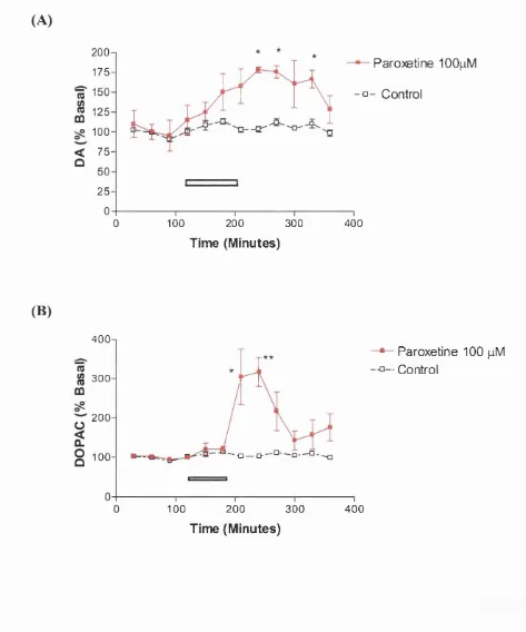
Figure 3.2 The effect of localised infusion of paroxetine on extracellular
A) DA and B) DOPAC in the frontal cortex. 90
Figure 3.3 The effect o f acutely administered clomipramine on extracellular
levels of A) DA and B) DOPAC in the frontal cortex. 91
Figure 3.4 The effect o f NMDA infusion in the frontal cortex on
extracellular A) DA and B) DOPAC in the frontal cortex. 93
Figure 3.5 Effect of acute paroxetine (Parox) on 100 pM NMDA-evoked
changes in extracellular A) DA and B) DOPAC in the frontal
cortex. 94
Figure 3.6 Effect of acute clomipramine (CLM) on 100 pM NMDA-
evoked changes in extracellular A) DA and B) DOPAC in
the frontal cortex. 96
Figure 3.7 Effect o f 7-day (sub-chronic) dosing of paroxetine (Parox) on
100 pM NMDA-evoked changes in extracellular A) DA and
B) DOPAC in the frontal cortex 97
Figure 3.8 Effect of 21-day (chronic) dosing of paroxetine (Parox) on
100 pM NMDA-evoked changes in extracellular A) DA and
B) DOPAC in the frontal cortex. 100
Figure 3.9 Effect o f 21-day (chronic) dosing of paroxetine (Parox,
10 mg/kg) with 48 hours ‘drug holiday’ on 100 pM
NMDA-evoked changes in extracellular A) DA and B) DOPAC in
the frontal cortex. 101
10 mg/kg) with 14 days ‘drug holiday’ on 100 pM NMDA-
evoked changes in extracellular A) DA and B) DOPAC in
the frontal cortex. 103
Figure 3.11 Effect o f 7-day (sub-chronic) dosing of clomipramine (CIM)
on 100 pM NMDA-evoked changes in extracellular A) DA
and B) DOPAC in the frontal cortex. 106
Figure 3.12 Effect o f 21-day (chronic) dosing o f clomipramine (CIM) on
100 pM NMDA-evoked changes in extracellular A) DA
and B) DOPAC in the frontal cortex. 107
Figure 3.13 Effect o f 14-day (chronic) dosing o f clomipramine (CIM) on
100 pM NMDA-evoked changes in extracellular A) DA
and B) DOPAC in the frontal cortex. 108
Figure 3.14 Effect o f 14-day (chronic) dosing o f clomipramine (CIM,
10 mg/kg) with 48 hours ‘drug holiday’ on 100 pM NMDA-
evoked changes in extracellular DA and DOPAC in the
frontal cortex. I l l
Figure 3.15 Effect o f 14-day (chronic) dosing o f clomipramine (CIM,
10 mg/kg) with 14 days ‘drug holiday’ on 100 pM NMDA-
evoked changes in extracellular A) DA and B) DOPAC in
the frontal cortex. 112
Figure 3.16 Schematic representation o f the interrelationship between
serotonergic, dopaminergic and noradrenergic transmission in
the frontal cortex at the presynaptic level: autoreceptors and
heteroreceptors 119
Figure 3.17 Sketch to show the effects o f chronic antidepressants
on DA transmission 131
Chapter 4
A) 5-HT and B) 5-HIAA in the frontal cortex. 136
Figure 4.2 The effect o f localised paroxetine infusion on extracellular
A) 5-HT and B) 5-HIAA in the frontal cortex. 137
Figure 4.3 The effect o f acutely administered clomipramine (CIM)
on extracellular A) 5-HT and B) 5-HIAA in the frontal cortex. 139
Figure 4.4 The effect o f NMDA infusion in the frontal cortex on
extracellular A) 5-HT and B) 5-HIAA in the frontal cortex. 140
Figure 4.5 Effect of acute paroxetine (Parox) on 100 pM
NMDA-evoked changes in extracellular A) 5-HT and B) 5-HIAA in
the frontal cortex 142
Figure 4.6 Effect o f acute clomipramine (CIM) on 100 pM
NMDA-evoked changes in extracellular A) 5-HT and B) 5-HIAA in
the frontal cortex. 143
Figure 4.7 Effect o f 7-day (sub-chronic) dosing o f paroxetine (Parox) on
100 pM NMDA-evoked changes in extracellular A) 5-HT and
B) 5-HIAA in the frontal cortex. 145
Figure 4.8 Effect o f 21-day (chronic) dosing o f paroxetine (Parox) on
100 pM NMDA-evoked changes in extracellular A) 5-HT and
B) 5-HIAA in the frontal cortex. 146
Figure 4.9 Effect o f 21 -day (chronic) dosing o f paroxetine (Parox,
10 mg/kg) with 48 hours ‘drug holiday’ on 100 pM NMDA-
evoked changes in extracellular A) 5-HT and B) 5-HIAA. 148
Figure 4.10 Effect o f 21-day (chronic) dosing o f paroxetine (Parox,
10 mg/kg) with 14 days ‘drug holiday’ on 100 pM NMDA-
evoked changes in extracellular A) 5-HT and B) 5-HIAA in
the frontal cortex. 150
Figure 4.11 Effect o f 7-day (sub-chronic) dosing of clomipramine (CIM)
on 100 pM NMDA-evoked changes in extracellular A) 5-HT
and B) 5-HIAA in the frontal cortex. 151
Figure 4.12 Effect o f 21 -day (chronic) dosing o f clomipramine (CIM)
on 100 pM NMDA-evoked changes in extracellular A) 5-HT
and B) 5-HIAA in the frontal cortex 154
100 pM NMDA-evoked changes in extracellular A) 5-HT
and B) 5-HIAA in the frontal cortex. 155
Figure 4.14 Effect o f 14-day (chronic) dosing o f clomipramine (CIM,
10 mg/kg) with 48 hours ‘drug holiday’ on 100 pM NMDA-
evoked changes in extracellular A) 5-HT and B) 5-HIAA in
the frontal cortex. 156
Figure 4.15 Effect o f 14-day (chronic) dosing of clomipramine (CIM,
10 mg/kg) with 14 days ‘drug holiday’ on 100 pM NMDA-
evoked changes in extracellular A) 5-HT and B) 5-HIAA in
the frontal cortex. 158
Figure 4.16 Sketch to show the effects o f chronic ADs on 5-HT transmission. 175
Chapter 5
Figure 5.1 The effect of CGP-40116 on paroxetine-induced changes
in the extracellular levels o f A) DA and B) DOPAC in
the frontal cortex. 183
Figure 5.2 The effect o f CGP-40116 on paroxetine-induced changes
in the extracellular levels of A) 5-HT and B) 5-HIAA in
the frontal cortex. 184
Figure 5.3 The effect of amantadine on paroxetine(Parox)-induced
changes in the extracellular levels o f A) DA and
B) DOPAC in the frontal cortex. 186
Figure 5.4 The effect o f amantadine (Aman) on paroxetine
(Parox)-induced changes in the extracellular levels of
A) 5-HT and B) 5-HIAA in the frontal cortex. 187
Figure 5.5 The effect o f budipine on paroxetine(Parox)-induced
changes in the extracellular levels of A) DA and B) DOPAC
in the frontal cortex. 189
Figure 5.6 The effect of budipine on paroxetine(Parox)-induced changes
in the extracellular levels o f A) 5-HT and B) 5-HIAA in the
frontal cortex. 190
in the extracellular levels o f A) DA and B) DOPAC in the
frontal cortex. 192
Figure 5.8 The effect of ifenprodil on paroxetine(Parox)-induced changes
in the extracellular levels o f A) 5-HT and B) 5-HIAA in the
frontal cortex. 193
Figure 5.9 The effect o f 7-day (sub-chronic) dosing o f paroxetine
(Parox) on amantadine induced changes in the extracellular
levels of A) DA and B) DOPAC in the frontal cortex. 195
Figure 5.10 The effect of 7-day (sub-chronic) dosing o f paroxetine
(Parox) on amantadine induced changes in the extracellular
levels o f A) 5-HT and B) 5-HIAA in the frontal cortex. 196
Table 1.1 Different types of depression: Severity (Data criteria for
depressive disorders in DSM IV) 30
Table 1.2 The historical development of antidepressants 31
Table 1.3 Side effects of antidepressants 37
Table 1.4 Distribution and function o f excitatory amino acid
receptors in the mammalian CNS 53
Table 2.1 The advantages and disadvantages of microdialysis in
comparison to other in vivo techniques 67
Table 2.2 In Vitro recovery of monoamines and their metabolites from
Standard solutions 76
Table 3.1 Summary o f chapter 3 results 113
Table 4.1 Summary o f chapter 4 results 159
Table 5.1 Summary o f chapter 5 results 197
AADC ACPC ACSF AMPA Aman ANOVA AP5 BDNF BBB cAMP CBT CGP40116 CIM CMS CNS COMT CREB CRH CSF DA
5, 7 DCKA
DOPAC ECT FC GABA GLU GLY GP HAM-D 5-HIAA HPA
Aromatic L-amino acid decarboxylase
1-aminocyclopropanecarboxylic acid
Artificial cerebrospinal fluid
a-amino-3-hydroxy-5-methyl-iosoxazole-4-proprionate
Amantadine
Analysis of variance
2-amino-5-phosphopentanoic acid
Brain-derived neurotrophic factor
Blood-brain barrier
Cyclic adenosine monophosphate
Cognitive behaviour therapy
(R)-(E)-2-amino-4-methyl-5-phosphono-3-pentanoic acid
Clomipramine
Chronic mild stress
Central nervous system
Catechol-O-methyltransferase
Cyclic adenosine 3%5’ monophosphate response element binding
protein
Corticotrophin releasing hormone
Cerebrospinal fluid
Dopamine
{^H}-5, 7 dichlorkynurenic acid
3 , 5-dihydroxyphenylacetic acid
Electroconvulsive therapy Frontal cortex y-aminobutyric acid Glutamate Glycine General practitioner
Hamilton depression rating scale
5-hydroxyindoleacetic acid
Hypothalamic-pituitary-adrenocortical axis
HPLC-ED HPLC with electrochemical detection
5-HT 5-hydroxytryptamine
5-HTP 5-hydroxytryptophan
I D. Inner diameter
i.p. Intraperitoneal
i.v. Intravenous
KA Kainate
KD Kilo dalton
LC Locus coeruleus
L-DOPA L-dihydroxyphenylalanine
LTP Long term potentiation
MAOI Monoamine oxidase inhibitor
MHPG 3-methoxy-4-hydroxy-phenylglycol
MK801 Dizocilpine
mRNA Messenger ribonucleic acid
NA Noradrenaline
Nacc Nucleus accumbens
NMDA N-methyl-D-aspartate
NRI Noradrenaline reuptake inhibitor
OB Olfactory bulbectomized
O.D. Outer diameter
8-OH DP AT 8-hydroxy-2-(di-n-propylamino) tetralin hydrobromide
Parox Paroxetine
PCP Phencyclidine
PDE Phosphodiesterase
PKA Protein Kinase A
PKC Protein Kinase C
RIMA Reversible inhibitors of monoamine oxidase type A
RN Raphe nuclei
s.c. Subcutaneous
S.E.M Standard error of the mean
SNRI Selective noradrenaline reuptake inhibitor
ST Striatum
Ti/2 Half-life
Tmax Maximum rate of absorbance
TCA Tricyclic antidepressant
VTA Ventral tegmental area
WAY 100635 N-{2-{4-(2mehoxyphenly)-l-piperazineyl}ethyl}-N-(2-
pyridil)cyclohaxanecarboxamide trihydrochloride
Chapter 1
1.0 Introduction
1.1 Depression
Depression is a potentially life-threatening disorder that affects between 1-12% of the
Western male and 3-25% o f the Western female population (Angst, 1998). It may occur
at any age from early childhood to late life. The impact o f this major psychiatric
disorder has been severely underestimated by traditional approaches that do not value
disability. If disability rather than death is used as a measure of socio-economic burden,
then major depression is the fourth highest most disabling ailment and has been
predicted to rank second only to heart disease by 2020 (Skolnick, 1999). The annual
direct and indirect costs o f depression are estimated to be in excess of £3 billion in the
United Kingdom and $43 billion in the United States. These figures include treatment,
loss o f earnings and productivity (Henry and Rivas, 1997).
Major depression is defined as a chronic state (>2 weeks) o f a patient suffering from at
least one core symptom and at least four secondary symptoms. The core symptoms are:
(i) lack of motivation and loss of interest in daily activities and (ii) the inability to
experience pleasure (anhedonia). The secondary symptoms are: (i) loss of appetite; (ii)
insomnia (increased amount and decreased latency of rapid eye movement (REM) sleep,
as determined by EEG measurement); (iii) motor retardation or agitation; (iv) feelings
of worthlessness or guilt; (v) continuous fatigue; (vi) learning difficulties and (vii)
suicidal thoughts [Diagnostic and Statistical Manual, 4^^ edition (DSM IV), American
Psychiatric Association, 1994].
There are two main types o f depression: unipolar and bipolar. Unipolar depression is
more common than bipolar depression and more often related to adverse circumstances.
This type o f depression is more common later in life and is often associated with
anxiety and aggression. In bipolar depression, mood and behaviour oscillate between
depression and mania. In the manic state, an individual portrays excessive exuberance,
enthusiasm and self-confidence. There is strong evidence for a hereditary link in the
condition. This type o f depression develops earlier in life and may have features in
1.2 Epidemiology
The World Health Organization (WHO) estimates that 5-10 % o f the population
(500 000 000 people) worldwide on any one day may be depressed. Creed (1993),
reported that over 80 million working days per year are lost in the UK through
depression. This is 30 times the number lost due to industrial disputes. As stated above,
the total costs o f depression on the UK economy is £3 billion and this is 1 % o f the
gross national product (Taylor, 2000).
Epidemiological studies in the UK initiated by Shepherd in the 1960s identified
substantial psychiatric morbidity in general care/primary care populations. The annual
prevalence rate is 5-10% o f the population or 30% of those seen in primary care.
Lifetime prevalence o f these disorders are estimated to vary between 20 and 50% of the
population.
1.2.1 Social epidemiology of depression.
Social (occupational) class, domestic position, sex and genetic predisposition are
factors that have been thought to contribute toward the manifestation of depression
(Bebbington, 1998).
There are conflicting reports on the direct link between social (occupational) class and
depression (Bebbington et al 1981 and Kessler et al, 1994). Generally, it is agreed that
the frequency of depression is greater in the less privileged. This is generally thought to
be a consequence o f the lack of advantages they face in the everyday world.
One o f the most obvious causes in the prevalence o f depression is directly related to
changes in domestic situations. These situations could be marital or relationship break
up, bereavement o f a loved one or being made unemployed (see Bruce, 2002). These
domestic changes are indeed stress-related events that trigger up to 50% of all
depression. Early life stress has been thought to play a role in increasing vulnerability to
depression in later life stages (Marano, 1999).
Several studies have concluded that females are more likely to suffer depression than
likely to report depressive symptoms (Sher et al, 2001). Men suffer a suicide rate 2-3
higher than women, a figure which may be artificially high due to the inability of males
to report their depressive condition (see Taylor, 2000).
There is a hereditary component for both unipolar and bipolar depression. The siblings
o f bipolar depressives show morbid risks o f bipolar depression o f 21% and o f unipolar
illness o f 0.5%. Similarly, the prevalence o f bipolar or unipolar depression in siblings of
unipolar depressives is equal to 0.5% and 12.6% respectively (Leboyer et al, 1998).
1.3 Causes of depression.
Depression is a difficult illness to define precisely as it is difficult to differentiate
between unpleasant feelings which are a normal consequence o f stressful events and the
state o f abnormal functioning which could be classified as a depressive illness.
Depression has psychological, environmental and biological roots. Different drugs such
as p- blockers, steroids, the contraceptive pill, opiates and L-DOPA have been shown to
cause depression (Nutt et al, 1997). Depression has also been linked to the
neuroendocrine system and chronic stress (Checkley, 1996) as well as the tryptophan
hydoxylase gene (Bellivier et al 1998), changes in the biochemical activity o f discrete
brain regions (Figure 1.1) (Drevets et al, 1997), and there is a direct link between stress
and the activation of the immune system and depression (see Leonard, 2000).
The areas o f the brain related to mood and emotion contain a high density of
monoaminergic neurones. According to the monoamine theory o f depression (see
section 1.7), which has formed the cornerstone o f research into the illness since the
1960Sythere is a deficit in transmission of noradrenaline, serotonin (Baldessarini, 1975)
and to a lesser extent, dopamine (Wilhier, 1983).
1.4 Clinical diagnosis of depression
Although depression is common in general practice and primary care, very few people
present clear-cut symptoms of depression. Many GPs feel there is a nervous component
to many physical disorders. After looking at recent life events, physical illness and
disorders. The current diagnostic criteria for this are stipulated in the Diagnostic and
Statistical Manual, 4* edition (DSM IV). Five or more of the following symptoms must
have been present nearly everyday during a 2 week period for a diagnosis of depression
may be made (Jackson et al, 1997).
1. Depressed mood for most of the day
2. Markedly diminished interest in or pleasure from normal activities.
3. Significant weight change (either loss or gain)
4. Insomnia or hypersomnia
5. Psychomotor agitation or retardation
6. Fatigue or loss of energy
7. Feelings o f worthlessness or excessive guilt
8. Reduced ability to concentrate
9. Recurrent thoughts o f death or suicide
10. Decreased eye contact, tearfulness, decreased libido and reduced self confidence.
Table 1.1 Different types of depression: Severity (Data criteria for depressive
disorders in DSM IV).
(Adapted from Freeman et al, 1997).
Severity Diagnosis Characteristics Treatment
Mild Adjustment disorder
Dysthymic disorder
Mild, depressive
symptoms for > 2 weeks,
following a stressful event.
Depression lasting > 2
years. Person feels
constantly negative.
Cognitive
behaviour therapy
Counselling,
Antidepressant
drugs
Moderate Major depression Overwhelming depressed
mood with significant
sleep and appetite
disturbances, including
weight loss.
Cognitive
behaviour therapy
ECT
Antidepressant
drugs
Severe Major depression with
melancholia
Total loss o f interest and
pleasure. Psychomotor
retardation.
Cognitive
behaviour therapy
ECT
Antidepressant
1.5 Treatment of depression
After the clinician has diagnosed a patient to be suffering from depression, a treatment
plan is drawn up. Treatment of depression varies among individuals and depression is
not solely treated by antidepressant drugs. There are other psychological treatments
used to combat depressive symptoms (discussed later).
In depression, there are 3 phases of treatment:
• Acute- This treatment will resolve symptoms.
• Continuation- This treatment will ensure the maintenance o f the response.
• Prophylaxis- This treatment will prevent relapse.
1.5.1 The pharmacological treatment of depression
There are several classes of drugs that act as antidepressants. Recently, a new class of
non-tricyclic antidepressants (TCAs) (see section 1.5.2.2 for TCAs).like drugs have
come into clinical use, which are as effective as TCAs but have fewer side effects.
These have been termed ‘atypical ADs’. Their therapeutic actions are usually explained
by the mechanism by which they enhance monoamine function in the brain. These
treatments, their classification and mechanism of actions are summarised in table 1.2
below.
Class
1957-70 TCAs
MAOIs
Tricyclic antidepressants
Non-selective monoamine oxidase inhibitors
1980-90 SSRIs Selective Serotonin reuptake inhibitors
1990-2000 RIMAs
SNRIs
SNARIs
Reversible inhibitors of monoamine oxidase
Serotonin and noradrenaline reuptake inhibitors
Selective Noradrenaline reuptake inhibitors
Table 1.2 The historical development of antidepressants (as categorised by Leonard
and Healy, 2000).
antidepressants which can be used in the treatment of depression (e.g. trazadone and
mianserin).
However, the 3 main classes of antidepressants used clinically are:
1. TCAs e.g. clomipramine and imipramine
2. SSRIs e.g. paroxetine and fluoxetine
3. SNRIs e.g. Venlafaxine
Changes in depression ANHEDONIA
Anorexia, sleep disturbance Decreased dopamnergic functicn Decreased drive and motivation
Decreased serotonergic function
DEPRESSION Decreased noradrenergic function
Decreased mocd Increased cholinergic function Increased number of
(3-and ou,- adrenoceptors
Increased ORF secr^ion
? Sleep disturbance Memory deficit
Anxiety
Hypercortisdaenia Decrease in cellular irrm inity (T cells, natural killer cells)
Increased probability of infections
Figure 1.1 Relationship between the biochemical changes in depression and the
mode of action of antidepressants (Adapted from Leonard and Healy, 2000).
1.5.2 Clinically used antidepressants:
1.5.2.1 Monoamine Oxidase Inhibitors (MAOIs)
In the 1950s it was found that isoniazid and its isopropyl derivative iproniazid, which
were used as antimycobacterials, induced euphoria (regarded as an adverse reaction) in
tubercular patients. It was subsequently found that these agents inhibit MAO, the main
enzyme that metabolises monoamines (Crane, 1956). These observations in large part
contributed to the foundation for the monoamine hypothesis o f depression. The
antidepressant action o f MAO-inhibitors (MAGI) results from the increase in synaptic
concentrations of amines due to increased monoamine release resulting from decreased
enzymatic breakdown. There are two generations o f MAOIs, subdivided on the basis of
their binding characteristics to MAO. Most of the older clinically available MAOIs are
unable to differentiate between the A or B isoform of MAO, which explains some of the
side effects seen with this class o f antidepressant. The first-generation MAOIs (e.g
phenelzine, isocarboxazid and tranylcypromine) are irreversible inhibitors o f MAO, and
are associated with a high toxicity profile. Their toxicity arises from their interaction
with primary amines, such as tyramine, contained in the diet (e.g. mature cheese,
marmite and pickled herrings). This ‘cheese reaction’ can lead to a sudden increase in
cardiac output and hypertension^may cause a cardiovascular crisis and even a stroke. In
contrast, the second-generation MAOIs (e.g. moclobemide) bind reversibly to MAOa,
and have an improved toxicity profile compared to the former agents. Although the first
developed irreversible, non-selective MAOIs are no longer the 1®^ choice in the clinic
because of their numerous side effects, several newly developed, clinically cleaner,
MAOIs remain valuable clinical tools today (for review see Thase et al, 1995).
1.5.2.2 Tricyclic antidepressants (TCAs)
Shortly after the discovery o f the antidepressant action of MAOIs a new class of
antidepressants was discovered; the TCA. TCAs are closely related and were initially
developed as potential neuroleptics. Imipramine, the first TCA, was found to have no
neuroleptic properties, but it proved effective in relieving the symptoms of depression.
Subsequently, minor structural modifications resulted in drugs such as clomipramine,
amitriptyline and desipramine. Contrary to the action o f MAOIs, TCAs increase
but rather by blocking the reuptake carriers which clear the neurotransmitters from the
synaptic cleft. The principal sites of action are the noradrenergic and the serotonergic
uptake carriers. Due to their superior safety, the TCAs have largely supplanted the
clinical use of MAOIs. Most side effects of both classes of antidepressants can be
attributed to the non-specific interactions with peripheral and central cholinergic,
adrenergic, dopaminergic, histaminergic and serotonergic receptors (Gareri et al., 2000)
(Refer to (Fig 1.3) TCAs have been shown to be as effective as more novel
antidepressants and may be more effective in the treatment of severe depression (Stahl,
1999). Clomipramine, as shown in Fig 1.3, displays highest potency at the 5-HT and NA
reuptake site. It is also effective in the treatment of obsessive compulsive disorder.
Unfortunately, high doses can lead to increased risk of seizures as with all TCAs.
1.5.2.3 Selective Serotonin reuptake inhibitors (SSRIs)
5-H T up site
5-HT terminal
5-HT receptor
Figure 1.2 Action of SSRIs on serotonin reuptake. SSRIs have no direct action on 5HT-receptors or on other monoamine transmitters.
Since the development o f the TCAs, research has focussed on developing agents that
share the reuptake properties o f the TCAs but lack the tricyclic structure, which is
believed to be a major determinant of TCA related non-specific receptor binding
characteristics. Among these are the selective reuptake inhibitors o f serotonin (SSRI).
SSRIs have revolutionised the treatment o f depression. In comparison to TCAs and
MAOIs, SSRIs have increased safety and tolerability with fewer side effects (Stahl,
1999). SSRIs act by selectively blocking the re-uptake o f 5-HT into the presynaptic
terminal (Figure 1.2 on previous page; Figure 1.3 below). Their side effects are well
tolerated and most are resolved in approximately 3 weeks, except sexual dysfunction
which may continue (Nutt et al, 1997). However, not all the effects o f the SSRIs on
sexual function are negative. Low acute doses o f the shorter acting SSRIs can be used to
treat premature ejaculation, which is accepted as a very common problem, not only in
those patients with depression or anxiety.
The use o f SSRIs have also been common in other known disorders which have a
depressive symptom and 5-HT component such as anxiety, obsessive compulsive
disorder (OCD) and panic syndrome. The effectiveness o f SSRIs in severe depression is
controversial (Hirschfeld and Schatzberg, 1994).
N A
-s e l e c t i v e
5 - H T
s e l e c t i v e
'4 n is o x e t i n e m a p r o t il i n e
^ --- d e s ip r a m in e
■4--- n o r t r ip t y lin e
^ --- im ip r a m in e
•4--- a m it r ip t y lin e
4 --- c lo m ip r a m in e
4 z i m e l id i n e
4--- t r a z o d o n e
4--- f l u o x e t i n e
-4--- p a r o x e t in e
- c i t a i op ram
Figure 1.3 Selectivity of various antidepressants for NA and 5-HT uptake carriers
1.5.2.4 Selective serotonin and noradrenaline reuptake inhibitors (SNRIs)
SNRIs differ structurally from the TCAs and generally have no direct action on the
muscarinic, histaminic or adrenergic receptors. Venlafaxine and milnacipran are the
only SNRIs currently available. SNRIs block the uptake o f both 5-HT and less so NA
and possibly DA (Bolden-Watson and Richelson, 1993). The specificity o f the reuptake
block and the degrees of side effects o f SNRIs are largely dose dependent. Low doses
block serotonergic function while medium doses block both serotonergic and
noradrenergic function and the highest doses block the reuptake o f all 3 monoamines
(see Roseboom and Kalin, 2000).
1.5.2.5 Selective noradrenaline reuptake inhibitors (SNARIs)
Research has led to the development of SNARIs such as reboxetine (Dostert et al, 1997)
which increases extracellular NA concentrations in the brain. SNARIs do not have the
typical side effects associated with TCAs as SNRIs lack affinity for 5-HT and DA
reuptake sites. SNARIs have low affinity for adrenergic, muscarinic and histaminergic
receptors (Burrows et al, 1998; Wong et al, 2000).
Although the development of these newer agents has improved the physicians’ choice
o f treatment and decreased treatment related side effects, there are still some key issues
that have not been resolved; therapeutic lag persists, and up to a third o f all patients
Table 1.3 Side effects of antidepressants
Antidepressant
Class
Side Effect
TCAs Consequence o f blockade o f muscarinic receptors (atropine
like effect):
dry mouth, blurred vision, raised intraoccular pressure, urinary
retention, constipation, tachycardia, confusion.
Consequence o f blockade o f a i adrenoceptors: orthostatic
hypotension, dizziness.
Consequence o f blockade o f Hi receptors: sedation, weight
gain
Reduced sexual dysfunction.
Cardiotoxicity, particularly in elderly patients and if taken in
overdose, arises from cardiac conduction block (quinidine-like
effect).
SSRIs Neurological side effects:
agitation, akathisia, anxiety, insomnia, sexual dysfunction
Vascular side effects:
Headache, migraine-like attacks
Gastrointestinal side-effects: nausea, vomiting, diarrhoea.
SNRIs Low dose: same as SSRI
Intermediate to high doses mediated by NA and DA as well as
5-HT.
Hypertension
Severe insomnia
Severe agitation
Severe nausea
1.6 Beyond pharmacological treatment
There are a number of alternative treatments that maybe used instead, or in combination
with, antidepressant drug therapy such as Electroconvulsive Shock therapy (ECS)
counselling and social intervention; cognitive behaviour therapy (CBT) and
Psychotherapy. In addition to the above treatments there are several complementary and
alternative therapies in the treatment of depression, such as exercise, acupuncture, and
relaxation therapy. These therapies are beyond the scope of this thesis.
1.6.1 Electroconvulsive shock therapy
Electroconvulsive therapy (ECT) has been practiced for over 60 years and is still used
today to successfully treat severe depression. ECT in man involves stimulation of the
brain or parts o f it by means o f electrodes placed on the head, with the patient slightly
anaesthetised and paralysed with neuromuscular blocking drugs so as to avoid physical
injury. ECT is occasionally used in hospitalised patients who do not respond to
conventional antidepressant therapy and/or have a high risk o f committing suicide
(Gareri. et al, 2000). Despite being very effective, it has several major disadvantages.
These include the levels of distress experienced by the patient undergoing this therapy
and chronic episodes of amnesia and cognitive abnormalities in addition to ECT being
substantially more expensive than pharmacotherapy.
Electroconvulsive therapy (ECT) still remains the treatment that is considered to be the
most potent in refractory depressive disorders. This treatment is usually restricted to
patients hospitalised for severe depression. An advantage o f this treatment is that it may
be used in the elderly as it maybe safer than antidepressant drugs. Rates of
responsiveness of up to 90% can be expected in delusional types of depression. There is
therefore the temptation to resort to ECT in any non-responsive condition. Generally,
ECT is a safe, effective treatment but there are risks o f memory impairment in
1.7 The monoamine theory of depression
Because o f the high prevalence o f depression the pharmaceutical industry have placed
great emphasis on unravelling its pathogenesis. This has resulted in several hypotheses
of depression pathophysiology. The initial hypothesis proposed by Schildkraut (1965)
suggested that depression was linked to a deficiency o f the noradrenergic
neurotransmitter system in the central nervous system (CNS). This theory contributed to
what became known as the monoamine theory o f depression. This hypothesis originated
from the observation that agents that deplete monoamines cause depressive symptoms.
This was first observed when a high proportion o f patients taking the rauwolfia alkaloid
‘reserpine’, for the treatment o f hypertension, developed symptoms o f depression
(Schildkraut, 1965). With ongoing research, it became evident that this theory had to be
revised to include a deficiency in central serotonergic (Praag and Korf, 1971) and to a
lesser extent, dopaminergic systems (Willner, 1983).
The monoamine hypothesis is supported by the fact that all clinically useful
antidepressant therapeutic agents act by increasing synaptic levels of monoamines e.g.
noradrenaline (NA), 5-hydroxytryptamine (5-HT) and dopamine (DA). However, this
theory does not explain the discrepancies between the rapid effects on monoamine
metabolism (i.e. release and reuptake caused by antidepressants) and the delayed onset
of clinical action, which usually takes up to several weeks to develop. An additional
drawback o f this theory is that approximately 30% o f patients do not respond to current
antidepressant therapies (Skolnick, 1999). These findings suggest that additional factors
contribute to the therapeutic mechanisms underlying antidepressant action.
1.8 Monoamine theory: Revised
The discrepancy between the onset o f neurochemical actions and the onset of clinical
action has led to the hypothesis that one or more adaptive changes must precede a
clinical antidepressant response (Vetulani, 1991; Duman et al, 1997). This hypothesis
was initially substantiated by the observation that activity o f central p-adrenoreceptors
in the cortex is decreased following chronic antidepressant treatment (Vetulani and
Sulser, 1975; Stanford et al, 1983). Subsequently, it has been shown that chronic
antidepressant treatment causes adaptive changes in the efficacy o f the monoaminergic
1980) and downregulation o f receptors (e.g. Pi, 5-HT2, Di) (Papp et al, 1994). These
observations have resulted in the monoamine hypothesis being revised to include the
adaptive changes caused by chronic antidepressant treatment. Currently, the generally
accepted mechanism o f antidepressant action is that as a result o f persistent elevated
synaptic monoamine levels, with concomitant receptor activation, there is an adaptive
decrease in receptor density or function. These adaptive changes are thought to underlie
the clinical action of antidepressant drugs. However it is still poorly understood how
these adaptive changes are linked to the clinical improvement.
Although there is general acceptance of the revised monoamine hypothesis, there are
several shortcomings. Firstly, there is not one general pattern o f neurological adaptive
change in any o f the monoaminergic systems that is shared by all antidepressant
treatments. This is illustrated by the fact that p-adrenoreceptor downregulation is seen
after treatment with tricyclic antidepressants (TCA’s) but not after treatment with all the
so-called selective serotonin reuptake inhibitors (SSRI’s) (Vetulani, 1991). Secondly,
there is a discrepancy between the time course of P-adrenoreceptor and 5-HT
dovmregulation and the clinical onset of antidepressant action (Duman et al, 1997).
Thirdly, attenuation of adrenergic functioning by p-adrenoreceptor antagonists fails to
elicit any antidepressant affects and can aggravate them (see Hirschfeld, 2000).
Based on these findings, it may be hypothesised that the clinical efficacy of
antidepressant drugs which alter monoaminergic transmission may act via a secondary
common signal transduction mechanism, which may be evoked by an adaptive change
in the monoaminergic neurotransmission system. Alternatively, the clinical efficacy of
antidepressant treatment may be mediated by an adaptive change in an, as yet,
uncharacterised neurotransmitter system.
1.9 Role of 5-HT in depression
5-HT has been implicated in the aetiology of depression. 5-HT is an indoleamine that
was first identified as a vasoconstrictor (Rapport, 1949), and was then discovered in the
CNS by Amin et al. (1954). The brain contains only 1-2% o f the body’s 5-HT. As 5-HT
does not cross the blood brain barrier (EBB), it is synthesised in the brain from the
hydroxylase, is responsible for converting L-tryptophan to 5-hydroxtryptophan
(5-HTP). 5-HTP is then rapidly decarboxylated to 5-HT by aromatic L-amino
decarboxylase (AADC) and then transported into storage granules within the
serotonergic neurones.
Serotonergic neuron
5-HTP < ■ L-T ryptophan
5-HT. MAO -►5-HIAA
c :
^ 5-HT^I
□ □□
5-HT receptors on ppstsynaptic tissueFigure 1.4 Biochemical events at serotonergic synapses. (5-HTP:
5-hydroxytryptophan; 5-HT: 5-hydroxytryptamine; 5-HIAA: 5-hydroxindoleacetic
acid).
The cell bodies of serotonin neurones are known to be restricted to an area lying in or
near the midline or raphe regions of the pons and the upper brain stem. Nine 5-HT
nuclei (B1-B9) have been described by Dahlsrom and Fuxe and recently
immunocytochemical localisation of 5-HT has also detected reactive cells in the area
postrema and in the caudal locus coeruleus as well as in and around the interpeduncular
nucleus. The more caudal groups project largely to the medulla and spinal cord. The
more rostral (intermediate) groups (raphe dorsalis, raphe medianus and centralis
superior), also called B7-B9, provides the extensive 5-HT innervation of the
telencephalon and diencephalon.
The main serotonergic pathways originate from the raphe nuclei and have projections to
the preffontal cortex, the hippocampus and the striatum (Figure 1.5). The firing rate of
these neurones is controlled by somatodendritic autoreceptors in the raphe nuclei
Romero and Artigas, 1997; Gobert and Millan, 1999). Additionally, presynaptic auto-
and heteroreceptors regulate the 5-HT release from nerve terminals (Fink et ah, 1996),
Gobert and Millan, 1999), The dorsal raphe distributes 5-HT terminals to areas
innervated by dopamine (e.g. the amygdala, basal ganglia and cortical areas), while the
median raphe innervates the hippocampus and cortex in a similar but more limited
distribution to NA (Azmitia and Segal, 1978).
D R ---Amygdala, Basal Ganglia and Cortex
MRN---Hippocampus and Cortex
Caudate ST
Hypothalamus
Raphe nuclei
Figure 1.5 Serotonergic pathway in rat brain. ST: stria terminalis; D and M: dorsal and medial superior raphe nuclei. (Adapted from Ganong, 1 9 9 9 )
Multiple receptors for serotonin in the CNS have been suggested by physiological and
molecular studies. In the last decade, a vast amount of new information has become
available concerning the various 5-HT receptor subtypes and their functional and
structural characteristics. Pre-synaptically, 5-HT receptors have been divided into
5-HTiA and 5 -H T ib /d subtypes. Post-synaptically, several receptors have been identified
(5-HTiA, iB/D, 2A, 2C, 3, 4, 6, i) (for review, see Barnes and Sharp, 1 9 9 9 ) . The presynaptic
5-HT]A receptors are termed as somatodendritic autoreceptors. These receptors appear
to possess a negative feedback mechanism that partially contributes to presynaptic 5-HT
release. Presynaptic 5-HT]b / d receptors are called terminal autoreceptors and regulate
5-HT release in a similar manner to the 5-HT]a receptors, though they only inhibit
terminal release without having an effect on cell firing (for review see Adell et al,
2002).
During the last decade, preclinical and clinical evidence has accumulated indicating the
involvement of the 5-HT system in the therapeutic action of anti depressant drugs (AD)
(Figure 1.6). Impairment of 5-HT synthesis leads to a transient reappearance of
depressive symptoms in patients in remission obtained with various SSRIs (Delgado et
al., 1990). On the other hand, tryptophan and lithium, which both increase 5-HT
function (Sharp et ah, 1991), can potentiate the therapeutic effect of ADs (de Montigny
et ah, 1983). Thus, there seems to be a clear association between the AD response and
enhanced 5-HT neurotransmission.
Antibulimic action
Antipanic action
Preffontal cortex
Hypothalamus
Midbrain raphe Hippocampus: limbic cortex
Antidepressant action
V
AntiOCD action
Basal ganglia
Figure 1.6 Schematic representation of the main serotonergic pathways involved in
the main therapeutic actions of the SSRIs (Adapted from Leonard and Healy, 2000)
1.10 Role of dopamine in depression
Dopamine (DA) synthesis, similar to all catecholamines in the CNS, originates from the
amino acid precursor tyrosine, which must be transported across the blood-brain barrier
into the DA neuron. The rate-limiting step in dopamine synthesis is the conversion of
L-tyrosine to L-dihydroxyphenylalanine (L-DOPA) by the enzyme tyrosine
hydroxylase. DOPA is subsequently converted to dopamine by aromatic L-amino
decarboxylase (AADC). Since tyrosine hydroxylase is the rate-limiting enzyme in the
biosynthesis of DA, this enzyme sets the pace for the formation of DA and is
particularly susceptible to physiological regulation and pharmacological manipulation.
Released DA is converted to dihydroxyphenylacetic acid (DOPAC) by intraneuronal
MAO after reuptake by the nerve terminal. Released DA is also converted to
homo vanillic acid (HVA), at an extraneuronal site through catechol-0-
methyltransferase (COMT).
The main dopaminergic pathways originate from cell bodies situated in the substantia
nigra (SN) and innervate the striatum, prefrontal cortex and the nucleus accumbens. The
A9 group of the SN mainly innervates the basal ganglia while the AlO group of the
ventral tegmental (VTA) mainly projects to the mesolimbic terminals (Figure 1.7).
A9 (SN) ---Basal Ganglia
AlO (VTA)--- Mesolimbic regions
Frontal cortex
Cingulate cortex
Substantia nigra Striatum
Nucleus accumbens
Hypothalamus Ventral tegmentum
Figure 1.7 Dopaminergic pathway in rat brain. MC: mesocortical system; NS: nigrostriatal system (Adapted from Ganong, 1999)
DA receptors mediate dopaminergic transmission within the CNS. DA synthesis and
release is influenced by neuronal dopaminergic activity. Postsynaptic dopamine
receptors include Di and Di subtypes and are present in the projection areas of the
midbrain DA neurons. They regulate the activity of neuronal feedback pathways e.g.
postsynaptic receptors in the striatum regulate communication pathways between
striatal neurones and DA cell bodies in the SN (see Starr, 1995; Tzscentke, 2001). D2
autoreceptors exist on most portions of DA cells and this determines their effects.
Activation of somatodendritic autoreceptors reduces the rate of neuronal firing whilst
terminal autoreceptors inhibit DA synthesis and release (see Starr, 1995; Tzscentke,
2 0 0 1).
Following chronic treatment with the TCA imipramine, it has been shown that there is a
selective downregulation of dopamine Di receptors in the mesolimbic system of rats
(Serra et al, 1990). However, this observation is not seen in AD-treated suicide
postmortems’ (Bowden et al, 1997). In general, it would appear that dopaminergic
transmission is increased by chronic antidepressant treatment, particularly in the
mesolimbic area (Vetulani, 1991). However, it remains unclear whether changes in DA
function are primary or secondary in depression.
1.11 Role of noradrenaline in depression
Noradrenaline (NA) synthesis is similar to that of DA. The final hydroxylation to
convert DA to NA is catalysed by the non-specific enzyme DA-P-hydroxylase. This
enzyme is restricted to NA producing cells. Similar to DA catabolism, MAO and
COMT are the 2 principal enzymes involved in NA breakdown.
Two major clusterings o f noradrenergic cell bodies have been described within the
brain. The first is the locus coeruleus (LC), a compact cell group (A6) within the caudal
pontine gray. The LC pathways terminate in the thalamus, cortex, amygdala,
hippocampus and hypothalamus (Moore and Bloom, 1979) and are extremely important
physiologically in the regulation o f learning, memory, sleep, adaptation, arousal and
stress (Leonard, 1997). The second group of cells (A l, A2, A3) lie outside the LC and
consist o f mainly descending fibres within the mesencephalon and spinal cord, although
the more anterior tegmental levels innervate the forebrain and diencephalon (for review