OPTIMISATION OF CHO CELL GROWTH AND RECOMBINANT
INTERFERON-y PRODUCTION
A thesis submitted to the University of London for the Degree of
DOCTOR OF PHILOSOPHY by
Paula Maria Lima e Castro
Department of Chemical & Biochemical Engineering
University College London
Torrington Place
London WC1E 7JE
October 1993
I'!"
LONDON
ABSTRACT
The optimisation of recombinant protein production by animal cell cultures is important for the economic feasibility of these processes. Simultaneously with product yield, product authenticity is a crucial aspect to consider as it may per se affect the therapeutic value of such proteins. More defmed culture media are being developed, particularly to ensure batch product consistency. A Chinese Hamster Ovary cell line (CHO 320) producing human interferon-y (IFN-y), a glycosylated protein, was chosen to investigate the effects of the culture environment on (I) cell growth, (2) product yield and (3) product authenticity.
A statistical approach was used to identify important culture components for cell growth and LFN-y production. When the concentration of the resulting positive variables was initially increased in culture, improvements of approximately 40% in both of these parameters were achieved; the glycosylation of JFN-y was not affected. The former analysis also indicated that different stimuli were required for growth and production. Fed-batch feeding of glucose and glutamine, components depleted early from culture, did not prolong cell growth or IFN-y production but the initial glycosylation pattern of WN-y was a function of glutamine concentration.
Bovine serum albumin (BSA) was shown to have important role(s) in culture and cell growth was not possible in its absence. Pluronic F68, alone or in combination with a lipid mixture or linoleic acid, was able to restore cell growth in low BSA (1 mg/ml) cultures. However, JFN-y production was significantly reduced and the extent of IFN-y glycosylation also changed. These effects were related to: (1) BSA concentration, (2) BSA type, and ultimately, (3) lipid composition of the culture.
The results reported in this thesis exhibit the necessity to consider the effects of the culture environment not only on cell growth and product yield but also on product authenticity throughout any optimisation process.
ACKNOWLEDGEMENTS
I would like to express my sincere gratitude to all those who have provided support throughout this work, at University College London and at the University of Kent at Canterbury, where Animal Cell Culture facilities were made available for my research. Special thanks go to my supervisors, Professor Alan T. Bull and Dr. Andrew P. Ison, for their guidance and constructive criticism during my research and in the preparation of this thesis.
I would also like to thank Dr. Nigel Jenkins for allowing me to use the facilities in his laboratory, Dr. Mike Bushell for helpful advice on the statistical methodology and Dr. John Clegg for the encouraging discussions during my research.
I am very grateful to all the members of the Animal Cell Biotechnology Group for making my time at UKC so enjoyable. In particular, I would like to mention Paul Hayter for his helpfuL suggestions in all aspects of my research and for reading and discussing substantial parts of the thesis. I would also like to mention Malcolm Gould, Lucy Gettings, Jerry Tong, David James and Lis Curling for their valuable advice and assistance.
The friendship and encouragement of many friends made my stay in England a period I will always cherish. A special thought goes to José Luz, Paulo Pinto, Helena Assis e Manuel Santos, Paulo Carvalho, Alison Hovey, Mark Calleja, Carlos Sá da Costa, Antonio Lobo and Kioko Kawamura.
A word of gratitude to my parents and my brothers and sisters for their constant support and love.
TABLE OF CONTENTS
Page
ABSTRACT 3
ACKNOWLEDGEMENTS 4
TABLE OF CONTENTS 5
TABLE OF TABLES 10
TABLE OF FIGURES 12
NOMENCLATURE 14
1- INTRODUCTION 17
1.1- Animal cells: a potential resource 17
1.1.1- General introduction 17
1.1.2- Why use animal cells? 18
1.1.3- Products obtained from animal cells in culture 20
1.2- Cultivation systems for animal cells 23
1.2.1- Kinetics of growth and product formation 23
1.2.2- Cultivation techniques 25
1.2.3- Hydrodynamic environment of the cells 28
1.2.3.1- Aeration and agitation 27
1.2.3.2- Additives for cell protection from fluid-mechanical damage 30
1.2.4- Temperature and pH 33
1.3- Cell nutritional environment- the culture medium 34
1.3.1- Development of serum-free medium 34
1.3.2- Cell requirements in serum-free medium 36
1.3.2.1- Energy sources 36
1.3.2.2- Amino acids 39
1.3.2.3- Lipids 40
1.3.2.4- Vitamins and minerals 41
1.3.2.5- Protein supplementation 42
1.3.3- Toxicity in animal cell culture 44
1.4- Production of glycoproteins 46
1.4.1 - Significance of glycosylation 47
1.4.2- The host and its environment 49
1.4.3- Interferon-y 50
1.5- Scope of the work 55
2- MATERiALS AND METHODS 57
2.1- Cell culture system 57
2.1.1- Cell line 57
2.1.2- Culture medium 57
2.1.2.1- Serum-free medium 57
2.1.2.2- Specific media supplementation 59
2.1.3- Cell culture methods 63
2.1.3.1- Cell maintenance 63
2.1.3.2- Shake-flask and fermenter cultures 64
2.1.3.3- Plackett-Burman experimental set-up 68
2.2- Analytical Methods 68
2.2.1 - Determination of CHO cell growth 68
2.2.2- IFN-y analysis 69
2.2.2.1- ELISA assay for IFN-ytitre 69
2.2.2.2- Analysis of JFN-'y glycosylation 71
2.2.2.2.1- Immunoprecipitation of IFN-y 71
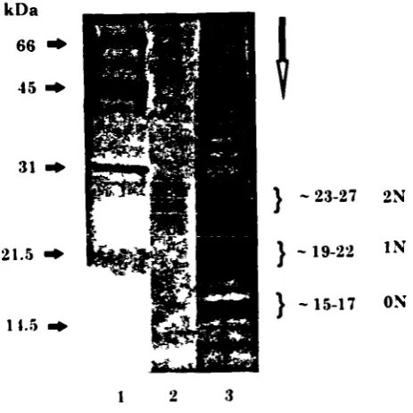
2.2.2.2.2- SDS-page electrophoresis 72
2.2.2.2.3- Silver staining 72
2.2.3- Metabolite analysis 75
2.2.3.1- Enzymatic determination of glucose 75
2.2.3.2- Enzymatic determination of lactate 76
2.2.3.3- Determination of ammonia 77
2.2.3.4- HPLC determination of amino acids 78
2.2.3.4.1- Sample and buffer preparation 79
2.2.3.4.2- Auto-sampler preparation 80
2.3- Experimental data analysis 81
3- STATISTICAL ANALYSIS OF THE CULTURE MEDIUM 83
3.1- Introduction 83
3.2- CHO cell growth and IFN-y production in serum-free medium 86 3.3- Statistical screening of the medium components:
the Plackett-Burman design 88
3.3.1- Experimental design 88
3.3.2- Experimental effects of variables 92
3.4- Cluster effect of positive and negative groups of variables 98 3.4.1- Cell growth and IFN-y production: titre and product quality 98
3.4.2- Analysis of the culture medium 101
3.4.3- The independent effect of BSA 107
3.5- The stimulating effect of the positive variables on CHO cell growth 110
3.5.1 - Effect of methotrexate on CHO cell growth 110
3.5.2- Fed-batch cultures with the positive group of components 112
3.6- Concluding remarks 115
4- FED-BATCH FERMENTER CULTURES 117
4.1- Introduction 117
4.2- Cell growth and IFN-y production in fermenter cultures 120
4.3- Glucose and glutamine fed-batch cultures 120
4.3.1- Glucose and glutamine profiles 122
4.3.2- Cell growth and IFN-'y production 127
4.3.3- IFN-y glycosylation patterns 133
4.4- Metabolic flux changes in batch and fed-batch cultures 135
4.4.1- Glucose and glutamine metabolism 137
4.4.2- Amino acid metabolism 138
4.5- Concluding remarks 141
5- ThE EFFECTS OF BSA, LIPIDS AND PLURONIC F68 ON CHO
CELL CULTURE 144
5.1- Introduction 144
5.2- Defmition of the culture conditions 147
5.3- Effect of reducing the BSA concentration of the culture medium 148 5.3.1- Possible substitutes for BSA- lipids and Pluronic F68 148 5.3.2- Influence of individual lipid components on the behaviour of
low-BSA cultures: Plackett-Burman statistical analysis 153
5.3.3- Linoleic acid substitution of the lipid mixture 159
5.3.4- Effects of fatty acid-free BSA in culture 167
5.3.5- The independent effects of Pluronic F68 and fatty acid-free BSA 172
5.4- Supplementation of control cultures with lipids and Pluronic F68 177
5.4.1 - Addition of lipids to control cultures 177
5.4.2- Addition of Pluronic F68 to control cultures 179
5.5- Effect of serum in culture 182
5.6- Concluding remarks 185
6- GENERAL DISCUSSION 188
6.1- The Plackett-Burman statistical design applied to the optimisation
of the cell culture composition 188
6.2- The cell culture environment 189
6.2.1 - CHO cell growth and JFN-y production enhanced by different stimuli 190 6.2.2- Influence of BSA on CHO cell growth and JFN-y production 193
6.2.2.1- Nutritional effect 194
6.2.2.2- "Protective" effect 194
6.2.2.3- Antioxidant effect 196
6.2.2.4- Cell structural integrity 196
6.2.3- Glycosylation of IFN-'y 198
6.2.3.1- Glycosylation of IFN-'y is not dependent on the rate of IFN-y
production 199
6.2.3.3- The extent of IFN-y glycosylation varies with the concentration
and type of BSA in culture 201
6.2.3.4- Serum affects the patterns of IFN-y 206
6.2.3.5- The impact of the media composition on the production
of glycoproteins 207
6.3- Nature of CHO cell growth limitation 209
7- CONCLUSIONS 211
8- SUGGESTIONS FOR FUTURE WORK 213
REFERENCES 215
TABLE OF TABLES
Page 1.1- Examples of products obtained from animal cells. 21
1.2- Recombinant products in clinical development. 22
1.3- Examples of recombinant protein production by different cultivation techniques 27 1.4- Protection of freely suspended animal cells from agitation and aeration damage. 31 1.5- Factors affecting the glycosylation pattern of recombinant proteins. 50 2.1- Cell culture medium supplements and final concentrations in RPMI 1640. 58 2.2- Solutions prepared to supplement the serum-free culture medium for the
statistical study. 61
2.3- Composition of buffers used for the IFN-y ELISA assay. 69
2.4- Procedure for the ELISA assay for IFN-y. 70
2.5- Buffers for immunoprecipitation of IFN-y. 71
2.6- Reagents required for the Bio-rad Silver Staining procedure. 73
2.7- Silver staining procedure for mini-gels. 73
2.8- Buffers for HPLC determination of derivatised amino acids. 79 2.9- Chromatography program for OPA amino acid assay. 81 3.1- Plackett-Burman matrix for the study of 23 variables with 24 experiments. 89 3.2- Components under study and their corresponding concentrations. 90 3.3- Cell growth and JFN-y production data for Plackett-Burman analysis. 91 3.4- Components and their effects on cell growth rate, viable cell production
and IFN-y titre. 94
3.5- Effects of the positive and negative variables on CHO cell growth and
IFN-y production. 98
3.6- Initial consumption rates of glucose and glutamine in batch control
cultures (I) and batch cultures containing the positive (+11) or negative (-II)
groups of variables. 106
3.7- Accumulation of lactate and ammonia in batch control cultures (I) and
batch cultures containing the positive (+11) or negative (-II) groups of variables. 106 3.8- Independent effect of the two positive group variables (amino acids plus
3.10- Effect of MTX and positive group on CHO cell growth. 112 3.11- Scheduled feeding of the positive group of variables to stirred batch cultures. 113 3.12- Effect of different feeding strategies of the positive group on CHO cell growth. 114 4.1- Feeding regime of glucose and glutamine to fermenter cultures. 122 4.2- Specific cell growth and IFN-y production rates in fermenter cultures. 127 4.3- Ammonia and lactate levels at cessation of cell growth in fermenter cultures 131
4.4- Metabolic quotients for glucose and glutamine in fed-batch cultures. 138
4.5- Amino acid initial consumption/production rates in fed-batch cultures. 139 5.1- Culture environments used in the present Chapter. 147
5.2- Addition of different combinations of a lipid mixture and Pluronic F68 to
1 mg/ml BSA cultures. 150
5.3- IFN-y production rates in cultures A, D and E. 151
5.4- Plackett-Burman matrix for the study of 7 variables. 153
5.5- Components under study and corresponding concentrations. 154
5.6- Cell growth and JFN-y data for Plackett-Burman analysis. 155
5.7- Components and their effects on CHO cell growth and JFN-y production. 157 5.8- Specific cell growth and IFN-y production rates in low-BSA cultures. 159 5.9- Intracellular and extracellular IFN-y accumulation in low-BSA cultures. 162
5.10- Initial JFN-y production rates in low-BSA cultures. 169
5.11- Proportions of the ON and 2N IFN-y glycoforms in low-BSA cultures. 175 5.12- Relative unit cost of IFN-y produced using different media. 187 6.1- Effect of several components on CHO cell growth and IFN-y titre and
glycosylation. 191
6.2- Effect of BSA and medium supplements on interferon-y production. 202
TABLE OF FIGURES
Page 1.1- Diagram of the major metabolic pathways of animal cells
(derived from Miller et al., I 989a). 37
1.2- N-linked oligosaccharide processing in mammalian cells
(derived from Goochee eta!., 1990). 48
1.3- Human interferon-y polypeptide sequence. 51
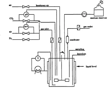
1.4- Biantennary N-glycans associated with 1FN-y (derived from Mutsaers eta!., 1986). 51 1.5- N-glycans associated with IFN-'y (derived from James eta!., 1993). 53 2.1- Process diagram for fermenter cultures under batch and fed-batch operation. 67 2.2- Analysis of JFN-y variants produced by CHO cells. 74 3.1- Kinetics of CHO cell growth and IFN-y production in stirred batch culture. 87 3.2- Glycosylation patterns of IFN-y obtained from a) control cultures and b) cultures
containing increased concentrations of the positive group of variables. 100 3.3- Cell growth in control batch cultures (I), and cultures containing the positive
group of nutrients (+11) or negative group of nutrients (-II). 102 3.4- Glucose and amino acid consumption/production in control batch cultures (I),
and cultures containing the positive group of nutrients (+11)
or negative group of nutrients (-II). 103
3.5- Effect of supplementing the culture with different sources of BSA on
CHO cell growth. 108
3.6- Synthesis of glycine from serine and tetrahydrofolate. 111 4.1- a) Cell growth and b) IFN-y production in control fermenter cultures and
cultures containing the positive group of variables. 121 4.2- Glucose concentration profile in fed-batch cultures a) FF1, b) FF2 and c) FF3. 124 4.3- Glutamine concentration profile in fed-batch cultures a) FF1, b) FF2 and c) FF3. 125 4.4- Cumulative consumption of a) glucose and b) glutamine in fed-batch cultures. 126 4.5- a) Cell growth, b) cell viability and c) IFN-'y production in fed-batch cultures. 128 4.6- Accumulation of a) ammonia and b) lactate in fed-batch cultures. 130 4.7- Percentage of each amino acid remaining in fed-batch cultures at the cessation
4.8- Glycosylation patterns of JFN-y obtained from fermenter cultures with
a,b) 2 mM initial glutamine and c,d)
0.5
mM initial glutamine concentration. 1344.9- Major routes of glucose metabolism. 136
4.10- Major routes of glutamine metabolism. 136
4.11- a) Glutamate and b) alanine production in fed-batch cultures. 142 4.12- Metabolism of serine and glycine in fed-batch cultures a) FF1, b) FF2 and c) FF3. 143
5.1-
a) Cell growth and b) 1FN-y production in cultures A, D and E. 1525.2-
a) Cell growth and b) WN-y production in cultures A, K, E, M and L. 1605.3-
Glycosylation patterns of JFN-y obtained in cultures A, K, E, M and L. 1635.4-
Analysis of IFN-y variants obtained in cultures A and L. 1655.5-
Initial production rates of the three IFN-y glycoforms in cultures A, K, E and L. 1655.6-
Accumulation of each IFN-yglycoform during cultures A, K, E and L. 1665.7-
a) Cell growth and b) IFN-y production in cultures A, N, 0, P. and Q . 1685.8- Glycosylation patterns of IFN-y obtained in cultures A, N, 0, P and Q. 170
5.9-
Initial production rates of the three IFN-'y glycoforms in cultures A, N, 0, P and Q . 1725.10-
Accumulation of each IFN-y glycoform during cultures A, N, 0, P and Q. 1735.11-
a) CHO cell growth and b) JFN-y production in cultures A, K, N and 0. 1745.12-
Glycosylation patterns of IFN-y obtained in cultures A, K, N and 0. 1765.13-
a) CHO cell growth and b) IFN-y production in5
mg/ml BSA culturessupplemented with I mi/l lipid mixture. 178
5.14-
Glycosylation patterns of IFN-y obtained in5
mglml BSA culturessupplemented with 1 ml/l lipid mixture. 179
5.15-
a) CHO cell growth and b) IFN-y production in 5 mg/mi BSA culturessupplemented with 0.1% Pluronic F68. 180
5.16-
Glycosylation patterns of IFN-y obtained in5
mg/mI BSA culturessupplemented with 0.1% Pluronic F68. 181
5.17-
a) CHO cell growth and b) JFN-'y production in serum supplemented cultures. 1835.18- Analysis of IFN-y variants obtained in serum-free and serum-supplemented
cultures. 184
5.19- Glycosylation patterns of IFN-y obtained in serum supplemented cultures. 184
NOMENCLATURE
AIDS Ala Arg Asn Asp a-kG BHK BSA Cl 27 CSF CD-4 CHO CMC CMP Cys Da DHFR DMEM DMSO Dol-P-P EBM EDTA EGF ELISA EPO ER FAF-BSA FCS FGF GDP Gic GIcNAcAcquired Immune Deficiency Syndrome alanine
arginine asparagine aspartate a- ketoglutarate
Baby Hamster Kidney cells bovine serum albumin Mouse Mammary cells cerebrospinal fluid
human T-4 differentiation antigen Chinese Hamster Ovary cells carboxymethycellulose cytidine monophosphate cystine
dalton
dihydrofolate reductase
Dulbecko's Modified Essential Medium dimethyl sulfoxide
dolychol-P-P
ephithelial basement membranes
[ethylenedinitrilol tetra-acetic, disodium salt epidermal growth factor
enzyme linked immunosorbant assay erythropoietin
endoplasmic reticulum
fatty acid-free bovine serum albumin foetal calf serum
fibroblast growth factor guanosine diphosphate glucose
Gin Glu Gly h HDL HPLC WN IFN-y - xN 'U Hdp His }IIV IGF IgG 1gM Iso IL Leu Lip mix LL-O Lys m Mab man MDCK MEM MES Met mPL-I MDC MW p OPA P glutamine glutamate glycine hour
high density lipoprotein
high performance liquid chromatography interferon
interferon-y glycosylated at x sites International Units
hydroxyproline histidine
human immunodeficiency virus insulin-like growth factor immunoglobulin 0 immunoglobulin M isoleucine iriterleukin litre leucine lipid mixture lipid-linked oligosaccharide lysine minute monoclonal antibody mannose
Madin-Darby Canine Kidney cells Minimum Essential Medium
2-[N-morpholino] ethanesulfonic acid methionine
mouse placental lactogen-I methotrexate
molecular weight
specific cell growth rate (h-I) orthophthalaldehyde
phosphate
PAGE PBS PDGF PEG Phe Pro pSV2 amm qglc qgln JFN lac RPM! rscu-PA SDS SE Ser 5V40 TCA Tris Thr
polyacrylamide gel electrophoresis phosphate buffered saline
platelet-derived growth factor polyethylene glycol
phenylalanine proline
mammalian expression plasmid vector
specific production rate of ammonia (pmo!IlO6cells/h) specific consumption rate of glucose (pmol/lO6cells/h) specific consumption rate of glutamine (nmol/lO6cells/h)
specific production rate of interferon-y (lUll 06 cells/h) specific production rate of lactate (pmoL'lO6cells/h) Roswell Park Memorial Institute medium
recombinant single chain urokinase-type plasminogen activator sodium dodecyl sulphate
standard error serine
Simian virus 40 tricarboxylic acid
2-amino-2-[hydroxymethylj 1 ,3-propandiol threonine
TNF tumour necrosis factor
t-PA tissue-type plasminogen activator
Try tryptophan
Tyr tyrosine
UDP uridine diphosphate
V variance
Val valine
1- INTRODUCTION
1.1- Animal cells: a potential resource
1.1.1- General introduction
The first reports on cultivation of animal cells date back to the beginning of the century. In 1907, Harrison suspended dissected nerve tissue from frog embryos in lymph fluid, allowed it to clot as a droplet on the underside of a microscope cover slip sealed with wax, and found that the embryonic nerve cells were able to grow for several weeks under these conditions (Harrison, 1907). The more recent area of cell culture technology started in the early 1950s with the work of Eagle, who analysed the nutritional requirements of animal cells in culture (Eagle, 1955a,b,c), and the number of available cell lines has increased significantly since this initial work was carried out.
The approach of using cells which naturally produced the product of interest has presented several limitations. The producer cells often could not be cultivated and the productivity of the cells was extremely low. The ability to fuse cells of different types emerged in the 1970s, making it possible to produce genetically stable hybridomas, capable of continuous secretion of specific monoclonal antibodies (Koler and Milstein, 1975). The development of the recombinant DNA technology also in the 1970s, with the expression of mammalian genes in bacteria, led to the production of mammalian proteins in bacterial cultures (Cohen et a!., 1973). This technology was then applied to mammalian cells and many types of cells were transformed to continuous cell lines capable of infinite growth capacity while others were genetically manipulated to produce selected products. The developments which occurred in recombinant DNA technology have made possible the production of many previously scarce human proteins in large quantities (Bebbington and Hentshell, 1985) and as a result several
recombinant mammalian cells capable of high specific production of selected compounds are now available.
The development of animal cell technology has been a continuous process and is the result of technological advances in different areas, such as bioreactor design, cultivation techniques, genetic engineering techniques, media design and protein concentration and purification techniques. Much effort is being dedicated to widen the knowledge of cellular physiology and protein processing, the mechanisms controlling gene expression and the activity of specific gene products. Development in the area of process control is also required.
1.1.2- 'Why use animal cells?
A detailed analysis of the biosynthetic capabilities of existent expression systems (bacteria, yeast, insect cells, animal cells) has been made by Bialy (1987). When a mammalian gene is introduced into an heterologous host, such as a bacterium, some aspects of the complex protein biosynthetic apparatus can be modified and they may have significant problems in modifying, over-expressing and folding foreign proteins. Bacteria are not able to perform post-translational modifications associated with mammalian cell proteins, like proteolytic cleavage, subunit association or addition reactions, especially glycosylations, phosphorylations, acylations and methylations. Inactive protein, altered or unexpected activity and increased difficulty in purification are some of the possible consequences. Yeasts constitute another alternative for the expression of some human proteins. They are able to perform post-translational modifications to secreted proteins (Smith et a!., 1985) although some of these may differ from those occurring in animal cells.
Another factor that needs consideration when comparing animal cell systems with bacterial systems is the usual absence of an effective secretion mechanism in the latter one. In bacterial systems, unless the product of the inserted gene is associated with a secretory component it will remain within the bacteria. The development of renaturation procedures for product recovery may be required, and this can limit the practicability of a bacterial production process, as it has been reported for the production of tissue-type plasminogen activator (t-PA) by Escherichia coli (Datar eta!., 1993). Proteins from animal cells are secreted to the medium, enabling product extraction to be made from culture supematants, which is much more convenient than extraction from the cell lysate, although it exposes the product to the possible harsh environment of the culture. In addition, endotoxins can be part of the bacterial lysate, and have to be completely removed before use of the desired protein (Butler, 1987).
The major drawbacks of using animal cells are still the cost of fermentation and the lower productivities achieved. The first point is particularly stressed when serum is used in the
culture medium. A comparative economic analysis of the recovery process of t-PA from either Chinese Hamster Ovary (CHO) cells or Escherichia coli has shown that the ratio of fermentation to recovery materials cost for CHO-tPA is 3:1 while for t-PA from Escherichia
coli the ratio is 1:7 (Datar et a!., 1993), highlighting the expensive mammalian cell culture
fermentations and the complex downstream processing required for products obtained from bacteria cultures.
Nevertheless, animal cells seem to produce the most structurally accurate human proteins, when compared to bacterial and yeast systems. Ability of performing post-translational modifications, accurate polypeptide folding, efficient assembly of subunits and secretion of proteins are the most significant aspects which enhance the importance of the use of animal cells.
1.1.3- Products obtained from animal cells in culture
A wide range of biological products are produced by animal cells in culture. Vaccines, monoclonal antibodies, growth factors, hormones, plasminogen activators and lymphokines are among the large group (Mizrahi, 1988; Leist et a!., 1990; Sanders, 1990; Birch, 1991). Examples of recent animal cell products, some of which are already in therapeutic use, are presented in Table 1.1. Sanders (1990) presented an extensive list of genes currently expressed in specific animal cell types.
(DHFR) and the product of interest in DHFR- cell lines, which present very high efficiency of
amplification and expression of recombinant genes (Kaufman and Sharp, 1982). A high degree
of similarity between oligosaccharide structures derived from CHO cells and human
glycoproteins has been reported for products such as interleukin-2 (Conradt et a!., 1989),
interferon-3i (Kagawa et a!., 1988), erythropoietin (Takeuchi et a!., 1988) and
tissue-plasminogen activator (Speilman et al., 1989). This makes CHO cells a desirable host for
recombinant therapeutic protein production.
Table 1.1- Examples of products obtained from animal cells.
Protein Cell line Reference
Tissue-plasminogen activator* CHO* Lubiniecki eta!., 1989; Spellman et a!.,l989 Mouse L Browne eta!., 1985
Erythropoietin* CHO* Takeuchi eta!., 1988
Interleukin-2 CHO Conradt et a!., 1989
Mouse T Yamada eta!., 1987
Human growth hormone* C127* Pavlakis and Hamer, 1983
CHO Friedman eta!., 1989
Factor Vifi CHO Kaufman eta!., 1988
BHK21 Pavirani eta!., 1987
Hepatitis B surface antigen CHO Michel eta!., 1985
Interferon-B C127 Zinn eta!., 1982
CHO McCormick eta!., 1984; Kagawa eta!., 1988
Interferon-y CHO Scahill eta!., 1983; Hayter eta!., 1991
Antithrombin
ifi
CHO Zettlemeissi eta!., 1987Monoclonal antibodies Mouse hybridomas PMA report, 1988
Recombinant antibodies CR0, Rhodes, 1989
mouse myeloma
Factor IX BHK Busby eta!., 1985
* cited in Birch (1991) as already in therapeutic use. CHO= Chinese hamster ovary cells; BHK21= Baby hamster kidney cells; C127= mouse mammary cells.
Pharmacologically active proteins for application in diagnosis and therapy are of special
interest and therapeutic products from animal cells are expected to make an increasing
commercial impact in the next five years (KJausner, 1993). Approximately 150 recombinant
proteins are at the present at some stage of clinical development (Table 1.2) and among them it
is estimated that about 100 represent novel substances, with no precedents in medical therapy
(Drews, 1993). It is anticipated that 30-40 products from this group will be successfully
marketed over the next 5-6 years.
Table 1.2- Recombinant products in clinical development.
Protein Number Selected Indications
Growth factors
e.g. TNF, CSF, EPO, FGF, PDGF
Hormones
e.g. insulin, IGF, relaxin
Interferons
Interleukins
Fibrinolytics
e.g. tPA
Vaccines
Recombinant proteins
Recombinant live vaccines
Recombinant monoclonal
antibodies
Soluble receptors
e.g. CD-4, IL-I-receptor
Others
e.g. Factor VIII, DNase
Total
27 Cancer, anemia, wound-healing, viral and bacterial infections, bone marrow transplantation
13 Diabetes, growth disorders, osteoporosis
11 Cancer, viral infections
19 Cancer
14 Cardiovascular diseases
28 Hepatitis B, AIDS, malaria, pertussis, typhus, influenza
22 6
11 Cancer, infections, inflammation
2 Inflammation, HIV-infection
18 Enzyme deficiencies
143
It is evident that the role of mammalian cell culture in the production of therapeutic proteins has significantly increased over the last years, and still other uses may be found in the future.
1.2- Cultivation systems for animal cells
The growth of animal cells, in contrast with microbial cells, is restricted to a narrow range of environmental conditions. These will determine the growth rate and maximum cell density that may be achieved, affecting the product yields obtained in the final culture medium. An appropriate cultivation system should provide cells with a suitable physical environment, but the choice requires an understanding of the cell growth characteristics and cell behaviour towards the surrounding environment. Together with the medium composition, environmental parameters such as dissolved oxygen, hydrodynamic forces, pH and temperature are among the most important factors to consider when designing a cultivation system for mammalian cells. Most of the effects on the growth and productivity of the cells are the result of complex interactions between those factors.
1.2.1- Kinetics of growth and product formation
The growth of animal cells in suspension culture follows a similar profile to that observed for most microorganisms. Distinct growth phases can be identified. During an initial stage, known as the lag phase, cells adapt to the fresh medium. After that, they begin an exponential growth phase, increasing in population with a typical doubling time of 18 to 24 hours. In a batch culture, the exponential growth is usually followed by a transition stage and subsequently cells reach a stationary phase, which is characterized by some kind of growth limitation, such as nutrient limitation, product inhibition or accumulation of toxic products.
Several parameters, such as medium composition and environmental parameters affect the cell
growth and productivity in culture. These are discussed in the following sections. The kinetics of cell growth, nutrient consumption and product formation in a batch culture are usually described mathematically as:
M X (1.1)
dt where,
x = cell concentration = cell specific growth rate
(1.2) dt Ys
where,
S = nutrient concentration
Ys = cell yield based on substrate ms = maintenance coefficient
=qp. X (1.3)
dt
where,
p = product concentration qp = specific productivity
1.2.2- Cultivation techniques
Animal cells can generally be classified according to their anchorage dependence. This is the first factor to consider for the design of a culture system. While anchorage-dependent cells need a surface to attach in order to grow, anchorage-independent cells can grow fully submerged in suspension culture. Most of the continuous cell lines (e.g. CHO, BHK21, hybridomas, myelomas) can be grown in suspension while most 'normal' cells, such as fibroblasts and some continuous cell lines, e.g. mouse C127, are anchorage dependent; a third group can either grow in suspension or attached to a surface, e.g. CHO and HeLa cells, which includes transformed cells derived from anchorage-dependent cell types (Hu and Dodge, 1985; Birch, 1991).
The subject of animal cell bioreactor technology has been extensively examined (Hu and Dodge, 1985; Adamson and Schmidli, 1986; Prokop and Rosenberg, 1989; Leist et al., 1990). Non-anchorage dependent cells can be grown in suspension, either floating free or immobilised in appropriate systems, e.g., microcarriers; anchorage dependent cells have to be attached to a surface. Suspension cultures are usually preferred. Homogeneity of the culture, reproducible and defmed culture conditions, simplicity of scale-up, reliable on-line analysis and process control are listed as the most important features accomplished by using suspension cultures. At the laboratory scale, suspension cultures can be performed in shaker flasks and spinner flasks. Anchorage-dependent cells are usually cultured in T-flasks and roller bottles. For scaling-up of suspension cultures stirred or air-lift bioreactors are normally used (Rhodes and Birch, 1988; Lubiniecki et al., 1989; Rhodes, 1989; Merchuk, 1990). More recent culture systems are based on the continuous passage of fresh medium through a bioreactor in which the cells are immobilised, known as perfusion cultures. These include hollow-fiber (Tharakan and Chau, 1986; Piret and Cooney, 1990) and porous microcarriers (Looby and Griffiths, 1990) bioreactors. The latter technology can be used in stirred tank bioreactors, and is applied
to both anchorage dependent and non-anchorage dependent cells (Fammiletti and Fredericks, 1988; Nilsson eta!., 1988; Smiley eta!., 1989; Schmidt eta!, 1992).
Stirred-tank reactors and air-lift reactors still remain the more widely used, due to the homogeneous environment they provide, their suitability to scale-up and process control and because substantial knowledge from traditional microbial fermentation literature is available.
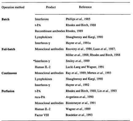
Different operation modes can be utilised for the cultivation of animal cells, which are generally classified as batch, fed-batch, continuous or perfusion cultures. Examples of use of each technique for the production of several biologicals are presented in Table 1.3. Each mode has its own advantages and the use of systems other than a batch culture is an attempt to overcome some of the limitations of that one.
this technique becomes inefficient if producing recombinant products that are growth
associated, as the immobilised cells inside the reactor are not in the growth phase.
Table 1.3- Examples of recombinant protein production by different cultivation techniques.
Operation method Product Reference
Batch Interferons Phillips eta!., 1985
t-PA Rhodes and Birch, 1988
Recombinant antibodies Rhodes, 1989
Lymphokines Shaughnessy and Kargi, 1990 Interferon-y Hayter eta!., 1991a
Fed-batch Monoclonal antibodies Reuveny eta!., 1986; Luan eta!., 1987; Miller et al., 1988; Rhodes and Birch, 1988 *Interferon.y Smiley eta!., 1989
Human IL-2 Lucki-Lang and Wagner, 1991
Continuous Monoclonal antibodies Ray eta!., 1989; Merten eta!., 1993 Lymphokines Shaughnessy and Kargi, 1990 Interferon-y Hayter eta!., 1992
Perfusion t-PA Rhodes and Birch, 1988; Lin eta!., 1993 rscu-PA Avgerinos eta!., 1990
Monoclonal antibodies BUntemeyer eta!., 1991 Human IL-2 Wagner eta!., 1989 Factor VIII Boedeker et a!., 1993
* indicates fed-batch in relation to the whole growth medium
Although the environment of the cells in a batch or fed-batch culture is continuously changing,
these processes have several advantages in terms of large-scale production, namely flexibility,
simplicity of plant and operation, clarity of batch definition for quality assurance and safety by
reducing the quantity of product at risk (Rhodes and Birch, 1988).
1.2.3- Hydrodynamic environment of the cells
1.2.3.1- Aeration and agitation
Oxygen is the terminal electron receptor in oxidative phosphorylation, being essential for energy generation. Supplying sufficient oxygen to the culture to allow cell growth and product biosynthesis is one of the design criteria for a bioreactor. For this, it is important to know the optimum oxygen level that should be provided. Fleischaker and Sinskey (1981) have compiled specific rates of oxygen uptake of several cultured cells. Below the critical dissolved oxygen tension, consumption is limited by oxygen availability (Lin and Miller, 1992) and above the critical oxygen tension varies with the glucose consumption rate (Frame and Hu, 1985; Miller
et a!., 1989a). Each cell type seems to have an optimum range of dissolved oxygen partial
pressures for growth. CHO cells cultured under low oxygen concentrations (Kurano et a!., 1 990a) had a specific growth rate half of its normal value, but cell growth and metabolism was found to be approximately constant between 50-100% air saturation. The studies conducted by Kilburn and Webb (1968) with mouse L cells have demonstrated the growth inhibitory effects of high oxygen levels and cultivation of cells out of the optimum range, either at lower or higher oxygen levels, limits their growth (Kilbum et a!., 1969). The production of oxygen-derived free radicals, which can damage cellular components such as lipids, DNA and proteins, may account for the toxic effect of oxygen (Lin and Miller, 1992). In fact, non-lethal levels of oxygen or H2O2 exposures may decrease the specific rate of production of t-PA by CHO cells by at least 50% (Lin et a!., 1993). They have also found that while CHO cell growth rate, cell titre and t-PA production rate were not affected by mild hypoxic conditions (50% reduction in specific oxygen consumption rate), the exposure of cells to anoxia led to a drastic decrease in cell titre and t-PA production rate.
0.2 mmolll at 37°C, and so it would be rapidly depleted from a high density cell culture, if not continuously replaced. Both agitation and aeration are required to maintain an adequate mixing of the culture and to provide enough oxygen to the cells. However, these are also the main sources of hydrodynamic shear stress in bioreactors and the mechanical-fluid forces generated may be of a magnitude that causes damage to the cells. Shear sensitivity has long been reported to vary between cell lines (Augenstein et a!., 1971) and culture parameters, such as agitation history of the cells, concentration of specific metabolites and cell growth phase (Petersen et a!., 1990; Ozturk and Palsson, 1991), with cells becoming more susceptible to shear in the stationary growth phase. A dependence of shear sensitivity on nutrient limitation, preventing maintenance of the structural integrity of the cell, has been suggested (Petersen et at., 1990). When considering the scale-up of cell culture processes, this parameter may become critical.
In shaker-flask cultures, agitation is simply provided by placing them in shaker incubators. In larger scale cultures, agitation is usually provided by mechanical means, e.g. impellers, paddles, coiled stiners, or in the case of air-lift bioreactors, the contents are pneumatically agitated by a stream of air (see Leist et a!., 1990). An important source of fluid shear in bioreactors is the agitation of the culture by such mechanical devices (Dodge and Hu, 1986; Peterson et a!., 1990). According to Bliem and Katinger (1988), the cell tolerance rates for agitation fall in the range of 100-200 revolutions per minute for industrial reactors (500-5000 1 batch reactors). However, Kurano et a!. (1990a) cultured CHO cells in small scale bioreactors at 400 rpm; adverse effects on specific cell growth rate were only noticed at 600 rpm. Kunas and Papoutsakis (1990) also reported that in a bubble vortex-free stirred tank bioreactor, the growth of hybridoma cells was not affected at 600 rpm agitation speed, concluding that cell damage in stirred and sparged bioreactors is mainly caused by the presence of bubbles.
As the agitation rate used is usually low to prevent damage of the cells, the low degree of fluid
mixing in the gas-liquid interface does not promote good oxygen transfer and as the ratio volume/surface increases head-space aeration becomes insufficient to supply the required oxygen. Several techniques have been developed to provide oxygen to cell cultures, e.g. use of surface aerators, air sparging (Aunins et al., 1986; Hu et a!., 1986; Prokop and Rosenberg, 1989). Air sparging provides good oxygen transfer, but excessive foaming and cell damage due to the shear force caused by rising air bubbles and bubble break-up have been reported (Handa-Corrigan eta!., 1989; Gardner eta!., 1990; Kunas and Papoutsakis, 1990; Papoutsakis, 1991b; Zhang et a!., 1992b). Sparged systems are usually used for the culturing cell lines which are more robust, such as BHK 21, Namalva, some CHO lines and several hybridomas, whereas microcarrier-based cultures and other cell lines seem to be predominately affected by
direct bubble sparging (Bliem and Katinger, 1988). A compromise between minimum physical
damage to the cells and maximum oxygen transfer from the gas phase to liquid medium has to be established for each culture system.
1.2.3.2- Additives for cell protection from fluid-mechanical damage
Several additives have been reported as "shear protectants" from agitation and aeration in animal cell cultures. Among them are serum, bovine serum albumin, carbohydrate polymers and pluronic alcohols. A list with several examples where this protective effect was involved is shown in Table 1.4. Papoutsakis (1991a) has recently reviewed this area.
biological mechanism, resulting from metabolic interactions rendering the cells more resistant
to shear were also reported (Michaels et a!., 1991; Smith and Greenfield, 1992). Leist et a!.
(1990) postulated that a biological/biochemical effect, probably due to serum proteins, is the
basis for the protection mechanism. Ramirez and Mutharasan (1991) partially attributed the
protective effect of serum to its property of decreasing the plasma membrane fluidity through
the transfer of cholesterol from lipoproteins to the plasma membrane. BSA seems to exert a
physical protective effect in nature (HUlscher and Onken, 1988; Smith and Greenfield, 1992),
by a mechanism similar to that proposed by Handa-Corrigan et a!. (1989) for Pluronic F68,
although a limited biological interaction has also been suggested (Smith and Greenfield,
1992).
Table 1.4- Protection of freely suspended animal cells from agitation and aeration damage.
Additive Cell line Reference
Serum Hybridomas Kunas and Papoutsakis, 1990; Handa-Corrigan eta!., 1989
Ramirez and Mutharasan, 1990; Ozturk and Palsson, 1991; Michaels eta!., 1991; Smith and Greenfield, 1992
Human melanoma Leist et a!., 1986
BHK-21 Handa-Corrigan et a!., 1989
BSA Hybridomas HUlscher and Onken, 1988; Takazawa eta!., 1988; Smith and Greenfield, 1992
Pluronic F68 Mouse IS Kilburn and Webb, 1968 BHK-21 Handa-Corrigan eta!., 1989
Hybridomas Gardner eta!., 1990; Ramirez and Mutharasan, 1990; Michaels eta!., 1991; Smith and Greenfield, 1992; Zhang eta!., 1992a,b; Lakhotia eta!., 1993
Insect cells Maiorella et a!., 1988; Murhammer and Goochee, 1988
PEG Hybridomas Michaels eta!., 1991
CMC Human melanoma Leist eta!., 1986
BSA- bovine serum albumin; PEG- polyethylene glycol; CMC-carboxymethylcellulose.
static cultures (Mizhari, 1975; Bentley eta!., 1989).
While much effort has been dedicated to the characterisation of the fluid-mechanical forces that cause damage to freely suspended cells, less work has been done on their actual effect on cell physiology. Recently, Lakhotia et a!. (1993) have reported a reduction in the content of specific surface proteins of the cells, CD13 and CD33, caused by an increase in agitation rate from 80 to 400 rpm with a simultaneous detrimental effect on cell growth. However, even at mechanical forces of a magnitude that did not cause any apparent cell damage (270 rpm), a reduction in the content of the same proteins was still observed. Pluronic F68 was shown to have a protective effect on the CD33 content per cell of cultures subjected to hydrodynamic stress but not on the content of CD13. It was suggested that the protective effect of Pluronic F68 is surface protein specific.
1.2.4- Temperature and pH
Temperature and pH are important extracellular parameters in culture. Animal cells are routinely grown at a temperature of 37°C but they can grow over a fairly wide range of temperatures, which allows the isolation of temperature sensitive mutants. Temperatures ranging from 34 to 40°C have been shown to allow the growth of CHO cells (Gottesman, 1985), and an optimum of 37°C has been reported (Kurano et a!., 1990a). Different cell lines have different pH optimum values for growth and production. CHO cells have been reported to grow from pH 7.0 to pH 8.2 in static cultures, but outside that range cells would not grow well (Gottesman, 1985). CHO cell growth in agitated cultures was shown to be limited to a more narrow range, pH 7.0 to pH 7.9 (Kurano et a!., 1990a), which was suggested to be related to an increase in shear sensitivity under agitation. Optimum culture pH was reported to be between 7.6-7.8 (Gottesman, 1985; Kurano et al., 1990a), but reported values for CHO cell cultures vary between 7.0 and 7.2 (Conradt eta!., 1989; Curling et a!., 1990).
1.3- Cell nutritional environment- the culture medium
The cell culture medium must provide the nutrients required by mammalian cells for the synthesis of cell biomass and products, and the additional function of maintaining suitable physiochemical conditions. This medium is far more complex and expensive than culture medium for microbial cells, based on an osmotically balanced mixture of vitamins, minerals and amino acids, often supplemented with animal serum and/or proteins. Quantitative studies on the effect of medium components on cell growth and product formation are essential for process development, as they vary widely with cell lines, culture systems and culture conditions. A good quantitative understanding of the nutrient requirements of the cell line to use is an important step for process optimisation.
1.3.1- Development of serum-free medium
as albumin and transferrin, attachment factors and also enzymes. Furthermore, serum proteins also seem to play an important role in providing physical protection to the cells against shear damage (see section 1.2.3.2) and serum-free media that support cell growth in shake-flask cultures may not be efficient in large scale fermenters, where cells are more exposed to mechanical stresses.
The development of serum-free media has not always been straightforward, mainly due to difficulties in isolating the growth promoting factors which are present in the serum and because each cell type has specific requirements. When growing cells in serum-free medium, several of its functions have to be replaced by supplementation with components able to perform them. The developments in the understanding of cell nutritional requirements have made possible the growth of many cell types in the complete absence of serum (Barnes and Sato, 1980; Barnes, 1987). Serum-free medium for hybridomas has been often reported (Murakami et a!., 1982; McHugh et a!., 1983; Glassy et a!., 1988; Takazawa et a!., 1988, Minamoto et a!., 1991). Cell lines usually used for the expression of recombinant protein, such as CHO cells (Hamilton and Ham, 1977; Gasser et a!., 1985; Hayter et a!., 1989; Ganne and Mignot, 1991; Ogata et a!., 1993), have been grown in serum-free medium.
Although several advantages arise from culturing cells in serum-free media, its apparent cost if supplemented with hormones and growth factors may become higher than serum supplemented media. However, serum-free media with low protein supplementation usually becomes more cost effective and does allow more efficient large scale operation for the production of biological products.
1.3.2- Cell requirements in serum-free medium
1.3.2.1- Energy sources
Glucose and glutamine are typically the main energy and carbon sources for most of the culture media. Glucose is usually included in the medium at a concentration of 5 to 20 mM and glutamine at a concentration varying from 0.7 to 5 mM. Figure 1.1 shows the major metabolic pathways of animal cells. The operation of particular metabolic pathways is dependent on the cell line and on the culture conditions (Eigenbrodt et a!., 1985; Mckeehan, 1986). Glucose is metabolised to pyruvate and lactate through the glycolytic pathway to produce energy (Eagle et al., 1958). Although glycolysis can supply cells with much of their energy requirements, its major role has been reported as the provision of anabolic substrates for biosynthesis (Hume et al., 1978, Wice et a!., 1981); it is a major source of carbon, providing ribose for nucleic acid synthesis (Zielke eta!., 1976, 1978). Glutamine is usually the most abundant amino acid in cell culture media, being typically metabolised to glutamate and ammonia (McKeehan, 1986). Its metabolism leads to different levels of energy production, depending on whether it is completely oxidised to CO2, or incompletely oxidised to either aspartate and lactate (Reitzer et a!., 1979). It is the major amino group donor for the synthesis of purines and pyrimidines, amino sugars, pyridine nucleotides and asparagine (McKeehan,
Glucose Biosynthesis
NADH +
NADH
(YCOLYS
ADH
I
t
AlP
-_ 'SI'—, -_
Serine Glycine
NAD NADH Amino ACidS
t
Pynivate Acetyl CoA
Lactate Glu
Biosyn11sis
/7
Atarñne
NAD(P)
Gln NAD NAD
TCA
Glu
NADH
I
t
.dminoacidNAD / I
NHi4r_J
a-ketoacid ADP
Glutamate
Oxidative
NAD + HOI-1
NH1.
qRNI2 Phosphorylation
R. X ATP
Glutainane
Carboxylic acid
Figure 1.1- Diagram of the major metabolic pathways of animal cells (derived from Miller eta!., 1989a).
Glucose and glutamine metabolism are complementary to the production of other metabolites (Zielke et a!., 1978). The relative amounts of energy provided to the cells by glucose and glutamine vary according to the cell line, If both nutrients are present in the culture medium, glutamine oxidation can contribute up to 30 to 50% of the cell energy requirements (Zielke et
al., 1984). Even at high glucose concentrations, it has been stated that glycolysis can only
supply 50% of the cell energy requirements, glutamine oxidation contributing 40% and pyruvate and fatty acid oxidation 10% (Eigenbrodt et a!., 1985). In the wild-type ovary cell line, in the presence of glucose, about 40% of the energy is supplied by glutamine (Donnelly and Scheffler, 1976). Despite this, cells can rely on the energy produced either by glycolysis, if
glutamine is limited, or by glutaminolysis, if carbohydrates are limited, and it was suggested that flux through neither pathways is compulsory for cell growth (Eigenbrodt eta!., 1985).
The availability of glucose can reduce the rate of glutamine utilisation by mammalian cells (Zielke et a!., 1978, Reitzer et a!., 1979, Lazo, 1981) and a decrease in glucose concentration has been shown to increase glutamine oxidation rates (Zielke et a!., 1984). However, additions of either glucose and glutamine to culture medium has been shown to increase the rates of
utilisation of both nutrients (Glacken et a!., 1986; Miller eta!., 1989a,b; Hayter et al., 1991a), decreasing the nutrient utilisation efficiency. It is important to consider this type of relations when designing a culture medium, in terms of providing glutamine and glucose.
1.3.2.2- Amino acids
Amino acids essentially act as precursors in the biosynthesis of proteins. They may also serve as an energy source, through oxidation to acetyl-CoA and tricarboxylic acid (TCA) cycle intermediates. Eagle, working with a mouse fibroblast and a human carcinoma cell line, reported the requirement of at least 13 amino acids for animal cell growth (Eagle, 1955a,b, 1959). These amino acids included arginine, cystine, glutamine, histidine, isoleucine, lysine, methionine, phenylalanine, threonine, tryptophan, tyrosine and valine and are most of the times considered essential for normal and established cell lines.
It was realised early that amino acid utilisation varies considerably with cell line. A high requirement of serine, glycine and proline was reported for normal human pituitary cells in the early 1960's (McCarthy, 1962). Culture medium must be designed in order to accommodate the needs of individual cell lines. As typical examples, the branched amino acids leucine, isoleucine and valine are consumed at high rates by some cell lines, namely Madin-Darby canine kidney (MDCK) cells (Butler and Thilly, 1982), human fibroblasts (Lambert and Pirt,
1975), hybndomas (Duval Ct a!., 1991) and CHO cells (Hayter et a!., 1991a). CHO cells are
proline auxotrophs, requiring the presence of this amino acid in the culture medium (Kao and Puck, 1967), while it is produced in culture by myeloma cells (Ljunggren and Haggström,
1992).
There is a strong interrelation between the metabolic pathways shown in Figure 1.1. Amino acid quantification in culture supernatants is a straightforward means of determining the metabolic requirements and status of the cells. Specific amino acid utilisation depends on the availability of the major energy sources in culture, e.g., specific consumption of leucme, isoleucine, valine and serine was shown to be higher at lower glutamine concentrations (Miller
cell lines has been reported (Butler and Thilly, 1982; Lanks, 1987; Miller et a!., 1989a,b, Duval et al., 1991; Hayter eta!., 1991a). Alanine accumulation may reflect the transamination of glutamate to pyruvate and its production rate seem to be regulated by both glucose and glutamine consumption rates (Miller et al., 1989a,b). Glycine and glutamate are a reflection of serine and glutamine metabolism, respectively.
Lately, more attention has been given to the concentration of amino acids in serum-free cultures and its importance for growth and production in animal cell cultures should be recognised for the development of any process. Due to the metabolic complexity between medium components, it is not possible to look at each component separately. Furthermore, no general rules can be made, as the amino acid metabolism varies between cell lines and even with the fermentation mode for the same cell line (BUntemeyer eta!., 1991).
1.3.2.3- Lipids
Lipids are included in most serum-free and/or protein-free medium formulations (Barnes and Sato, 1980; Glassy et a!., 1988) and several lipids, e.g. cholesterol, oleic acid, linoleic acid and certain phospholipids, have been shown to stimulate the growth of several cell lines (Darfier and Insel, 1982; Kovar and Franek, 1986; Sato et a!., 1987; Minamoto et a!., 1991; Jayme, 1991). Serum and serum albumin are major sources of lipids in the culture medium and when using serum-free and protein-free media they can be supplemented to the medium in the form of isolated plasma lipoprotein fractions, free fatty acids complexed to serum albumin or fatty acid/phospholipid microemulsions (Schmid et a!., 1991). Pluronic F68 has also been used as an emulsifying agent (Jayme, 1991).
Quantitative data available on the subject is scarce (Schmid eta!., 1991) and although analysis of total fatty acids was shown to be an unreliable indicator of cell culture performance, individual quantitative analysis has allowed the identification of critical lipids for certain cell culture applications (Jayme, 1991). Most of the studies on the influence of lipids on culture performance focus on cell growth and production data are most of the times unavailable.
1.3.2.4- Vitamins and minerals
Addition of vitamins and trace elements is common practise in serum-free cultures. Several forms of water-soluble vitamins (vitamin B complex), such as riboflavin and thiamine, were found to be essential for the growth of some cell lines, e.g. mouse L and HeLa cells (Eagle,
1955c). Supplementation of vitamins to most media is mostly made based on those early
nutritional studies and not much work has been done lately on the consumption of vitamins by cultured cells. Biotin, choline, folic acid, inositol, niacinamide, pantothenic acid, pyridoxine, riboflavine and thiamine are present in almost all basal media formulations. The lipid-soluble vitamins, A, D, E and K, are required by higher animal species, but they are not normally used in culture media. A more recent study on the consumption and stability of four vitamins,
ascorbic acid, nicotinamide, choline and thiamine (Kurano et a!., 1990c) in CHO cell culture has shown that while nicotinamide and choline were stable in culture medium, ascorbic acid and thiamine were not. Furthermore, ascorbic acid did not appear to be required for the growth of the CHO lines but growth limitation by choline at high cell densities was suggested.
At least 15 trace elements have been reported as essential or beneficial for the growth of several cell lines (Nielsen, 1981). Among these are copper, iron, manganese, selenium and zinc. Knowing the specific cell requirements for these nutrients is very important when using serum-free medium and specific supplementations have been developed according to the cell line. Some inorganic ions are essential for cell growth, as reported by Eagle (1955c): sodium, potassium, calcium, magnesium, chloride and phosphate and they are all present in basal media formulations.
1.3.2.5- Protein supplementation
The proteins insulin, transferrin and bovine serum albumin are usually included in most serum-free media formulations (Barnes and Sato, 1980; Glassy eta!., 1988), in order to replace some of serum functions. The function of the latter ones is mainly related to their binding capacities of several components, e.g., lipids (albumin) and trace elements (transferrin).
exerting a stimulatory effect on cell proliferation. Most cell lines under serum-free conditions require its presence to proliferate (Iscove and Melchers, 1978; Murakami et al., 1982; Kovar and Franek, 1984). However, the concept that transferrin would never be replaced in the medium seems less likely as the replacement of transferrin by others sources of iron such as e.g., ferric citrate (Kovar and Franek, 1987; Scheneider, 1989) and iron gluconate (Minamoto et al., 1991) has been reported.
Unlike insulin and transferrin, BSA is usually included in the medium at relatively high concentration, 1-5 mg/mi. BSA may serve as a carrier for metals, lipids and hormones (Barnes and Sato 1980; Iscove eta!., 1980) and it has been shown to strongly inhibit copper-stimulated peroxidation and generation of free hydroxyl radicals from systems containing copper and H202, preventing toxicity by oxidation of unsaturated fatty acids in the membranes (Daffier and Insel, 1983; Halliwell, 1988). It may also provide the cells with protection from shear damage (see section 1.2.3.2). It is most likely that its stimulating properties are due to the effects of unidentified molecules bound to it. Because BSA is derived from a somewhat raw material, bovine plasma and serum, even the purified fraction will reflect some of this natural variability. It is thus a component that may be a major source of variability in cell culture media.
There are several advantages in removing proteins from the culture media and efforts towards the development of protein-free medium are being made. In fact, the lower the level of exogenous proteins in the crude product the easier the downstream processing process. Also, these proteins may represent a possible source of contamination. Complete replacement of those proteins is not however a very easy task and protein-free medium is not necessarily more cost effective, specially if it is supplemented with hormones and lipid precursors. Each production system will always have to be analysed individually, as there is a clear dependence on the cell line and culture method. Successful protein-free media for the production of
monoclonal antibodies by hybridomas have been reported by Kovar and Franek (1987), Schneider (1989), Darfier (1990) and Fike et a!. (1991). The former ones are quite complex and use hormone supplements (hydrocortisone, oestradiol, progesterone) while the detailed formulation for the latter one is only available for FDA purposes, but the authors do not refer to any hormone supplementation. While Darfler (1990) mostly reported cell growth in static cultures, the others included suspension cultures in their investigations, which is an important consideration for the scaling-up of the process. Production of recombinant interleukin-2 by suspension cultures of BHK 21 cells in a rather simple protein-free medium has also been reported (Lucki-Lange and Wagner, 1991). Clonal growth of the parental CHO-KI in protein-free medium has been long achieved by Hamilton and Ham (1977), but suspension cultures would most likely behave differently. Furthermore, the level of contaminants existent in the chemicals at that time would probably be a problem in reproducing the same conditions. Substantial work is still required in order to routinely cultivate recombinant cells in totally protein-free medium.
1.3.3- Toxicity in animal cell culture
Ammonia and lactate are the usually reported potential toxic compounds accumulated in animal cell culture. As stated in section 1.3.2.1, ammonia mainly derives from the metabolism of glutamine by glutaminase activity while lactic acid is a result of the catabolism of glucose by anaerobic glycolysis.
found for some hybridoma cell lines (Reuveny et a!., 1986; Hassel et a!., 1991) while L-mouse cells have been found to be inhibited by concentrations as low as 0.5 mM (Ryan and Cardin, 1966). On the other hand, the accumulation of lactic acid in culture may exceed the buffering capacity of the medium, leading to a decrease in the culture pH and to suboptimal conditions for cell growth (Reuveny et a!., 1986) but usually no inhibition is found up to 25 mM lactate. Concentrations as high as 40 mM did not any effect on the growth of several hybridoma cell lines (Hassel et al., 1991).
The sensitivity of CHO cells to lactate and ammonium was investigated by Schlaeger and Schumpp (1989), who reported inhibition constants (50% growth inhibition) of 8-10 mM ammonia and ca. 75 mM lactate. The work of Hayter et a!. (1991a) with CHO cells gave support to the previously reported results, showing that cell growth is not inhibited by ammonium concentration of 2 mM although inhibitory effects were seen at 4.5 mM, and cells seemed to be relatively insensitive to lactate as concentrations of 17.5 mM were reported to have little effect on cell growth. Kurano et a!. (1990b) also found that CHO cell growth was not affected by lactate itself and an inhibition constant of about 7.5 mM was found for ammonia.
An interactive effect between ammonia and lactate inhibition has been suggested by Hassel et al. (1991). They found that at lower concentrations of lactate, the apparent toxicity of ammonia decreased. Reuveny ef a!. (1986) also found that the addition of lactate to culture medium stimulated the growth of some hybridomas. Ammonia inhibition was also reported to be pH dependent (Doyle and Butler, 1990), e.g., as the culture pH was reduced from 7.8 to 6.8 the initial concentration of ammonium chloride causing 50% growth inhibition shifted from 4 mM to 7.6 mM.
Some strategies have been proposed to overcome the problem of ammonia and lactate
accumulation in culture medium, when they become the bottleneck of the process. The use of slowly utilised carbohydrates results in less lactate production (Eagle et a!., 1958; Imamura et a!., 1982). The glucose concentration in the medium is known to affect the lactate production: the rate of glucose utilisation is higher at higher glucose concentrations (Zielke, 1978; Glacken et a!., 1986; Miller et a!., 1989a; Hayter et al., 1991a), increasing the flux of glucose carbon through glycolysis to lactic acid. The same rational is applied to glutamine utilisation and consequent ammonia formation (Glacken et a!., 1986; Miller et a!., 1988; Hayter et a!., 1991a). Controlled feeding of glucose and glutamine into the culture medium, in order to keep their concentration within acceptable limits has been shown to be successful in reducing lactate and ammonia production (Glacken et a!., 1986; Ljunggren and Haggstrom, 1990). The accumulation of metabolic end products can be also limited by perfusion culture (Glacken et a!., 1986; Glacken, 1988), but this may not be economically viable. Kurano et a!. (1990b) cultured CHO cells in glutamine-free and asparagine supplemented medium, reducing ammonia production by 40%. More recently, in-situ removal of ammonia through electrical means has been reported (Chang et a!., 1993). It has also been reported that potassium ion can be used as an ammonia detoxifying agent in murine myeloma cell cultures, by inhibition of inward transport of ammonium ions (Martinelle and Haggstrom, 1993).
1.4- Production of glycoproteins
different types of glycosylation, N-glycosylation and 0-glycosylation, but here we will only refer to the former.
1.4.1- Significance of glycosylation
Glycosylation is one of the several post-translational modifications of human proteins. It may have a major role on the biological properties of a protein, often dictating its therapeutic value. Biological properties, such as plasma clearance rate, antigenicity, immunogenicity, specific activity, solubility and resistance to proteolytic attack, may all be affected by the oligosaccharide structures of a glycoprotein (Goochee et a!., 1991). The importance of glycosylation for biological function and stability has however to be evaluated for each protein individually. While N-glycosylation is essential for full biological activity in vivo of EPO (Delorme et a!., 1992) and dramatically increases the functional anticoagulant activity of human Protein C (Grinnell et a!., 1991), it has also been reported that an increased glycosylation of recombinant antithrombin causes a decrease in the affinity for heparin (Björk eta!., 1992) and decreases the specific activity of t-PA (Einarsson eta!., 1985).
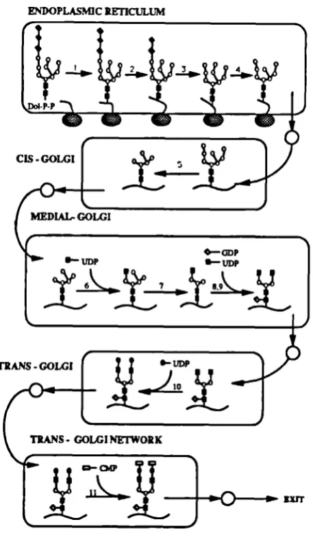
A review on the assembly of N-linked oligosaccharides has been written by Kornfeld and Komfeld (1985). A diagram with the glycosylation pathway is shown in Figure 1.2. The oligosaccharides are covalently attached to the polypeptide chain through an asparagine residue, at the consensus sequence Asn-X-Ser/Thr. Each step in the glycosylation pathway is cataiysed enzymaticaily. Initially, a Glc3Man9GlcNAc2 structure is synthesised on a polyisoprenoid derivative, the dolichol-P-P (GIc3Man9G1cNAc2-P-P-dol) and transferred en bloc to the asparagine residues of the nascent polypeptide chain in the endoplasmic reticulum (ER). Several "trimming" reactions are then catalysed by exoglycosidases in the ER to yield high mannose N-linked structures. These high mannose structures are variably modified in the Golgi compartments by several exoglycosidases and glycosyltransferase reactions, leading to
TRANS - GOLGI
TRANS - GOLGI NETWORK
the formation of "complex type" oiigosaccharide structures, terminated by sialic acids. Not all
the potential sites for glycosylation are used in a protein, leading to forms that differ in the
number of oligosaccharide chains attached. Variation in the terminal sugars and chain
branching may also occur and ultimately affect protein behaviour in vivo (Maiorella et al.,
1993).
ENDOPLASMIC RETICULUM
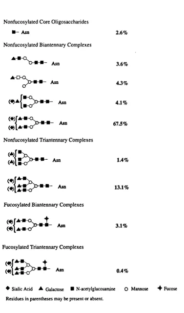
S 0. ina.. •- N tyIucs.nIa. 5. ñea.. S . adcs, . IIc add
1.4.2- The host and its environment
How to control the variation of the oligosaccharide structures remains unclear, but several studies on the factors which may affect that variation have been carried out. It is now well recognised that the glycosylation profile of a given glycoprotein is host-dependent. Host-dependent oligosaccharide structures have been reported for IFN-B (Utsumi et al., 1989), t-PA (Parekh et a!., 1989b) and EPO (Goto et a!., 1988). The degree of similarity between oligosaccharide structures derived from CHO cells and human glycoproteins has been found to be quite high. This has been reported for several products, such as IFN-Bl (Kagawa et a!., 1988), EPO (Takeuchi et al., 1988) and t-PA (Speilman et a!., 1989). This fact seems to put CHO cells in a very favourable position when selecting a cell line for the synthesis of recombinant human proteins. It has to be pointed out that CHO cells have a limited capacity of
y- carboxylation and an inability for oligosaccharide suiphation (Goochee et a!., 1991).
Glycosylation patterns also vary with the extracellular environment of the cells and more attention now is being given to this subject. Some examples reported in the literature are shown in Table 1.5.
As the glycosylation profile of a protein may significantly affect its therapeutic value, glycosylation analysis throughout the development of production processes for such proteins becomes a major concern. The development of new strategies and instrumentation to allow a rapid defmition of the glycosylation status is a major area of research (Parekh and Pate!, 1992; James et a!., 1993).