78:3 (2016) 315–320 | www.jurnalteknologi.utm.my | eISSN 2180–3722 |
Jurnal
Teknologi
Full Paper
T
HE
E
FFECT OF
L
ASER
I
RRADIATION ON THE
V
IABILITY OF
H
UMAN
B
REAST
C
ANCER
C
ELL
,
MDA-MB-231
Aishah Badruzzaman
a,b, Noriah Bidin
a,b*, Siti Pauliena Mohd
Bohari
ca
Laser Center, Ibnu Sina Institute for Scientific & Industrial
Research, Universiti Teknologi Malaysia, 81310 UTM Johor Bahru,
Johor, Malaysia
b
Physics Department, Faculty of Science, 81310 UTM Johor,
Malaysia
c
Faculty of Bioscience and Medical Engineering, Universiti
Teknologi Malaysia, 81310 UTM Johor Bahru, Johor, Malaysia
Article history
Received
15 August 2015
Received in revised form
15 November 2015
Accepted
30 December 2015
*Corresponding author
noriah@utm.my
Graphical abstract
Abstract
This study compared the effects of different sources of laser phototherapy on the cell viability of the in vitro human breast cancer cell lines. Laser phototherapy is used in the breast cancer clinical treatment, despite the limited safety information of laser irradiation effect on the cancer cell behavior. This study contributed on the development of guidelines for safer laser usage in treating breast cancer and minimising the possibility of activating postmastectomy lymphedema. Cancer cell viability and morphology were studied in this research. Human breast cancer (MDA-MB-231) cell lines were cultured for 24 hours in CO2 incubator and irradiated with different laser sources and number of pulse. The viability of cancer cells were assessed by MTT assay 24 hours after laser irradiation. The result showed that MDA-MB-231 cell viability increased after being irradiated by excimer 248 nm laser. However, the cancer cell viability slightly decreased after irradiation by both Nd:YAG 1064 and 532 nm. Although certain doses of laser may affect the MDA-MB-231 cell viability, additional laser exposures had nearly no effect. The research shows that Nd:YAG 1064 nm more effective in lowering cancer cell survivability than 532 nm and 248 nm. Further in vivo studies are needed for better understanding on the mechanism of laser-tissue interaction and improve the laser usage safety in photothermal therapy.
Keywords: Photothermal laser, MDA-MB-231, Excimer, postmastectomy lymphedema
Abstrak
mekanisma interaksi laser-tisu dan meningkatkan keselamatan penggunaan laser dalam terapi photothermal.
Kata kunci: Fototerapi laser MDA-MB-231, Excimer, postmastectomy lymphedema
© 2016 Penerbit UTM Press. All rights reserved
1.0 INTRODUCTION
The clinical application of a thermal laser with low power outputs [1, 2] has been used now for many years in laser phototherapy to treat numbers of disease including epidermal abrasions, osteoarthritis and recently breast cancer-related lymphedema [3-5]. Both physical and psychological will get impacts of the untreated lymphoedema especially on an individual’s well-being. The signs and symptoms of lymphoedema are limb deformation and swelling, immobilization, restricted shoulder mobility, burning, elevated skin temperature, fibrosis, and social isolation [3, 6-9]. 30% of patients treated for breast cancer will have lymphoedema of the arm as it was one of the main side effects following breast cancer radiation therapy or surgery [6]. For the present time, there still no definite treatment for lymphoedema, although standard treatments for lymphedema include pressotherapy, exercise, manual lymphatic drainage, compression bandaging and skin care. However these treatments are generally applied in combination of each other known as Complete Decongestive Therapy (CDT) and it is very expensive, time consuming, require qualified medical professionals, less accepted by patients and limited effectiveness [10-12].
In previous studies [13-15], double blind and
medicine-controlled trials have shown the
effectiveness of Low Level Laser Therapy (LLLT) in
improving the postmastectomy lymphedema
symptoms. The treatments consisted of radiation with
904 nm wavelength, energy density of 1.5 J/cm2, from
10 to 17 points distributed in the affected area and the number of sessions ranged from 8 to 36 . A significant reduction in the size of the affected arm, volume of extracellular fluids and the tissue hardness, as well as improved the shoulder mobility and grip strength [16, 17].
Powell et al. [18] used four different cell line, human breast adenocarcinoma (MCF-7), human breast ductal, carcinoma with melanomic genotypic traits (MDA-MB-435S), immortalized human mammary epithelial (SVCT and Bre80hTERT), to study the effect of three different laser source, 780 nm continuos (50 mW), 830 nm continuos (30 mW) and 904 nm pulsed (90 mW) with range 1 to 3 laser exposure on cells proliferation. The result showed, although certain doses of laser will increase cell proliferation, however multiple doses exposures had either no effect or showed negative dose-response relationships. There was no sign of malignant modification and transformation of cells by
laser phototherapy was observed under the applied conditions.
In other research [19], Schartinger et al. used human
bronchial epithelial cells (BEAS-2B), human gingival fibroblasts and oral squamous cell carcinoma cells (SCC-25) to determine the cell proliferation after irradiate by GaAlAs, diode laser at 660 nm with power output 350 mW for 15 min and 3 repetition exposure in subsequent days. The result shows, there was increasing in cell proliferation for human gingival fibroblasts but decreasing for non-neoplastic epithelial cells and oral carcinoma cells SCC-25. However in SCC-25 cells, there is a proapoptotic effect of laser phototherapy was observed.
Gao et al., [20] studied the effect of two low power
laser irradiation (LPLI) of He–Ne laser, 632.8 nm and 5 mw at fluence of 0.8 J/cm2 and high fluence LPLI of
He–Ne laser, 632.8 nm and 40 mw at fluence of 60
J/cm2 on ASTC-a-1 cells. The study showed low fluence
of LPLI will increase the cell proliferation and activated the protein kinase Cs (PKCs) in the cells. However, the study reported that high fluence of LPLI was decrease the cell viability and PKCs activity but increases the cell apoptosis.
These finding encourage more research on the application of laser phototherapy in cancer treatment such as chemotherapy and radiotherapy induced BCRL and oral mucositis. Few safety studies on the usage of laser in cancer treatment have been undertaken, although it has been by numbers of researches suggested that laser phototherapy has no effect on disease recurrence or survival in cancer patients [21,22] and it been suggested by Werneck et al.
[23]‘‘clinically, irradiating a cancer lesion may result in increased tumor progression’’. Although researches suggests that the growth of cancer cell in culture can be stimulated by laser phototherapy, and the clinical application laser in cancer treatment is increasing. The effect on cancer cells in vivo requires more critical review on the safety and efficiency of the laser application.
2.0 EXPERIMENTAL
2.1 Chemicals and Cell Line
MDA-MB-231, human breast cancer cells were a kind gift on Dr. Pauliena Bohari (Faculty of Biosciences and Medical Engineering, Universiti Teknologi Malaysia). Cells were grown in Dulbecco’s Modified Eagle’s Medium (DMEM), with 10% FBS, Fetal Bovine Serum and 1% Penicillin-Streptomycin Liquid and kept at 37℃ in
incubator with 5% CO2. The viability of the cells were measured with MTT assay, 3-(4,5-dimethylthiazol-2-yl)-2,5-diphenyltetrazolium bromide.
2.2 In Vitro Treatment Protocol
MDA-MB-231 cells were seeded in 24-well plate with 6×10⁴ seeding density and incubated for 24 hours.
After 24 hours, the medium for all cells were changed with new medium to make sure all cell have enough food and not to die because of the insufficient supplement. After the medium were renewed, cells were treated by the lasers and incubated for another 24 hours before the cell viability determined by the MTT assay.
2.3 Laser treatment
The treatments have been performed with two different lasers. The Excimer laser, which available with average powers up to 35 W with KrF at 248 nm and Nd:YAG laser, which have power up to 500W at 1064 nm and 532 nm. For Excimer laser, KrF at 248 nm, a single pulse with constant frequency 20 Hz was used and the energy was varied in range 1 to 6 mJ delivered to the cells. The laser pulses were manually started using external trigger. A pulse of Nd:YAG laser at both 1064 and 532 nm were varied in energy from 200 to 1200 mJ. The three different wavelength lasers were set with constant energy but the number of pulses was increase from 1 to 6 pulses. 24 hours before treatment, cells were seeded in 24-wellplate with seeding density 6×10⁴. For laser irradiation, the
24-wellplates were placed on a stationary platform and the 4 treatments were carried out 24 hours away from each other and the experiment was repeated 3 times. The treated samples were compared with controls cancer cells maintained in the same conditions, without laser irradiation.
Figure 1 The viability (percentage of total cells) of human breast cancer cell, MDA-MB-231 after irradiated by (a)Excimer KrF 248nm (b)Nd:YAG 532nm (c)Nd:YAG 1064nm lasers with different energy
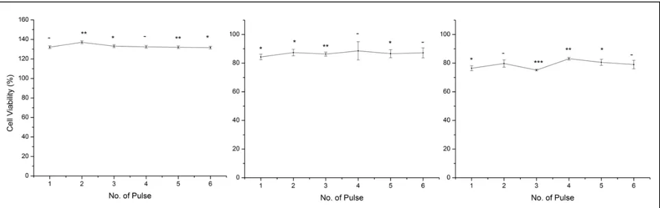
Figure 2 The viability (percentage of total cells) of human breast cancer cell, MDA-MB-231 after irradiated by (a)Excimer KrF 248nm (b)Nd:YAG 532nm (c)Nd:YAG 1064nm lasers with different number of pulse
Figura 3 The morphology of the human breast cancer cell MDA-MB-231 (a)control without treatment (b)treated by Excimer KrF 238nm, (c) treated by Nd:YAG 532nm, and (d)treated by Nd:YAG 1064nm
2.4 Cell Viability
The viability of cell MDA-MB-231 were determine by MTT(3-(4,5-dimethylthiazolyl-2)-2,5-diphenyltetrazolium bromide) assay. Once the laser treatment were done, the medium of each well were change with new medium and 5mg/mL MTT was added and the all the MTT procedures were done in the dark as it very sensitive to light. The MDA-MB231 cells were incubated for 4 hours at 37℃. After 4 hours, the absorbance of
the cells were measured at 577 nm using micro plate reader. The percentage of total cells was calculated from the absorbance value and analyzed.
3.0 RESULTS AND DISCUSSION
This experiment investigates the effects of different laser wavelength commonly used in clinical practice of laser phototherapy on human breast cancer cell MDA-MB-231. In the clinical treatment setting, breast cancer patients usually receive multiple treatments with the same laser device. Therefore, we also investigated the effects of repeated exposures doses on the human breast cancer cell line. The usage of specific wavelength and dose of laser phototherapy resulted in either stimulation or inhibition to no changes in cell growth.
3.1 The Effect of Laser Wavelength and Energy
The effect of laser treatment on cell viability of breast cancer cell, MDA-MB-231 was analyzed. The result showed in Figure 1 (a) after being irradiated by Excimer 248 nm laser, the cell viability of MDA-MB-231 increased up to 157%. However, the cell percentage was decrease as the laser energy was increased. This finding consistent with Zainudin et al. [24] which it was reported that the irradiation of F2 excimer laser 157 nm was improved the spreading, attachment and growth
of human corneal limbal epithelial HLE cells. In other research, Olivia Serdarevic et al. [25] reported at the 248-nm wavelength, the thermal effects of laser become more dominant therefore; all the fungal elements will be ablated. These ranges of laser wavelength are suitable for healing treatment acting as sterilization agent.
The result in Figure 1 (b) & (c) shows after irradiate by both Nd:YAG at 1064nm and 532nm the percentage of total cell are lesser compare to the control untreated breast cancer cells. However, when the cells were treated by Nd:YAG with same energy 200mJ but different wavelength, it shows that at wavelength 1064nm, there are more cell death which have only 74% of cell survived after the treatment compare to the cell treated by 532 nm which have 84%. As the laser energy of both 532 and 1064 nm Nd:YAG laser were increased, the percentage of cell survive were decreased.
Base on literature, there are records on therapeutic effects by low-level of laser in range of visible to near-infrared wavelengths, 500 – 1000 nm. The therapeutic effects caused by laser phototherapy, such as increasing cellular activity not only influenced by the laser wavelength, doses of exposure, power density , as well as the laser frequency of treatment and type of injury and tissue condition of the breast cancer patient
[26].
The mechanism of laser-tissue interaction was explained by Welch et al. [27]. In laser photothermal reactions, the laser light will absorb by cell chromophores and will convert to heat. This causes a rise in temperature of the cells and the heat will diffuse through the cell causing a rise in temperature in the surrounding cell. If the heat is high enough, it will result in denaturization, and even vaporization. The damage done to the cell depends on the temperature that is reached.
3.2 The Effect of Number of Laser Pulse on Cell Viability
Although using different source of laser will increase and decrease the MDA-MB-231 cell viability, additional pulses of laser exposures had nearly no effect on the result as shown in Figure 2 (a) (b) and (c). When MDA-MB-231 cells were treated with more than one exposure of laser irradiation at three different wavelengths, there were no significant changes on viability of the cells. This result proved the study conducted by Powell et al. as they reported that no change in proliferation of the breast cancer cell MCF-7 when irradiated with more than one exposure doses of laser [28].
from the laser light. If the electric field is very high, a small region of plasma will occur with high electric fields, associate with dielectric breakdown, the formation of shockwave, and tissue rupture [29].
4.0 CONCLUSION
Although these results does not show any significant laser-induced modification of cancer cell behaviour, further in vivo studies with high methodology quality are needed to better understand on the action mechanism of laser and improve the safely applying laser therapy to prevent and minimize the postmastectomy lymphedema.
Acknowledgement
We wish to thank to Gavornment of Malaysia through FRGS grant vote 4F543 and MyBrain 15 for the scholarship to the first author. We also thanks Faculty of Biosciences and Medical Engineering, Universiti Teknologi Malaysia (UTM) for providing facilities and services.
References
[1] Rubinov, A. N. 2003. Physical Grounds For Biological Effect
Of Laserradiation. J. Physics D: Appl. Physics. 36: 2317-2330.
[2] Hawkins, D. H., Tech, M. Abrahamse, H. 2006. The Role Of
Laser Fluence In Cell Viability, Proliferation, And Membrane Integrity Of Wounded Human Skin Fibroblasts Following
Helium-Neon Laser Irradiation. Laser Surg. Med. 38: 74-83.
[3] Carati, C. J., Anderson, S. N., Gannon, B. J. and Piller, N.B.
2003. Treatment Of Postmastectomy Lymphedema With Low-Level Laser Therapy: A Double Blind,
Placebo-Controlled Trial. Cancer. 98: 1114-1122.
[4] Gur, A., Cosut, A., Sarac, A. J., Cevik, R., Nas, K. Uyar, A.
2003. Efficacy Of Different Therapy Regimes Of Low-Power Laser In Painful Osteoarthritis Of The Knee: A Double-Blind
And Randomized-Controlled Trial. Laser Surg. Med. 33:
330-338.
[5] Hopkins, J. T., McLoda, T. A., Seegmiller, J. G. and Baxter, G.
D. 2004. Low-Level Laser Therapy Facilitates Superficial Wound Healing In Humans: A Triple-Blind, Sham-Controlled
Study. J. Athl. Training. 39: 223-229.
[6] Carati, C. J., Gannon, B. J. and Piller, N. B. 2004. Low-Level
Laser As A Treatment Option For Postmastectomy
Lymphedema. Am. J. Oncol. 3: 2-7.
[7] Oliver, G. and Detmar, M. 2002. The Rediscovery Of The
Lymphatic System: Old And New Insights Into The Development And Biological Function Of The Lymphatic
Vasculature. Genes Dev. 16: 773-783.
[8] Kaviani, A., Fateh, M., Nooraie, R. Y., Alinagi-Zadeh, M.
Ataie-Fashtami, L. 2006. Low-Level Laser Therapy In
Management Of Postmastectomy Lymphedema. Laser
Med. Sci. 21: 90-94.
[9] Hayes, S., Cornish, B. Newman, B. 2006. Prevalence And
Cumulative Burden Of Lymphoedema Between 6 To 18 Months Following Treatment For Breast Cancer In Brisbane
Women. 6th Australasian Lymphology Association
Conference. 106-110.
[10] Casley-Smith, J. 1992. Modern Treatment Of Lymphoedema.
I. Complex Physical Therapy: The First 200 Australian Limbs.
Australasian Journal of Dermatology. 33(2): 61-8.
[11] Harris, S. R., Hugi, M. R., Olivotto, I. A., Levine, M. 2001.
Clinical Practice Guidelines For The Care And Treatment Of
Breast Cancer: 11. Lymphedema. Canadian Medical
Association Journal. 23: 164(2): 191-9.
[12] Rodrick, J. R., Poage, E., Wanchai, A., Stewart, B. R.,
Cormier, J. N., Armer, J. M. 2013. Complementary, Alternative, And Other Non-Complete Decongestive Therapy (CDT) Treatment Methods In The Management Of
Lymphedema. A Systematic Search And Review. American
Academy of Physical Medicine and Rehabilitation. 6:
250-274.
[13] Carati, C. J., Anderson, S. N., Gannon, B. J., Piller, N. B. 2003.
Treatment Of Postmastectomy Lymphedema With Low-Level Laser Therapy. A Double Blind, Placebo-Controlled
Trial. Cancer. 98(6): 1114-1122.
[14] Ahmed Omar, M. T., Abd-El-GayedEbid, A., El Morsy, A.M.
2011. Treatment Of Postmastectomy Lymphedema With Laser Therapy: Double Blind Placebo Control Randomized
Study, Journal of Surgical Research. 165: 82-90.
[15] Kozanoglu, E., Basaran, S., Paydas, S., Sarpel, T. 2009.
Efficacy Of Pneumatic Compression And Low-Level Laser
Therapy In The Treatment Of
Postmastectomylymphoedema: A Randomized Controlled
Trial. Clinical Rehabilitation. 23(2): 117-124.
[16] Zainuddina, Traian, V., Chirilaa, Zeke Barnarda, Gregory S.,
Watsonf, ChiongTohc, IdrissBlakeyc, Andrew K. Whittakera, David J. T., Hill. 2011. F2 Excimer Laser (157 nm) Radiation Modification And Surface Ablation Of PHEMA Hydrogels And The Effects On Bioactivity: Surface Attachment And Proliferation Of Human Corneal Epithelial Cells Radiation.
Physics and Chemistry. 80(2): 219-229
[17] Richard W. Darrell, Ronald R. Krueger, Stephen L. Trokel.
1985. Excimer Laser Therapy for Experimental Candida
Keratitis Olivia Serdarevic. American Journal of
Ophthalmology. 99(5): 534-538
[18] Powell, K., Low, P., McDonnell, P. A., Laakso, E. L., Ralph, S. J.
2010. The Effect Of Laser Irradiation On Proliferation Of Human Breast Carcinoma, Melanoma, And Immortalized
Mammary Epithelial Cells. Photomedicine And Laser
Surgery. 28(1): 115-123.
[19] Schartinger, V. H., Galvan, O., Riechelmann, H., Dudás, J.
2012. Differential Responses Of Fibroblasts, Non- Neoplastic Epithelial Cells, And Oral Carcinoma Cells To Low Level
Laser Therapy. Support Care Cancer. 20(3): 523-529.
[20] Gao, X., Chen, T., Xing, D., Wang, F., Pei, Y., Wei, X. 2006.
Single Cell Analysis Of PKC Activation During Proliferation
And Apoptosis Induced By Laser Irradiation. Journal of
Cellular Physiology. 206(2): 441-448.
[21] Mikhailov, V. A., Denisov, I. N., Frank, G. A. and Voltchenko,
N. N. 2000. Results Of Treatment Of Patients With Second- To Third-Stage Breast Cancer By Combination Of Low-Level Laser Therapy (LLLT) And Surgery: Ten-Year Experience.
Proc. Soc. Photo. Opt. Instrum. Eng. 4166: 40.
[22] Lenhard, R. E., Osteen, R. T., and Gansler, T. 2001. Clinical
Oncology. Atlanta GA. The American Cancer Society.
25-30.
[23] Werneck, C. E., Pinheiro, A. L. B., Pacheco, M. T. T., Soares,
C. P., and De Castro, J. L. F. 2005. Laser Light Is Capable Of Inducing Proliferation Of Carcinoma Cells In Culture: A
Spectroscopic In Vitro Study. Photomedicine and Laser
Surgery. 23: 300-303.
[24] Halsted W. 1894. The Results Of Operations For The Cure Of
Cancer Of The Breast Performed At The Johns Hopkins
Hospital From June, 1889 To January, 1894. Ann Surg. 20(5):
497-555.
[25] Silvia, E. M., Lilian, V. R., Mônica F. F., Angélica, R. A.,
Camila, T. V. 2014. Treatment Of Upper Limb Lymphedema
With Low-Level Laser: A Systematic Review.
FisioterapiaemMovimento. 27(4): 663-74
[26] Karu T. I .1987. Photobiological Fundamentals Of Low-Power
Laser Therapy. IEEE J Quantum Electron. 23(10): 1703-17.
[27] Welch, Jorge, A. J., Torres, H., Wai-Fung Cheong. (1989).
Laser Physics and Laser-Tissue Interaction. Texas Heart
[28] Harris, S. R., Hugi, M. R., Olivotto, I. A. Levine, M. 2001. Clinical Practice Guidelines For The Care And Treatment Of
Breast Cancer: 11. Lymphedema. Can. Med. Assoc. J. 164:
191-199.
[29] Mgbonyebi, O. P., Russo, J., Russo, I. H. 1999. Roscovitine
Induces Cell Death And Morphological Changes Indicative
Of Apoptosis In MDA-MB-231 Breast Cancer Cells. Cancer
Research. 59(8): 1903-10.
[30] Serge, H., Yves, L., Marc, P. 2000. Oleate Activates
Phosphatidylinositol 3-Kinase and Promotes Proliferation and Reduces Apoptosis of MDA-MB-231 Breast Cancer Cells,
Whereas Palmitate Has Opposite Effects. Cancer Research.
60: 6353– 6358.
[31] Keiko, T., George, L. 2004. Disruption Of Mitochondria During Tocotrienol-Induced Apoptosis In MDA-MB-231 Human
Breast Cancer Cells. Biochemical Pharmacology. 67: