of SpsD, a Surface Protein from
Staphylococcus
pseudintermedius,
with Host Extracellular Matrix
Proteins
Giampiero Pietrocola1, Joan A. Geoghegan2, Simonetta Rindi1, Antonella Di Poto1, Antonino Missineo1, Valerio Consalvi3, Timothy J. Foster2, Pietro Speziale1*
1Department of Molecular Medicine, Institute of Biochemistry, Pavia, Italy,2Department Microbiology, Moyne Institute of Preventive Medicine, Trinity College, Dublin, Ireland,3Department of Biochemical Sciences ‘‘A. Rossi Fanelli’’, University of Rome ‘‘La Sapienza’’, Roma, Italy
Abstract
Staphylococcus pseudintermedius, a commensal and pathogen of dogs and occasionally of humans, expresses surface proteins potentially involved in host colonization and pathogenesis. Here, we describe the cloning and characterization of SpsD, a surface protein ofS. pseudintermediusreported as interacting with extracellular matrix proteins and corneocytes. A ligand screen and Western immunoblotting revealed that the N-terminal A domain of SpsD bound fibrinogen, fibronectin, elastin and cytokeratin 10. SpsD also interfered with thrombin-induced fibrinogen coagulation and blocked ADP-induced platelet aggregation. The binding site for SpsD was mapped to residues 395–411 in the fibrinogenc-chain, while binding sites in fibronectin were localized to the N- and C-terminal regions. SpsD also bound to glycine- and serine-rich omega loops within the C-terminal tail region of cytokeratin 10. Ligand binding studies using SpsD variants lacking the C-terminal segment or containing an amino-acid substitution in the putative ligand binding site provided insights into interaction mechanism of SpsD with the different ligands. Together these data demonstrate the multi-ligand binding properties of SpsD and illustrate some interesting differences in the variety of ligands bound by SpsD and related proteins fromS. aureus.
Citation:Pietrocola G, Geoghegan JA, Rindi S, Di Poto A, Missineo A, et al. (2013) Molecular Characterization of the Multiple Interactions of SpsD, a Surface Protein fromStaphylococcus pseudintermedius,with Host Extracellular Matrix Proteins. PLoS ONE 8(6): e66901. doi:10.1371/journal.pone.0066901
Editor:Brian Stevenson, University of Kentucky College of Medicine, United States of America
ReceivedMarch 7, 2013;AcceptedMay 12, 2013;PublishedJune 21, 2013
Copyright:ß2013 Pietrocola et al. This is an open-access article distributed under the terms of the Creative Commons Attribution License, which permits unrestricted use, distribution, and reproduction in any medium, provided the original author and source are credited.
Funding:The support by Fondazione CARIPLO ‘[Vaccines 2009-3546]’ to P. Speziale is acknowledged. TJF acknowledges the support of Science Foundation Ireland [grant number 08/IN.1/B1845]. The funders had no role in study design, data collection and analysis, decision to publish, or preparation of the manuscript.
Competing Interests:The authors have declared that no competing interests exist.
* E-mail: [email protected]
Introduction
Staphylococcus pseudintermediusis a commensal and opportunistic pathogen of companion animals, especially dogs [1,2], mainly causing skin infections such as pyoderma as well as surgical wound infections, urinary tract infections and otitis externa. Cases of infections in humans have occasionally been reported [3–6]. Methicillin-resistant S. pseudintermedius occurs widely [7,8]. The complete genome sequences of two isolates ofS. pseudintermediusare available [9,10]. The strains are predicted to encode many putative virulence factors including toxins, extracellular enzymes such as lipases and proteases and surface proteins designatedS. pseudintermediussurface proteins A-R (SpsA-R) [11] some of which are known to promote adhesion of the bacterium to desquamated skin epithelial cells (corneocytes) [12–14] and to components of the extracellular matrix [11,15].
One such surface protein that is likely to be important in skin colonization and virulence is SpsD. The presence of SpsD on the bacterial cell surface promotes adhesion to fibrinogen (Fbg), fibronectin (Fn) and cytokeratin 10 (K10). Immunoglobulin G specific for SpsD occurs in dogs with pyoderma indicating that the protein is expressed during infection [11]. SpsD has many features that are typical of staphylococcal surface proteins called microbial
surface components recognizing adhesive matrix molecules (MSCRAMMs) that are related to clumping factors (Clf) and fibronectin binding proteins (FnBPs) ofS. aureus[11].
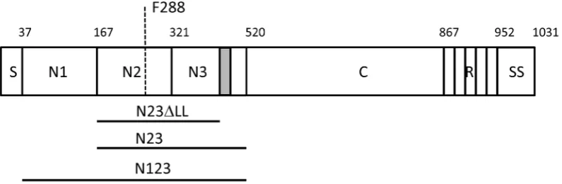
The primary translation product of SpsD has an N-terminal secretory signal sequence and a C-terminal sorting signal comprising an LPXTG motif for sortase A-mediated linkage to peptidoglycan, a hydrophobic membrane-spanning domain and positively charged residues. The N-terminus of the mature protein comprises an A domain, part of which has sequence similarity to the A domains of ClfA and FnBPs (Figure 1).
in place byb-strand complementation with absheet in subdomain N2. Structural and mechanistic studies strongly suggest that the N2N3 subdomains of FnBPA and FnBPB bind to the Fbgcchain by the same mechanism. However differences exist in the repertoire of ligands bound in addition to Fbg, with ClfA recognizing complement factor I from human serum [19,20], FnBPA and FnBPB both additionally binding to elastin [17,21,22] and subdomains N2N3 of FnBPB also binding to fibronectin [23]. Clumping factor B is another MSCRAMM ofS. aureuswith an A domain that is similar in structure and function to ClfA but which binds different ligands to ClfA and FnBPs by the dock, lock and latch mechanism [24]. ClfB binds to the glycine and serine-rich omega loops that occur in the C-terminal tail of cytokeratin
10 and throughout the corneocyte envelope protein loricrin [25,26]. It also binds to a related sequence in theaC region of the achain of Fbg [24,27].
Located distally to the A domains of FnBPA and FnBPB is an extended unfolded region comprising 11 or 10 tandemly repeated domains, respectively, that bind to the N-terminal type I modules of Fn by the tandemb-zipper mechanism [28,29]. In ClfA and ClfB this region is occupied by multiple repeats of the dipeptide Ser-Asp which have no known ligand binding function [30].
[image:2.612.60.458.62.192.2]SpsD has been reported to promote bacterial adhesion to Fbg, K10 and Fn. In this study we set out to dissect SpsD and to localize and characterize its ligand binding region(s). We identified a region that is most closely related to the A domain of FnBPB ofS. Figure 1. Schematic diagram of SpsD.The A domain modelled on the structure of ClfA spans residues 37–519 following the secretory signal sequence S. This is followed by a connecting region C (residues 521–867) and a repeat region R in strain ED99. The number of repeats varies from strain to strain resulting in proteins of slightly different sizes. A sorting signal (SS: LPXTG motif, hydrophobic domain and positively charged residues) occurs at the extreme C-terminus. The minimum ligand binding domain construct N2N3 spans residues 167–519. Residues comprising the ‘lock’ and ‘latch’ (grey box residues 490–499) are indicated (N23DLL). Residue F288 that is predicted to lie within the ligand binding trench is indicated by the dashed line.
doi:10.1371/journal.pone.0066901.g001
Figure 2. Binding of SpsD proteins to extracellular matrix or plasma proteins.(A), ligands were coated onto the surface of microtiter plates (1mg/well) and incubated with 17.5 nanomoles of SpsD37–519or SpsD167–519, followed by mouse anti-His IgG and HRP-conjugated rabbit anti-mouse
IgG.A, absorbance. (B), purified, unreduced human Fbg, Fn,a-elastin, mouse K10, collagen type II, laminin and BSA were subjected to SDS-PAGE, transferred to nitrocellulose filter and then incubated with SpsD167–519, followed by incubation with mouse anti-SpsD37–519and HRP-conjugated
rabbit anti-mouse antibodies. Molecular mass markers are indicated (kilodaltons). doi:10.1371/journal.pone.0066901.g002
[image:2.612.63.552.444.669.2]aureusthat bound to these ligands and we provide insights into the ligand binding mechanism.
Materials and Methods
Bacterial Strains and Growth Conditions
Escherichia colistrain TOPP3 (Stratagene, La Jolla, CA, USA) was used as host for expression of recombinant proteins. E. coli
strain TOPP3 was grown in Luria broth (LB) containing ampicillin (100mg/ml).S. pseudintermediusstrain 326 was isolated from canine pyoderma [15]. Staphylococcal cells were grown for the indicated times at 37uC in BHI (Brain Heart Infusion) broth (BD, Sparks, MD, USA) with shaking.
Proteins
Fibronectin was purified from human plasma as previously reported [31]. The N-terminal fragment of Fn (N29) containing the five N-terminal type I modules, and the gelatin-binding domain (GBD) consisting of four type I modules and two type II modules were isolated following the protocol reported by Zardi
et al [32]. The recombinant fragment corresponding to residues 607–1265 consisting of the first seven type III modules, the region including the amino acid residues 1266–1908 containing the extra domain B (EDB) plus type III modules 8–13 and the initial region of module 14, and C-terminal fragment 1913–2477 containing one type III module, the IIICS module, three type I modules and the site for interchain disulfide linkage of Fn were purchased from R&D Systems (Minneapolis, MN, USA). Human Fbg and a-elastin, a heterogeneous mixture of soluble peptides obtained from human lung, were obtained from Calbiochem (Merck, Darmstadt, Germany). Fbg was further purified on a gelatin-Sepharose column to remove contaminating Fn. The a-elastin preparation has a molecular weight of 10–60 kDa and runs as a smear on SDS-PAGE.
Bovine, porcine, ovine and canine Fbg as well as bovine serum albumin (BSA) were from Sigma (St Louis, MO, USA). CultrexH
murine sarcoma basement membrane laminin was purchased from Trevigen (Gaithersburg, MD, USA). The synthetic peptide corresponding to the C-terminus of the Fbgcchain (residues 395– 411) and the scrambled peptide were a generous gift by Dr I. Margarit (Novartis Diagnostics & Vaccines, Siena, Italy).
DNA Manipulation
DNA encoding the SpsDN1–N3 subdomains (SpsD37–519), the
SpsDN2–N3 region (SpsD167–519) and SpsD N2–N3 lacking
C-terminal residues (SpsD167–498) were amplified by PCR using
chromosomal DNA isolated from S. pseudintermedius 326 as template. All oligonucleotides were purchased from Integrated DNA Technologies (Leuven, Belgium). To amplify DNA encoding the N1–N3 domain, primer SpsD N1 forward (59 -GGCGGATCCGCTTCAGAAGT-39) and SpsD reverse (59 -GAGCTGCAGTTAACTATATCC-39) were used. To amplify the N2–N3 region, the primers SpsD N2 forward (59 -GATG-GATCCGGAACAGATGT-39) and SpsD reverse were used. To amplify DNA encoding SpsD167–498the primers SpsD N2 forward
and SpsD DLL reverse (59
-GTAGAAAGCAACACCAT-TATCCC-39) were used. Restriction enzyme cleavage sites BamHI and PstI were incorporated at the 59ends of the primers to facilitate cloning into the pQE30 expression plasmid (Qiagen, Chatsworth, CA, USA). Restriction enzymes were purchased from New England Biolabs (Hertfordshire, UK). The presence of appropriate DNA sequences was confirmed by sequencing (GATC Biotech, Konstanz, Germany). Amino-acid substitution F288A was introduced by site-directed mutagenesis using overlapping complementary primers (59
-CTTTATCTGGTTTAATAGC-TAAGTTCAATGC and 59
GCATTGAACTTAGCTAT-TAAACCAGATAAAG-39. The pQE30 plasmid containing DNA encoding amino acids SpsD167–519 was used as template.
Products were digested with DpnI to eliminate parental DNA and transformed intoE. coli. The F228A amino-acid substitution was verified by DNA sequencing (Source Biosciences, Nottingham, UK).
Expression and Purification of Recombinant Proteins Recombinant SpsD proteins were expressed from pQE30 plasmid inE. coli TOPP3. Overnight starter culture was diluted 1:50 in LB containing ampicillin (100mg/ml) and incubated with shaking until the culture reached A600 0.6–0.8. Recombinant
protein expression was induced by addition of isopropyl 1-thio-b-D-galactopyranoside (0.1 mM) and continued for 4 h. Bacterial cells were harvested by centrifugation, and frozen at 280uC. Recombinant proteins were purified from cell lysates by Ni2+ affinity chromatography on a HiTrap chelating column (GE Healthcare). Protein purity was assessed by SDS-PAGE. Recom-binant Bbp270–599[33], ClfA40–559[34], ClfB201–542[26], CNA30–
531 [35], FnBPB163–480 [23] and SasG50–428 [36] were each
previously expressed with 6 x His N–terminal affinity tags usingE. colivectors and purified by Ni2+-chelate chromatography. Mouse cytokeratin 10 (residues 1–570, MK101–570), the N-terminal
‘‘head’’ domain (MK101–145) and the C-terminal ‘‘tail’’ region
(MK10454–570) proteins were prepared as reported previously [25].
GST fusion proteins containing the human Fbgc-chain sequence (GST-Fbgc395–411), human Fbga-chain sequence (GST-Fbga316–
367) and the human K10 C-terminal ‘tail’ region (GST-HK10544–
563) were described previously [26,37]. GST fusion proteins were
purified on a GSTrap FF purification column (GE Healthcare) according to the manufacturer’s instructions.
SDS-PAGE and Western Blotting
[image:3.612.61.296.59.195.2]SDS-PAGE was performed with a 10% polyacrylamide gel. The gels were stained with Coomassie Brilliant Blue (BioRad, Hercules, CA, USA). For the Western blot assay, human Fbg and Fn were subjected to SDS-PAGE and then electroblotted onto a nitrocel-lulose membrane (GE Healthcare). The membrane was treated with a solution containing 5% (w/v) dried milk in PBS, washed, and incubated with SpsD167–519 (5mg/ml) for 1 h at 22uC.
Figure 3. Dose-dependent binding of SpsD167–519to Fbg from
different species.Microtiter wells were coated with human, canine, ovine, bovine and porcine Fbg overnight. The wells were probed with SpsD167–519followed by incubation with mouse anti-His-IgG and
HRP-conjugated rabbit anti-mouse IgG. Values represent the means of triplicate samples6S.E. This experiment was performed three times with similar results.
Following additional washings with 0.5% (v/v) Tween 20 in PBS (PBST), the membrane was incubated for 1 h with PBST containing an anti-His tag mouse monoclonal antibody (7E8) (2mg/ml). After several washings in PBST, the membrane was
[image:4.612.57.553.56.551.2]incubated for 45 min with HRP-conjugated rabbit anti-mouse IgG. Finally, the membrane was treated with ECL detection reagents 1 and 2 according to the procedure recommended by the Figure 4. SpsD recognizes Fbgcchain.(A), human Fbg was subjected to SDS-PAGE in reducing conditions and then stained with Coomassie Blue. (B), Fbg was subjected to SDS-PAGE in reducing conditions, transferred to a nitrocellulose membrane and Fbg chains detected by incubation of the membrane with anti-mouse Fbg IgG and then HRP-conjugated rabbit anti-mouse IgG. (C), Fbg was subjected to SDS-PAGE under reducing conditions, electroblotted onto nitrocellulose filter and probed with SpsD167–519followed by incubation with mouse anti-SpsD IgG and
HRP-conjugated rabbit anti-mouse IgG. (D) and (E), effect of the Fbg C-terminalcchain peptide on the binding of SpsD167–519protein to immobilized Fbg.
Surface-coated human (D) or canine (E) Fbg was incubated with SpsD167–519in the presence of increasing concentrations of the 17- mer synthetic
peptide of thecchain. The effect of a scrambled peptide on Fbg-SpsD167–519interaction is also reported as control; the assay was performed as in (C).
(F), dose-dependent binding of SpsD167–519protein to immobilized GST fused to the Fbg C-terminus ofcchain. Microtiter wells were coated with
GST-Fbgcchain and probed with increasing concentrations of SpsD167–519. Complex formation was detected by addition of mouse anti- 6-His IgG
followed by HRP-conjugated rabbit anti-mouse IgG. Values are the means6S.E of triplicate wells and are representative of one experiment. This experiment was performed three times with similar results.
doi:10.1371/journal.pone.0066901.g004
manufacturer (GE Healthcare) and exposed to an X-ray film for 30–60 s.
Antibodies
Mouse polyclonal antisera against SpsD37–519, ClfB44–542 and
human Fbg were generated by injecting BALB/c mice intraper-itoneally five times at 1-week intervals with 50mg of the purified protein. The antigen was emulsified with an equal volume of complete Freund’s adjuvant for the first immunization, followed by three injections in incomplete adjuvant. The mice were bled, and the sera were tested for reactivity to the purified antigen using ELISA and Western blot. The antibodies were purified by affinity chromatography on Protein A/G-Sepharose columns according to the recommendations of the manufacturer (GE Healthcare). Horseradish peroxidase (HRP)-conjugated rabbit anti-mouse antibody was from Dako (Glostrup, Denmark). 7E8 is an in-house generated monoclonal antibody recognizing recombinant His tagged proteins. This study was carried out in strict accordance with the recommendations in the Guide for the Care and Use of Laboratory Animals of the Italian Ministry of Health according to the European Union guidelines for the handling of laboratory animals. The protocol was approved by the local authorithies (‘‘Comitato Etico per la Sperimentazione Animale’’ of the University of Pavia, Italy) (Permit Number: 2–2012). The animals were sacrified under lidocaine anesthesia, and all efforts were made to minimize suffering.
Binding of SpsD Proteins to Extracellular Matrix Proteins by Solid Phase Binding Assay
To screen binding of SpsD to extracellular matrix proteins, 1mg of human Fbg, human Fn, human lung elastin, murine K10, bovine nasal septum type II collagen, murine sarcoma basement
membrane laminin and BSA were coated onto microtiter wells overnight at 4uC in 0.1 M carbonate buffer. The wells were washed, blocked with 2% BSA for 1 h at 22uC and incubated with SpsD167–519 or SpsD37–519 (17.5 nanomoles/well). Complex
formation was detected with mouse anti-His tag antibody 7E8 (0.2mg/well), followed by addition of peroxidase-conjugated rabbit anti-mouse IgG (1:1000) (Dako). Binding of SpsD167–519
to Fbg from different species in ELISA format was performed by incubating 1mg of the recombinant bacterial protein with each surface-coated Fbg type (1mg/well) and detecting the bound protein as reported above.
Surface Plasmon Resonance (SPR)
All experiments were performed at 25uC on a BIAcore X instrument (Pharmacia Biosensor AB, Uppsala, Sweden). Purified human and canine Fbg,a-elastin or human Fn were covalently immobilized on the dextran matrix sensor chip surface (CM5 chip) by using a protein solution (30mg/ml in 50 mM sodium acetate buffer, pH 5.5 for human and canine Fbg or 50 mM sodium acetate buffer, pH 4.0 for human Fn) in a 1:1 dilution with N-hydroxy-succinimide and N-ethyl-N’-(3-dimethylaminopropyl)-carbodiimide hydrochloride. The excess active groups on the dextran matrix were blocked with ethanolamine (1 M), pH 8.5 [38]. On another flow cell, the dextran matrix was treated as described above but without any ligand to provide an uncoated reference flow cell. The running buffer used was PBS containing 0.005% (v/v) Tween 20. Increasing concentrations of SpsD proteins were passed in succession over the ligand and reference surfaces without regeneration at the flow rate of 5ml/min in running buffer.
To perform SPR experiments with GST-HK10544–563, goat
[image:5.612.59.553.60.294.2]anti-GST antibody IgG (30mg/ml) (GE Healthcare) was diluted in Figure 5. SpsD interferes with Fbg biological activities.(A), SpsD protein inhibits fibrin clot formation. Increasing amounts (1.25–10mM) of SpsD167–519were preincubated with human Fbg prior to the addition ofa-thrombin. Polymer formation was measured as a change in turbidity at
350 nm with time. The action of 10mM Bbp270–599and BSA are shown as a positive and negative controls, respectively. The effect of 10mM SpsD167– 519on canine fibrin clot formation is also reported. (B), SpsD protein inhibits ADP-induced platelet aggregation. Washed human platelets (9610
7
) were preincubated in an aggregometer with the indicated concentrations of SpsD167–519, ClfA40–560and CNA30–531in the presence of 200mg/ml Fbg
and 1 mM CaCl2at 37uC for 5 min and then added with 10mM ADP. Platelet aggregation is expressed as the percentage of light transmission. The
10 mM sodium acetate buffer, pH 5.0, and immobilized on carboxy-derivatized sensor chip. GST-HK10544–563(500 nM) was
passed over a flow cell, while GST alone was passed over the other flow cell to provide a reference surface. Increasing concentrations of SpsD proteins were flowed over the surface of both flow cells without regeneration at a rate of 5ml/min.
Sensorgrams from three sets of data for each of the protein concentrations were collected and averaged for each ligand. All of the sensorgram data presented were subtracted from the corresponding data from the reference flow cell. The data were analyzed using the BIAevaluation software version 3.0. A plot of the level of binding (response units) at equilibrium against concentration of ligand was used to determine theKD.
Inhibition of the SpsD-Fbg Interaction
A solution of Fbg (10mg/ml) was used to coat microtiter wells overnight at 4uC. The wells were washed three times with PBST and blocked with 2% (w/v) BSA in PBS for 1 h at 22uC. The wells were washed and 100ml SpsD proteins (10mg/ml) added after preincubation for 1 h at 22uC with a range of concentrations of a
synthetic peptide comprising the 17 C-terminal residues of thec chain of Fbg or a scrambled peptide. The plates were incubated for 1 h at 22uC and bound protein detected by incubation with anti-His tag mouse 7E8 monoclonal antibody (0.2mg/well) for 1 h at 22uC, followed by horseradish peroxidase-conjugated rabbit anti-mouse IgG serum. The percentage inhibition was calculated relative to the level of Fbg-bound protein detected in the absence of the inhibitor peptide.
Inhibition of Fibrin Clot Formation
Thrombin-catalyzed Fbg clotting was evaluated by incubating 150ml of 3mM Fbg in 20 mM Tris/HCl, pH 7.4 containing 200 mM NaCl and 5 mM CaCl2 with the indicated
concentra-tions of SpsD167–519, Bbp270–569 or BSA and 50ml of thrombin
[image:6.612.60.556.59.403.2](1.0 NIH unit/ml). Clot formation was measured as a change in turbidity with time related to the amount of the scattered light at 350 nm in a Jasco, V-630 spectrophotometer (Jasco, Tokyo, Japan).
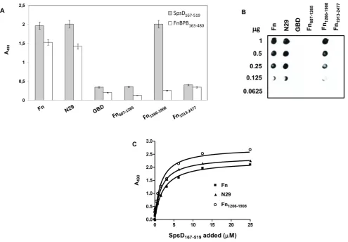
Figure 6. Localization of SpsD binding sites in Fn.(A), native Fn, the N29, the GBD, the recombinant fragments Fn607–1263, Fn1266–1908and the
C-terminal region Fn1913–2477of Fn were immobilized onto a microtiter plate and then incubated with of SpsD167–519. Bound protein was detected by
addition to the wells of anti-SpsD37–519mouse antibody, followed by HRP-conjugated rabbit anti-mouse antibody. Binding of FnBPB163–480to Fn and
its fragments reported as control was detected by addition to the wells of anti-FnBPB163–480rabbit IgG, followed by HRP-conjugated goat anti-rabbit
antibody. (B), different amounts of Fn and Fn fragments (1000-62.5 ng) were dotted onto a nitrocellulose membrane, probed with SpsD167–519(1mg/
ml) and complexes detected by addition of mouse anti-SpsD37–519IgG, followed by HRP-conjugated rabbit anti-mouse IgG. (C), surface-coated Fn, the
N29 fragment and the region including the aa residues 1266–1908 were incubated with increasing concentrations of SpsD167–519and complex
formation detected as reported in (A). Results shown in panels A and C are the mean values of triplicate samples6S.E. The experiments were repeated three times with similar results.
doi:10.1371/journal.pone.0066901.g006
Inhibition of Platelet Aggregation
300ml (96107) washed platelets were preincubated in a Born aggregometer (Chrono-Log Havertown, PA, USA) with either buffer or the indicated concentrations of SpsD167–519or region A
of ClfA or CNA in the presence of 200mg/ml Fbg and 1 mM CaCl2at 37uC for 5 min. Then 10mM ADP was added to trigger
platelet activation and aggregation. The rate and extent of platelet aggregation was detected as the percentage of light transmission and presented as aggregation tracings.
SpsD Binding to Immobilized Fn Fragments by ELISA and Dot Blot
Fn fragments were used to identify binding sites of SpsD. Fn (1mg/well) and Fn fragments (250 ng/well) were immobilized onto microtiter wells and then incubated with indicated concen-trations of SpsD167–519. Bound protein was detected using an
anti-SpsD37–519 mouse antibody followed by HRP-conjugated rabbit
anti-mouse IgG. For dot blot experiments, different amounts of Fn and Fn fragments (1000- 62.5 ng) were dotted onto a nitrocellulose membrane, probed with SpsD167–519 (1mg/ml) and complexes
detected as described above.
Results
Region A of SpsD
The sequence of the entirespsDgene ofS. pseudintermediusED99 is known. Here we describe the A domain of SpsD from strain 326 which was obtained following PCR amplification of genomic DNA. The A domains of SpsD from ED99 and 326 have the same number of residues and are 80% identical. They are most closely related to the A domain of FnBPB (40% and 42% identity to isoform I, respectively) [39] and they have 23% and 27% identity respectively to the A domain of ClfA fromS. aureusNewman [40]. A 3D molecular model based on the crystal structure of ClfA allowed us to predict the coordinates of subdomains N1, N2 and N3 (Figure 1).
The SpsD A domain (N1N2N3 region) comprising residues 37– 519 (SpsD37–519) and the putative minimum ligand binding
truncate comprising residues 167–519 (N2N3, SpsD167–519) were
cloned and expressed with 6 x His affinity tags.
Binding of Recombinant SpsD to Extracellular Matrix Proteins
Previous work has demonstrated that SpsD can promote bacterial adhesion to Fbg, Fn and K10 [11]. Here we sought to determine if the ligand binding ability resides in the N-terminal domain of the protein. An ELISA type ligand-binding assay was used to study the interaction of the recombinant SpsD proteins with surface-coated extracellular matrix proteins. SpsD37–519and
SpsD167–519bound to immobilized Fbg, Fn and K10, as well as to
a-elastin, while no interaction was observed with collagen, laminin or BSA (Figure 2A). The interaction of Fbg, Fn, K10 anda-elastin with the recombinant SpsD167–519 protein was confirmed by
Western ligand blotting where host proteins were electroblotted onto a nitrocellulose filter, probed with SpsD167–519and complex
formation detected with a monoclonal antibody targeting the His-tag. Conversely, no interaction was observed when SpsD167–519
was incubated with membrane-bound collagen type II, laminin or BSA (Figure 2B). Identical results were obtained with SpsD37–519
(data not shown). Together these results suggest that the minimal region of SpsD required for binding to Fbg, Fn, K10 and elastin is the N2N3 subdomain. The absence of signal when Fbg, Fn, a-Figure 7. Binding of SpsD167–519to immobilized cytokeratin 10 proteins.(A), SpsD167–519was incubated in wells coated with recombinant
mouse K101–570, K101–145and K10454–570. (B), SpsD167–519was added to ELISA wells coated with GST-human K10544–563, (HK10544–563); or GST alone.
Bound SpsD167–519was detected by addition of mouse anti-SpsD37–519IgG, followed by HRP-conjugated rabbit anti-mouse IgG. (C), GST-HK10544–563
or GST alone were subjected to SDS-PAGE, electroblotted onto nitrocellulose filter and probed with SpsD167–519followed by incubation with mouse
[image:7.612.58.550.60.227.2]anti-SpsD IgG and HRP-conjugated rabbit anti-mouse IgG. doi:10.1371/journal.pone.0066901.g007
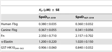
Table 1.Summary ofKD6SE values of SpsD proteins for extracellular matrix components.
KD(mM)±SE
SpsD37–519 SpsD167–519
Human Fbg 0.38060.035 0.36060.032 Canine Fbg 0.36760.055 0.34160.056
Fn 2.35060.710 2.15760.702
a-Elastin 1.20060.220 1.02060.150
GST-HK10544–563 0.90660.060 0.84060.032
[image:7.612.62.297.611.721.2]elastin and K10 were tested for binding to His-tagged S. aureus
MSCRAMM SasG50–428 either in ELISA or Western blotting,
further demonstrates the specificity of SpsD167–519interaction with
host ligands (data not shown).
Next we determined the ability of SpsD167–519 protein to
recognize Fbg molecules obtained from different animals. Increasing amounts of SpsD167–519 were added to surface-coated
human, porcine, canine, bovine and ovine Fbg and the binding was measured. SpsD167–519bound to each of the different types of
Fbg with similar affinities with exception of the ovine protein (Figure 3).
SpsD Binds the C-terminus of thec-chain of Fbg Western ligand blotting was employed to determine if SpsD bound to thea-,b- orc- chain of Fbg. Human Fbg was reduced with dithiothreitol and the three chains separated by SDS-PAGE (Figure 4A). Efficient transfer to a control nitrocellulose membrane was shown by probing with anti-Fbg IgG (Figure 4B). Another membrane was incubated with recombinant His-tagged SpsD167–
519and when probed with a monoclonal antibody recognizing the
His-tag only thec-chain reacted (Figure 4C).
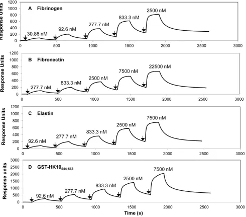
ClfA, FnBPA and FnBPB are known to bind to a short sequence at the extreme C-terminus of thec-chain of Fbg while ClfB binds to repeat number 5 of theachain. To determine if SpsD bound the same c chain residues as ClfA, the ability of a 17 residue synthetic peptide comprising the C-terminal residues of the Fbg c-Figure 8. Surface Plasmon Resonance analysis of the interaction of SpsD167–519with Fbg, Fn,a-elastin and cytokeratin 10.Human Fbg (A), Fn (B) anda-elastin (C) were immobilized onto the surface of dextran chips. GST-human K10544–563, (D) was captured onto a sensor chip coated
with goat anti-GST IgG. Representative sensorgrams display binding of SpsD167–519to and dissocation from Fbg, Fn,a-elastin and GST-human K10544– 563, in a single cycle kinetics assay. Binding was measured as response units (RU) against time. The affinities were calculated from curve fitting to a
plot of the RU values against concentrations of SpsD. Arrows indicate the time at which SpsD167–519was injected. The data shown is representative of
3 individual experiments.
doi:10.1371/journal.pone.0066901.g008
[image:8.612.62.553.62.493.2]chain to inhibit binding of SpsD167–519 to immobilized Fbg was
tested. The peptide strongly inhibited the binding of SpsD to human and canine Fbg dose-dependently while a scrambled peptide had no effect (Figure 4D and 4E).
Finally, direct binding of SpsD to the Fbg c-chain peptide coupled to GST that had been immobilized on the surface of an ELISA plate was tested. As shown in Figure 4F, SpsD bound dose-dependently and saturably to GST-Fbgc395–411 with a half
maximum binding concentration of 0.3260.02mM but not at all to the GST control. Thus, SpsD binds to the same C-terminal Fbgc-chain residues as ClfA and the FnBPs.
Region A of SpsD Interferes with Fibrin Clot Formation and Platelet Aggregation
Several adhesins not only interact with the target molecule in the host but also neutralize and even disrupt the biological function of the target. For example, ClfA, although it does not interfere with catalytic activity of thrombin, by binding to the C-terminus ofcchain it affects fibrin clot formation by abrogating fibrin monomer polymerization and cross-linking of Fbgcchains [41]. Therefore, we examined the effect of SpsD on fibrin clot formation. Human Fbg was preincubated with different concen-trations (1.25–10mM) of SpsD167–519 prior to the addition of
thrombin. In these conditions SpsD inhibited coagulation as effectively as Bbp270–599, a recently identified Fbg-binding protein
fromS. aureus[33] (Figure 5A). Conversely, no effect was observed when Fbg was preincubated with 10mM BSA. Consistent with the
indication that SpsD recognizes the C-terminus of thecchain of canine Fbg, we also observed that preincubation of canine Fbg with 10mM SpsD resulted in complete inhibition of clot formation.
It is well known that the C-terminus of Fbgcchain contains the platelet integrin GPIIb/IIIa recognition sequence (QAGDV411) [42] and that a monoclonal antibody that recognizes thecchain C-terminus of Fbg exerts inhibitory effects on platelet aggregation [43]. Thus, SpsD was tested for its impact on ADP-induced platelet aggregation. As depicted in Figure 5B, ADP-stimulated platelet aggregation was inhibited in a dose-dependent manner by SpsD. In contrast, the collagen-binding domain of the S. aureus
adhesin CNA (CNA30–531), did not affect the process. Together
these data demonstrate that SpsD could act as a modulator of thrombus formation.
Binding Sites in Fibronectin for SpsD
To locate the binding site(s) in Fn for the A domain of SpsD, the binding of SpsD167–519to the Fn N29 domain, the gelatin binding
domain and the segments spanning residues 607–1265, 1266– 1908 and 1913–2477 was tested. Native Fn and the purified fragments were immobilized onto microtiter wells, probed with SpsD167–519and complex formation detected with anti-SpsD IgG.
A strong signal was observed when SpsD167–519 was incubated
with full length Fn, the N29 fragment and the segment spanning the residues 1266–1908 (Fn1266–1908), while the other fragments
[image:9.612.62.551.58.369.2]failed to bind (Figure 6A). A similar assay was performed with Figure 9. Binding of SpsD167–519, SpsD167–519F288A and SpsD167–519latch mutant to immobilized Fbg, Fn, a-elastin and K10.
Microtiter wells were coated with human Fbg (A), Fn (B),a-elastin (C) and K10 (D) in a bicarbonate buffer overnight. The wells were probed with increasing concentrations of SpsD proteins followed by incubation with mouse anti SpsD37–519IgG and HRP-conjugated rabbit anti-mouse IgG.
Values are the means6S.E of triplicate wells and are representative of one experiment. This experiment was performed three times with similar results.
FnBPB163–480and confirmed its ability to bind to full length Fn
and the N29 fragment and not to the C-terminal Fn1266–1908
fragment [23]. The binding of SpsD to the two different regions of Fn was confirmed by incubating the Fn fragments with SpsD167–
519in a dot blot assay (Figure 6B). Finally, as shown in Figure 6C,
binding of SpsD167–519 to both the N29 and the Fn1266–1908
fragment was concentration-dependent and saturable. The affinity for each domain appeared to be similar with half maximum binding concentrations of 2.2160.30mM for N-terminal domain and 1.9760.3mM for Fn1266–1908.
SpsD Binds to the K10 Tail Region
ClfB binds to the C-terminal tail region of K10 [25]. To determine if SpsD binds to the same domain, recombinant proteins comprising full length murine K10 along with the N-terminal head and C-N-terminal tail regions, were immobilized into microtitre wells and tested for their ability to support SpsD binding. SpsD167–519bound to full length K10 and the tail region
dose-dependently and saturably and with half maximal binding concentrations of 1.0260.25 and 1.260.1mM, respectively (Figure 7A).
ClfB is known to bind to glycine (G)-serine (S)-rich domains within the omega loop regions in the C-terminal tail of K10, in loricrin and in the a-chain of Fbg [26]. To determine if SpsD could bind to the same sequences, recombinant SpsD167–519was
tested in solid phase binding assays with GST-HK10544–563. The
SpsD protein bound dose-dependently and saturably but failed to bind to the GST (Figure 7B). This interaction was confirmed by Western blot (Figure 7C). However, in contrast to ClfB, SpsD did not interact with Fbg a chain (Figure S1). Consistent with this, GST-Fbga316–367 inhibited binding of ClfB, but not that of
SpsD167–519, while GST-Fbgc395–411 blocked the interaction of
SpsD167–519but not that of ClfB with Fbg (Figure S2). Thus SpsD
bound the same GS-rich omega loop in K10 as ClfB but surprisingly failed to recognize the Fbgachain.
Measurements of Dissociation Constants of SpsD for Host Ligands by Surface Plasmon Resonance
Binding of SpsD167–519 to human Fbg, Fn,a-elastin or
GST-HK10544–563immobilized on a sensor chip was analyzed by SPR
using BIAcore X. Increasing concentrations of SpsD167–519were
passed over the surface of each ligand-coated chip: in these conditions SpsD167–519bound to the ligand in a dose
dependent-manner. From analysis of the equilibrium binding data, the dissociation constant (KD) for the interaction between SpsD167–519
and Fbg was determined to be 0.36060.032mM (Figure 8A and Table 1). SpsD167–519 showed a dissociation constant value for
canine Fbg comparable to that reported for human Fbg (Table 1). SpsD167–519bound Fn with aKDof 2.1660.70mM, indicating that the affinity of the recombinant SpsD protein for Fn was lower than that exhibited for Fbg (Figure 8B and Table 1). TheKDvalues of SpsD167–519 for a-elastin and GST-HK10544–563 were
1.0260.15 and 0.8460.032mM, respectively (Figure 8, panels C and D and Table1). Very similarKDvalues were obtained when SpsD37–519was passed over sensor chips coated with these proteins
(Table 1).
Ligand Binding by SpsD167–519Mutants
One goal of this work was to determine whether the N2N3 subdomains of SpsD bind Fbg by the same mechanism as the A domains of ClfA and FnBPs. Our previous work demonstrated that the C-terminal residues of the N3 subdomain of FnBPB were crucial for its interaction with Fbg [23]. As SpsD interacts with the
Fbg c-chain with a high affinity, it was proposed that the C-terminal 20 residues of the N3 subdomain of SpsD form the locking and latching segment and play a similar role in the interaction of SpsD with Fbg. To test this, a recombinant truncate of the SpsD167–519protein, which lacked the predicted locking and
latching segment (SpsD167–498:DLL, Figure 1), was expressed and
its ability to bind to immobilized Fbg was analysed by ELISA. No detectable interaction was observed when increasing concentra-tions of SpsD167–499 were incubated with Fbg-coated microtiter
wells (Figure 9A), while a small but remarkable reduction in elastin binding was noticed (Figure 9C). In contrast, no reduction in binding was observed when increasing amounts of SpsD167–499
were incubated with surface-coated Fn or K10 (Figure, 9 panels B and D). To investigate further if SpsD binds to the ligands at same or different sites, an amino acid substitution was introduced in SpsD167–519at residue F288. F288 is predicted to be located within
the ligand binding trench and is in an equivalent position to the crucial residues F314 of FnBPB and Y338 of ClfA. SpsD F288A binding to Fbg and elastin was greatly reduced showing that F288 is indeed important for ligand binding and supports the notion that both Fbg and elastin bind to the same or overlapping regions of SpsD. Conversely, a small reduction was observed when SpsD F288A was tested for binding to Fn or K10.
Discussion
The ability of S. aureus cells to adhere to components of the extracellular matrix is promoted by several distinct surface proteins called MSCRAMMs. The mechanistic basis of many of the interactions has been very well characterized by structural analysis and by probing function with appropriate variant mutant proteins. By molecular modelling and mutagenesis it is possible to infer/predict that proteins with similar structural organization bind their ligands by the same mechanism as those that have been investigated in detail.
The A domain of ClfA is the archetype of a family of proteins that fold into three subdomains called N1, N2 and N3, the last two of which are the absolute minimum required for ligand binding by the ‘‘dock, latch and lock (DLL)’’ mechanism [18]. The A domain of ClfA binds to the c-chain of Fbg by DLL and it does not interact with Fn, elastin or K10 [44–46]. In contrast, the A domain of ClfB binds by DLL to thea-chain of Fbg and also to K10. In both cases the DLL mechanism was shown by X-ray crystal structure and supported by the loss of binding by mutants that had substitutions in the ligand binding trench (trench mutants) or which lack the C-terminal lock-latch domain (LL mutants) [24]. The Fn binding proteins FnBPA and FnBPB are composed of two distinct ligand binding domains. Their A domains are most closely related to ClfA and bind to the Fbgc-chain [16,23]. In contrast to ClfA, both FnBPA and FnBPB also bind to elastin [21] and in the case of FnBPB, Fn [23]. Binding to Fbg and elastin most likely occurs by DLL as indicated by studies with mutant proteins [17]. Ten or 11 tandemly repeated domains at the C-termini of FnBPA and FnBPB bind to Fn with high affinity by the tandembzipper mechanism [29].
individual repeats are shorter than, and bear no similarity to, the Fn-binding repeats of FnBPs [11].
The recombinant A domain of SpsD was shown here to bind to thec-chain of Fbg (and not to thea-chain), to K10, to Fn and also to elastin. There are intriguing similarities to and differences from the MSCRAMM A domains ofS. aureus.
To investigate if the DLL mechanism is involved in ligand binding, DLL and trench mutants of the N2N3 construct were expressed and purified. Binding to Fbg c-chain was completely abrogated in the SpsD DLL and trench mutants indicating that binding to this ligand occurred by DLL. However SpsD also bound to K10 which is a defining property of ClfB, while ClfB binds to thea-chain and not to thec-chain of Fbg [27]. The ligand peptide sequences that bind to the trench are completely dissimilar. Indeed structural biology showed that the Fbga-chain peptide and the K10 omega loop peptide bound to the same part of the ligand binding trench of ClfB and a consensus motif was identified [24]. Therefore it was intriguing to discover that the DLL and trench mutants of SpsD bound to K10 with the same affinity as the wild-type while similar mutants of ClfB were previously shown to be defective in binding to both ligands [24]. We can only conclude that SpsD binding to K10 does not occur by DLL and propose a distinct K10 binding site in N2N3.
Like its closest relative FnBPB, SpsD also binds to elastin. This most likely occurs by DLL because the trench and the DLL mutants were both defective in binding to this ligand. Although a similar investigation was not carried out with FnBPB, detailed studies with the A domain of FnBPA supported the DLL mechanism for elastin binding [17]. This confirms the close relationship of the A domain of SpsD to those of the FnBPs.
It was not clear if the binding of FnBPB A domain to Fn occured by DLL because theDLL truncate bound Fn with the same affinity as the wild-type while a trench mutant was defective in both Fbg and Fn binding. It seemed that the Fn binds to FnBPB in the trench region but that lock latch aspect of DLL is not involved [23]. The SpsD A domain shares with FnBPB the ability to bind Fn but with the difference that it bound to two distinct domains within the host protein while FnBPB only recognized the N29 fragment [23]. This might explain why both the SpsDDLL and trench mutants bound Fn while theDLL mutant of FnBPB was defective.
A domains can be stitched onto distinct ‘‘stalks’’ (B repeats, Sdr repeats, Fn-binding repeats, or to a complete different sequence in SpsD). The DLL binding mechanism in N2N3 regions has evolved to binds to different ligands. Indeed even the orientation of a peptide ligand can vary [24]. Many have acquired the capacity to bind ligands by mechanisms other than DLL although the binding
of a large folded ligand might occlude the trench region and block DLL mechanism of binding to other ligands.
In conclusion, SpsD appears to be the most promiscuous staphylococcal adhesin so far identified and possibly is a factor involved in S. pseudintermedius colonization of the host. In future studies the three-dimensional structure of the apo-protein and ligand bound SpsD may provide molecular details on these multiple interactions and give additional insights to better understand the role played by this microganism in human and animal pathogenesis.
Supporting Information
Figure S1 Binding of SpsD167–519 or ClfB201–542 to Fbg
and Fbgachain.Increasing concentrations of SpsD167–519 (A)
or ClfB201–542(B) were incubated in wells coated with 500 ng/well
human Fbg, GST in fusion with theachain peptide (aa 316–367) or GST alone. Bound ligand was detected by addition of mouse anti-SpsD37–519 IgG or anti-ClfB45–542 IgG followed by
HRP-conjugated rabbit anti-mouse IgG. Results shown in the panels are the mean values of triplicate samples 6 S.E. The experiments were repeated three times with similar results.
(TIF)
Figure S2 Effect of Fbg recombinant chains on the binding of SpsD167–519 or ClfB201–542 to surface-coated
Fbg. 500 ng SpsD167–519 (A) or ClfB201–542 (B) mixed with
increasing amounts of GST-taggedachain, GST in fusion with the C-terminus of thecchain, or GST alone were added to and incubated with microtiter wells coated with Fbg (500 ng/well). Bound ligand was detected by addition of mouse anti-SpsD37–519
IgG or anti-ClfB45–542 IgG followed by HRP-conjugated rabbit
anti-mouse IgG. Results shown in the panels are the mean values of triplicate samples. Error bars show the standard deviation. The experiments were repeated three times with similar results. (TIF)
Acknowledgments
The authors are grateful to Dr. Sofia Giorgetti and Prof. G. Merlini for assistance and advice with BIAcore measurements and Dr. Lina Cipolla for performing platelet aggregation experiments.
Author Contributions
Conceived and designed the experiments: GP JAG TJF PS. Performed the experiments: GP JAG SR ADP AM VC. Analyzed the data: GP JAG TF PS. Contributed reagents/materials/analysis tools: GP JAG TJF PS. Wrote the paper: GP JAG TJF PS.
References
1. Perreten V, Kadlec K, Schwarz S, Gro¨nlund Andersson U, Finn M, et al. (2010) Clonal spread of methicillin-resistantStaphylococcus pseudintermediusin Europe and North America: an international multicentre study. J Antimicrob Chemother 65: 1145–1154.
2. Weese J, van Duijkeren E (2010) Methicillin-resistantStaphylococcus aureusand Staphylococcus pseudintermediusin veterinary medicine. Vet Microbiol 140: 418–429. 3. Stegmann R, Burnens A, Maranta CA, Perreten V (2010) Human infection associated with methicillin-resistant Staphylococcus pseudintermedius ST71. J Antimicrob Chemother 65: 2047–2048.
4. Gerstadt K, Daly JS, Mitchell M, Wessolossky M, Cheeseman SH (1999) Methicillin-resistant Staphylococcus intermedius pneumonia following coronary artery bypass grafting. Clin Infect Dis 29: 218–219.
5. Kempker R, Mangalat D, Kongphet-Tran T, Eaton M (2009) Beware of the pet dog: a case ofStaphylococcus intermediusinfection. Am J Med Sci 338: 425–427. 6. Campanile F, Bongiorno D, Borbone S, Venditti M, Giannella M (2007)
Characterization of a variant of the SCCmec element in a bloodstream isolate of Staphylococcus intermedius. Microb Drug Resist 13: 7–10.
7. Onuma K, Tanabe T, Sato H (2012) Antimicrobial resistance ofStaphylococcus pseudintermediusisolates from healthy dogs and dogs affected with pyoderma in Japan. Vet Dermatol 23: 17–22.
8. Laarhoven LM, de Heus P, van Luijn J, Duim B, Wagenaar JA, et al. (2011) Longitudinal study on methicillin-resistant Staphylococcus pseudintermedius in households. Plos One 6: e27788.
9. Tse H, Tsoi HW, Leung SP, Urquhart IJ, Lau SK, et al. (2011) Complete genome sequence of the veterinary pathogenStaphylococcus pseudintermediusstrain HKU10–03, isolated in a case of canine pyoderma. J Bacteriol 193: 1783–1784. 10. Ben Zakour NL, Bannoehr J, van den Broek AH, Thoday KL, Fitzgerald JR (2011) Complete genome sequence of the canine pathogen Staphylococcus pseudintermedius. J Bacteriol 193: 2363–2364.
11. Bannoehr J, Ben Zakour NL, Reglinski M, Inglis NF, Prabhakaran S, et al. (2011) Genomic and surface proteomic analysis of the canine pathogen Staphylococcus pseudintermedius reveals proteins that mediate adherence to the extracellular matrix. Infect Immun 79: 3074–3086.
13. McEwan NA, Kalna G, Mellor D (2005) A comparison of adherence by four strains ofStaphylococcus intermediusandStaphylococcus hoministo canine corneocytes collected from normal dogs and dogs suffering from atopic dermatitis. Res Vet Sci 78: 193–198.
14. Bannoehr J, Brown JK, Shaw DJ, Fitzgerald RJ, van den Broek AH, et al. (2012) Staphylococccus pseudintermediussurface proteins SpsD and SpsO mediate adherence to ex vivo canine corneocytes. Vet Dermatol 23: 119–124.
15. Geoghegan JA, Smith EJ, Speziale P, Foster TJ (2009) Staphylococcus pseudintermedius expresses surface proteins that closely resemble those from Staphylococcus aureus. Vet Microbiol 138: 345–352.
16. Wann ER, Gurusiddappa S, Ho¨o¨k M (2000) The fibronectin-binding MSCRAMM FnbpA ofStaphylococcus aureusis a bifunctional protein that also binds to fibrinogen. J Biol Chem 275: 13863–13871.
17. Keane FM, Loughman A, Valtulina V, Brennan M, Speziale P, et al. (2007) Fibrinogen and elastin bind to the same region within the A domain of fibronectin binding protein A, an MSCRAMM of Staphylococcus aureus. Mol Microbiol 63: 711–723.
18. Ponnuraj K, Bowden MG, Davis S, Gurusiddappa S, Moore D, et al. (2003) A ‘‘dock, lock, and latch’’ structural model for a staphylococcal adhesin binding to fibrinogen. Cell 115: 217–228.
19. Hair PS, Ward MD, Semmes OJ, Foster TJ, Cunnion KM (2008)Staphylococcus aureusclumping factor A binds to complement regulator factor I and increases factor I cleavage of C3b. J Infect Dis 198: 125–133.
20. Hair PS, Echague CG, Sholl AM, Watkins JA, Geoghegan JA, et al. (2010) Clumping factor A interaction with complement factor I increases C3b cleavage on the bacterial surface of Staphylococcus aureus and decreases complement-mediated phagocytosis. Infect Immun 78: 1717–1727.
21. Roche FM, Downer R, Keane F, Speziale P, Park PW, et al. (2004) The N-terminal A domain of fibronectin-binding proteins A and B promotes adhesion ofStaphylococcus aureusto elastin. J Biol Chem 279: 38433–38440.
22. Keane FM, Clarke AW, Foster TJ, Weiss AS (2007) The N-terminal A domain of Staphylococcus aureus fibronectin-binding protein A binds to tropoelastin. Biochemistry 46: 7226–7232.
23. Burke FM, Di Poto A, Speziale P, Foster TJ (2011) The A domain of fibronectin-binding protein B ofStaphylococcus aureuscontains a novel fibronectin binding site. FEBS J 278: 2359–2371.
24. Ganesh VK, Barbu EM, Deivanayagam CC, Le B, Anderson AS, et al. (2011) Structural and biochemical characterization ofStaphylococcus aureusclumping factor B/ligand interactions. J Biol Chem 286: 25963–25972.
25. Walsh EJ, O’Brien LM, Liang X, Ho¨o¨k M, Foster TJ (2004) Clumping factor B, a fibrinogen-binding MSCRAMM (microbial surface components recognizing adhesive matrix molecules) adhesin ofStaphylococcus aureus, also binds to the tail region of type I cytokeratin 10. J Biol Chem 279: 50691–50699.
26. Mulcahy ME, Geoghegan JA, Monk IR, O’Keeffe KM, Walsh EJ, et al. (2012) Nasal Colonisation byStaphylococcus aureusDepends upon Clumping Factor B Binding to the Squamous Epithelial Cell Envelope Protein Loricrin. Plos Pathog 8: e1003092.
27. Walsh EJ, Miajlovic H, Gorkun OV, Foster TJ (2008) Identification of the Staphylococcus aureusMSCRAMM clumping factor B (ClfB) binding site in the alphaC-domain of human fibrinogen. Microbiology 154: 550–558.
28. Schwarz-Linek U, Werner JM, Pickford AR, Gurusiddappa S, Kim JH, et al. (2003) Pathogenic bacteria attach to human fibronectin through a tandem beta-zipper. Nature 423: 177–181.
29. Meenan NA, Visai L, Valtulina V, Schwarz-Linek U, Norris NC, et al. (2007) The tandem beta-zipper model defines high affinity fibronectin-binding repeats withinStaphylococcus aureusFnBPA. J Biol Chem 282: 25893–25902. 30. Hartford O, Francois P, Vaudaux P, Foster TJ (1997) The dipeptide repeat
region of the fibrinogen-binding protein (clumping factor) is required for
functional expression of the fibrinogen-binding domain on the Staphylococcus aureuscell surface. Mol Microbiol 25: 1065–1076.
31. Speziale P, Visai L, Rindi S, Di Poto A (2008) Purification of human plasma fibronectin using immobilized gelatin and Arg affinity chromatography. Nat Protoc 3: 525–533.
32. Zardi L, Carnemolla B, Balza E, Borsi L, Castellani P, et al. (1985) Elution of fibronectin proteolytic fragments from a hydroxyapatite chromatografy colum. A simple procedure for the purification of fibronectin domains. Eur J Biochem 146: 571–579.
33. Vazquez V, Liang X, Horndahl JK, Ganesh VK, Smeds E, et al. (2011) Fibrinogen is a ligand for theStaphylococcus aureusmicrobial surface components recognizing adhesive matrix molecules (MSCRAMM) bone sialoprotein-binding protein (Bbp). J Biol Chem 286: 29797–29805.
34. O’Connell DP, Nanavaty T, McDevitt D, Gurusiddappa S, Ho¨o¨k M, et al. (1998) The fibrinogen-binding MSCRAMM (clumping factor) ofStaphylococcus aureushas a Ca2+-dependent inhibitory site. J Biol Chem 273: 6821–6829.
35. Rich RL, Deivanayagam CC, Owens RT, Carson M, Ho¨o¨k A, et al. (1999) Trench-shaped binding sites promote multiple classes of interactions between collagen and the adherence receptors, alpha(1)beta(1) integrin andStaphylococcus aureuscna MSCRAMM. J Biol Chem 274: 24906–24913.
36. Roche FM, Massey R, Peacock SJ, Day N, Visai L, et al. (2003) Characterization of novel LPXTG-containing proteins ofStaphylococcus aureus identified from genome sequences. Microbiology 149: 643–654.
37. Geoghegan JA, Ganesh VK, Smeds E, Liang X, Ho¨o¨k M, et al. (2010) Molecular characterization of the interaction of staphylococcal microbial surface components recognizing adhesive matrix molecules (MSCRAMM) ClfA and Fbl with fibrinogen. J Biol Chem 285: 6208–6216.
38. Johnsson B, Lo¨fa˚s S, Lindquist G (1991) Immobilization of proteins to a carboxymethyldextran-modified gold surface for biospecific interaction analysis in surface plasmon resonance sensors. Anal Biochem 198: 268–277. 39. Burke FM, McCormack N, Rindi S, Speziale P, Foster TJ. (2010)
Fibronectin-binding protein B variation inStaphylococcus aureus. BMC Microbiol 10: 160. doi: 10.1186/1471-2180-10-160.
40. McDevitt D, Francois P, Vaudaux P, Foster TJ. (1995) Identification of the ligand-binding domain of the surface-located fibrinogen receptor (clumping factor) ofStaphylococcus aureus. Mol Microbiol 16: 895–907.
41. Liu CZ, Shih MH, Tsai PJ (2005) ClfA(221–550), a fibrinogen-binding segment ofStaphylococcus aureusclumping factor A, disrupts fibrinogen function. Thromb Haemost 94: 286–294.
42. Kloczewiak M, Timmons S, Hawiger J (1983) Recognition site for the platelet receptor is present on the 15-residue carboxy-terminal fragment of the gamma chain of human fibrinogen and is not involved in the fibrin polymerization reaction. Thromb Res 29: 249–255.
43. Jirouskova´ M, Smyth SS, Kudryk B, Coller BS (2001) A hamster antibody to the mouse fibrinogen gamma chain inhibits platelet-fibrinogen interactions and FXIIIa-mediated fibrin cross-linking, and facilitates thrombolysis. Thromb Haemost 86: 1047–1056.
44. Hawiger J, Kloczewiak M, Bednarek MA, Timmons S (1989) Platelet receptor recognition domains on the alpha chain of human fibrinogen: structure-function analysis. Biochemistry 28: 2909–2914.
45. McDevitt D, Nanavaty T, House-Pompeo K, Bell E, Turner N, et al. (1997) Characterization of the interaction between theStaphylococcus aureusclumping factor (ClfA) and fibrinogen. Eur J Biochem 247: 416–424.
46. Ganesh VK, Rivera JJ, Smeds E, Ko YP, Bowden MG, et al. (2008) A structural model of theStaphylococcus aureusClfA-fibrinogen interaction opens new avenues for the design of anti-staphylococcal therapeutics. PLoS Pathog 4: e1000226.