Aus der Medizinischen Klinik und Poliklinik III - Großhadern
der Ludwig-Maximilians-Universität München Direktor: Prof. Dr. med. W. Hiddemann
Deciphering the genetic heterogeneity in Acute Myeloid Leukemia:
Association of gene mutations with distinct chromosomal aberrations
Dissertation
zum Erwerb des Doktorgrades der Humanbiologie (Dr. rer. biol. hum.)
an der Medizinischen Fakultät der Ludwig-Maximilians-Universität zu München
vorgelegt von Luise Hartmann
aus Hannover
2
Mit Genehmigung der Medizinischen Fakultät der Universität München
Berichterstatter: Prof. Dr. Karsten Spiekermann
Mitberichterstatter: Prof. Dr. Elke Holinski-Feder
Priv. Doz. Dr. Ursula Zimber-Strobl Priv. Doz. Dr. Michael Albert Mitbetreuung durch den
promovierten Mitarbeiter: Dr. Philipp Greif
Dekan: Prof. Dr. med. dent. Reinhard Hickel
3
Eidesstattliche Versicherung
Hartmann, Luise
Ich erkläre hiermit an Eides statt, dass ich die vorliegende Dissertation mit dem Thema:
‚Deciphering the genetic heterogeneity in Acute Myeloid Leukemia: Association of gene mutations with distinct chromosomal aberrations’
selbständig verfasst, mich außer der angegebenen keiner weiteren Hilfsmittel bedient und alle Erkenntnisse, die aus dem Schrifttum ganz oder annähernd übernommen sind, als solche kenntlich gemacht und nach ihrer Herkunft unter Bezeichnung der Fundstelle einzeln nachgewiesen habe.
Ich erkläre des Weiteren, dass die hier vorgelegte Dissertation nicht in gleicher oder in ähnlicher Form bei einer anderen Stelle zur Erlangung eines akademischen Grades eingereicht wurde.
München, den
4
5
Table of contents
I. Zusammenfassung (Summary in German)………...……. P.6
II. Summary……….…...………...…………. P.7
III. Abbreviations……….………...…...……...………. P.8
IV. Tables and Figures……….………...……….……. P.9
1. Introduction
1.1. Acute Myeloid Leukemia (AML)………...…………..………. P.10 1.2. Chromosomal alterations in AML………..………...…..….. P.12 1.3. The mutational landscape of AML………..………...….. P.15
2. Specific aims and questions……….………...……….….. P.17
3. Summary of results
3.1. Paper I:Characterization of AML with trisomy 13……….…...…. P.18 3.2. Paper II: ZBTB7A mutations in t(8;21) positive AML…...…….. P.19
4. Conclusion and outlook………...……….…...……….…….. P.20
5. References………...………….……….……….….. P.22
6. Acknowledgements………...………...……….………..….. P.29
7. Curriculum vitae………...……….……….. P.30
Appendix:………....………..….….. P.32
6
I.
Zusammenfassung
Das Hauptziel der vorliegenden Dissertation ist die genetische Charakterisierung von zytogenetischen Subgruppen der Akuten Myeloischen Leukämie (AML). Grundlage dieser kumulativen Dissertation sind die beiden aufgeführten Publikationen, die in renommierten Fachzeitschriften erschienen sind (Impact-factor von Blood in 2014: 10.452; aktueller Impact-factor von Nature Communications: 11.470):
- Herold, T., K. H. Metzeler, S. Vosberg, L. Hartmann, C. Röllig, F. Stölzel,
S. Schneider, M. Hubmann, E. Zellmeier, B. Ksienzyk, V. Jurinovic, Z. Pasalic, P. M. Kakadia, A. Dufour, A. Graf, S. Krebs, H. Blum, M. C. Sauerland, T. Büchner, W. E. Berdel, B. J. Wörmann, M. Bornhäuser, G. Ehninger, U. Mansmann, W. Hiddemann, S. K. Bohlander, K. Spiekermann and P. A. Greif (2014). "Isolated trisomy 13 defines a homogeneous AML subgroup with high frequency of mutations in spliceosome genes and poor prognosis." Blood 124(8): 1304-1311.
-
Hartmann, L., S. Dutta, S. Opatz, S. Vosberg, K. Reiter, G. Leubolt, K. H.Metzeler, T. Herold, S. A. Bamopoulos, K. Bräundl, E. Zellmeier, B. Ksienzyk, N. P. Konstandin, S. Schneider, K. P. Hopfner, A. Graf, S. Krebs, H. Blum, J. M. Middeke, F. Stölzel, C. Thiede, S. Wolf, S. K. Bohlander, C. Preiss, L. Chen-Wichmann, C. Wichmann, M. C. Sauerland, T. Büchner, W. E. Berdel, B. J. Wörmann, J. Braess, W. Hiddemann, K. Spiekermann and P. A. Greif (2016). "ZBTB7A mutations in acute myeloid leukaemia with t(8;21) translocation." Nat Commun 7: 11733.
In beiden Arbeiten wurden Genmutationen identifiziert, die spezifisch bei AML Patienten mit bestimmten chromosomalen Veränderungen auftreten: SRSF2 Mutationen bei Patienten mit Trisomie 13 und ZBTB7A Mutationen bei Patienten mit t(8;21) Translokation.
7
II.
Summary
The main objective of this dissertation is the genetic characterization of cytogenetic subgroups of acute myeloid leukemia (AML). This cumulative dissertation is based on two articles that were published in leading scientific journals (impact factor of Blood in 2014: 10.452; recent impact factor of Nature Communications: 11.470):
- Herold, T., K. H. Metzeler, S. Vosberg, L. Hartmann, C. Röllig, F. Stölzel,
S. Schneider, M. Hubmann, E. Zellmeier, B. Ksienzyk, V. Jurinovic, Z. Pasalic, P. M. Kakadia, A. Dufour, A. Graf, S. Krebs, H. Blum, M. C. Sauerland, T. Büchner, W. E. Berdel, B. J. Wörmann, M. Bornhäuser, G. Ehninger, U. Mansmann, W. Hiddemann, S. K. Bohlander, K. Spiekermann and P. A. Greif (2014). "Isolated trisomy 13 defines a homogeneous AML subgroup with high frequency of mutations in spliceosome genes and poor prognosis." Blood 124(8): 1304-1311.
- Hartmann, L., S. Dutta, S. Opatz, S. Vosberg, K. Reiter, G. Leubolt, K. H.
Metzeler, T. Herold, S. A. Bamopoulos, K. Bräundl, E. Zellmeier, B. Ksienzyk, N. P. Konstandin, S. Schneider, K. P. Hopfner, A. Graf, S. Krebs, H. Blum, J. M. Middeke, F. Stölzel, C. Thiede, S. Wolf, S. K. Bohlander, C. Preiss, L. Chen-Wichmann, C. Wichmann, M. C. Sauerland, T. Büchner, W. E. Berdel, B. J. Wörmann, J. Braess, W. Hiddemann, K. Spiekermann and P. A. Greif (2016). "ZBTB7A mutations in acute myeloid leukaemia with t(8;21) translocation." Nat Commun 7: 11733.
In both studies, gene mutations were found that occur specifically in AML patients with distinct chromosomal aberrations: SRSF2 mutations in patients with trisomy 13 and ZBTB7A mutations in patients with t(8;21) translocation.
8
III.
Abbreviations
2-DG 2-Deoxy-D-glucose AML Acute myeloid leukemia CBF Core binding factor
9
IV.
Tables and Figures
Table 1: WHO 2008 classification of acute myeloid leukemia
Table 2: MRC AML risk classification according to chromosomal aberrations Table 3: Recurrently mutated genes in AML
Figure 1: Normal hematopoiesis and acute myeloid leukemia
Figure 2: Cytogenetic results from the Medical Research Council (MRC) trials Figure 3: The core binding factor (CBF) complex
Figure 4: Molecular pathogenesis of AML
10
1. Introduction
1.1. Acute myeloid leukemia (AML)
Clinical characteristics
Acute myeloid leukemia (AML) is a hematopoietic malignancy characterized by excessive growth of clonal myeloid progenitor cells. The term ‘leukemia’ was coined in the 19th century by Rudolf Virchow, based on his observations of ‘white blood’
(Kampen, 2012).
Common symptoms of AML include anemia, bleeding and frequent infections. The diagnosis is based on cytomorphological assessment of bone marrow and peripheral blood. AML is mostly a disease of the elderly, with a median age of >65 years at diagnosis (Juliusson et al, 2012; Wang, 2014). A combination of daunorubicin and cytarabine (the so-called ‘3+7’ regimen) is the standard initial treatment for AML and results in remission, i.e. reduction of bone marrow blast counts to <5%, in 40-80% of patients (Burnett et al, 2011). However, a high proportion of patients will eventually relapse and become non-responsive to further therapy approaches. The five-year survival rate for adult AML can be as low as 10% (Burnett et al, 2011). Importantly, it was shown that remission and survival rates highly depend on clinical (e.g. age) and biological factors (e.g. karyotype, gene mutations), allowing for risk stratification and treatment adjustment such as consideration of allogeneic stem cell transplantation for suitable patients with high risk disease (Estey and Döhner, 2006; Döhner et al, 2010). Initially, AML was classified based on cytomorphology. In 1976, the French-American-British (FAB) co-operative group proposed the so-called FAB classification which recognizes eight subtypes (M0- M7) with respect to cell type and differentiation (Bennett et al, 1976). Later, with better understanding of AML pathogenesis, a more refined classification established by the World Health Organization (WHO) also included biological and cytogenetic factors (Vardiman et al, 2009).
Table 1: WHO 2008 classification of acute myeloid leukemia (Vardiman et al, 2009)
Acute myeloid leukemia
Acute myeloid leukemia with recurrent genetic abnormalities Acute myeloid leukemia with myelodysplasia-related changes Therapy-related myeloid neoplasms
11 Leukemogenesis
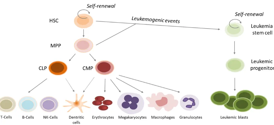
Normal hematopoiesis follows a tightly regulated hierarchy (Figure 1). Hematopoietic stem cells (HSC) reside in the bone marrow and have self-renewal capacities but can also differentiate into all blood cell types. Upon stimulation, HSCs differentiate to multipotent progenitors (MPP) which are still able to generate all kinds of mature blood cells but have lost self-renewal capacity (Fiedler and Brunner, 2012). The common lymphoid progenitors (CLP) and common myeloid progenitors (CMP) give rise to the mature cells of the lymphoid lineage (T-cells, B-cells, NK-cells) or the mature cells of myeloid lineage (erythrocytes, megakaryocytes, macrophages, granulocytes), respectively (Kondo et al, 1997; Akashi et al 2000). Differentiation and commitment to cell lineage fates have been demonstrated to highly depend on the expression of specific combinations of transcription factors (Tenen, 2003; Wilson et al, 2010; Pouzolles et al, 2016).
It was shown that AML derives from early progenitor cells (Bonnet and Dick, 1997). Ddifferentiation of myeloid progenitors is blocked and the cells proliferate unrestrictedly, leading to accumulation of clonal immature precursor cells in the bone marrow and consecutive suppression of normal hematopoiesis.
12
The transformation of normal HSCs or MPPs to leukemic blasts is a multi-step process driven by sequential leukemogenic events (reviewed by Horton and Huntly, 2012). These events are commonly alterations of the genome. In consequence, characterization of genomic lesions in AML is essential to understand the pathogenesis of AML and ultimately to enable the development of tailored, more effective therapies.
1.2. Chromosomal alterations in AML
Recurrent cytogenetic alterations, i.e. structural or numerical chromosomal abnormalities, in AML were already described more than 40 years ago by pioneering work of Janet Rowley and others (reviewed by Freireich et al, 2014). The discovery of recurring balanced translocations between chromosomes 8 and 21, termed t(8;21)(q22;q22), in AML was the first translocation to be described in human cancers and is considered a milestone in our understanding of cancer genetics (Rowley, 1973). In approximately 50-60% of AML patients, abnormal karyotypes can be detected and as shown in Figure 2, the diversity of cytogenetic abnormalities is rather high.
13
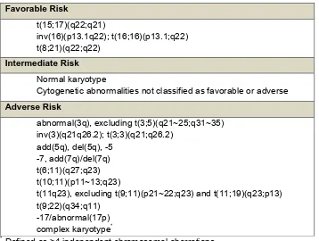
Despite this complexity, the prognostic impact of the most common chromosomal abnormalities has been assessed through efforts of numerous study groups (overview in Burnett et al, 2011), leading to the widely used risk classification established by the European Leukemia Network (ELN) and Medical Research Council (MRC).
Table 2: MRC AML risk classification according to chromosomal aberrations (Grimwade et al, 2010)
Favorable Risk
t(15;17)(q22;q21)
inv(16)(p13.1q22); t(16;16)(p13.1;q22) t(8;21)(q22;q22)
Intermediate Risk Normal karyotype
Cytogenetic abnormalities not classified as favorable or adverse Adverse Risk
abnormal(3q), excluding t(3;5)(q21~25;q31~35) inv(3)(q21q26.2); t(3;3)(q21;q26.2)
add(5q), del(5q), -5 -7, add(7q)/del(7q) t(6;11)(q27;q23) t(10;11)(p11~13;q23)
t(11q23), excluding t(9;11)(p21~22;q23) and t(11;19)(q23;p13) t(9;22)(q34;q11)
-17/abnormal(17p) complex karyotype*
* Defined as >4 independent chromosomal aberrations
Besides assessing their prognostic impact, understanding the underlying mechanisms how chromosome abnormalities arise and how they contribute to leukemogenesis is of great importance.
14
The precise molecular mechanism which causes chromosomal translocations remains elusive. Studies showed that homologous recombination, non-homologous end joining and chromosome fragile sites potentially trigger the formation of translocations (reviewed by Aplan, 2006). Moreover, it was shown that chromosome segregation errors during mitosis can lead to translocations as well (Janssen et al, 2011). In general, oncogenic translocations lead either to novel fusion genes (Hermans et al, 1987; de Thé et al, 1991) or juxtaposition of regulatory elements from one translocation partner to the other, resulting in aberrant gene expression (ar-Rushdi et al, 1983; Gröschel et al, 2014). The functional consequences of many chromosomal rearrangements have been subject to intensive studies. The recurrent translocation t(8;21)(q22;q22), for example, leads to the chimeric RUNX1/RUNX1T1 gene (also known as AML1-ETO) (Erickson et al, 1992). RUNX1 is an important transcription factor for regulation of hematopoiesis (Tanaka et al, 1995; Okuda et al, 1996) and part of the so-called core binding factor (CBF) complex. Through fusion with RUNX1T1, normal function of RUNX1 in the CBF complex is disturbed, preventing transcription of CBF target genes important for myeloid differentiation, and thereby leading to disruption of normal hematopoiesis and inactivation of tumor suppressor genes (Westendorf et al, 1998; Goyoma and Mulloy, 2011).
Figure 3: The core binding factor (CBF) complex (adapted from Solh et al, 2014). (A) The CBF consists of 2 subunits. RUNX1 and CBFB form a complex known to initiate transcription of genes involved in myeloid differentiation. (B) The t(8;21) translocation leads to the RUNX1/RUNX1T1 fusion and, via recruitment of additional factors, to inactivation of CBF target genes.
15
1.3. The mutational landscape of AML
Besides microscopically detectable chromosomal alterations, gene mutations in AML have also been intensively investigated. Initially, gene mutations were identified based on candidate approaches or serendipitously. For example, AML samples were screened for NRAS mutations based on the observation that this oncogene is mutated in other types of cancer (Bos et al, 1985). NPM1 mutations, which occur in approximately 25-35% of AML patients, were discovered after detection of aberrant cytoplasmic localization of the protein. It was shown that in most cases an insertion of 4 bases lead to a frame shift in the region encoding the C-terminus of NPM1, thereby truncating the protein and leading to loss of a nuclear localization signal and consequently abnormal sub-cellular localization (Falini et al, 2005).
With the introduction of next generation sequencing (NGS) technologies (reviewed by Welch and Link, 2011), the number of known recurrently mutated genes in AML has increased tremendously. In fact, the first human cancer genome to be completely sequenced was from a patient with AML (Ley et al, 2008). Shortly after, DNMT3A mutations were described by the same research group (Ley et al, 2010), followed by the discovery of several other novel gene mutations in AML such as BCOR (Grossmann et al, 2011), GATA2 (Greif et al, 2012), RAD21 (Dolnik et al, 2012) and ASXL2 (Micol et al, 2014). Through high-throughput sequencing approaches, these and other mutations have been studied by several groups with regards to their frequency and prognostic significance (reviewed by Larsson et al, 2013; Meyer and Levine, 2014; Döhner et al, 2015). An overview of the most common recurrently mutated genes in AML is shown in Table 3.
Table 3: Recurrently mutated genes in AML (according to Döhner et al, 2015). ITD= Internal tandem duplication, PTD= Partial tandem duplication
Mutated gene Frequency
NPM1 FLT3-ITD DNMT3A
NRAS
25-35% 20% 18-22%
15%
TET2 7-25%
CEBPA 6-10%
RUNX1 5-15%
ASXL1 5-17%
IDH1; IDH2 7-14%; 8-19%
KIT <5%
KMT2A-PTD 5%
16
Development of AML is believed to be a multistep process that requires the sequential acquisition of several mutations. Based on studies of CBF leukemia, it was proposed that these mutations would fall into two distinct categories (Speck and Gilliland, 2002). Class I mutations (for example in FLT3, KIT and NRAS) enhance proliferation and survival, predominantly through constitutively activated signaling pathways. In contrast, class II mutations result in impaired differentiation of hematopoietic progenitor cells and often affect transcription factors such as RUNX1 or GATA1/2. Mutations of both classes are likely necessary to develop full-blown leukemia.
In the last years, with the discovery of numerous novel gene mutations, this model had to be revised. Functional analyses demonstrated that several mutations do not accurately fit in class I or II but can be categorized in other functional groups. DNMT3A, for example, encodes a DNA methyltransferase and DNMT3A mutations lead to global changes of the DNA methylation pattern (Russler-Germain et al, 2014). Likewise, TET2 and IDH1/2 mutations have also been associated with epigenetic changes (Figueroa et al, 2010). In consequence, new functional classifications of
gene mutations in AML have been suggested as shown in Figure 4 (Thiede, 2012).
17
2. Specific aims and questions
AML is an exceedingly heterogeneous disease on the genetic level (Grimwade et al, 2016; Papaemmanuil et al, 2016; Metzeler et al, 2016). Probably, we will not identify two individuals with AML that are characterized by exactly the same genetic alterations. However, since associations between gene mutations and certain chromosomal aberrations have already been shown, e.g. KIT mutations in AML with t(8;21) or inv(16) (Beghini et al, 2000; Care et al, 2003) and TP53 mutations in AML with complex karyotype (Haferlach et al, 2008), it is worth investigating cytogenetic subgroups of AML in order to identify further patterns of mutational co-occurrence and thereby decipher the genetic heterogeneity. Furthermore, it is of great interest to study the impact of these mutations on a clinical and functional level. Can we improve risk stratification if we include information about gene mutations? Are co-occurring gene mutations just bystanders or how do they contribute to the AML phenotype? This information might be particularly valuable for the design of novel targeted therapies.
The studies presented in this thesis aimed (I) to investigate the mutational landscape of selected cytogenetic subgroups and (II) to evaluate clinical and functional consequences of identified mutations.
18
3. Summary of results
Paper I: Characterization of AML with trisomy 13
Herold T, Metzeler KH, Vosberg S, Hartmann L, Röllig C, Stölzel F, et al. Isolated
trisomy 13 defines a homogeneous AML subgroup with high frequency of mutations in spliceosome genes and poor prognosis. Blood. 2014
Trisomy 13 (+13) as sole aberration is a rare cytogenetic finding in AML with an incidence of <1%. According to ELN and MRC risk stratification, patients with isolated +13 fall into the intermediate risk group. However, previous studies indicated adverse clinical outcome for AML patients with +13.
The aims of the presented study were (I) clinical characterization, (II) mutational profiling and (III) gene expression analysis of AML patients with +13.
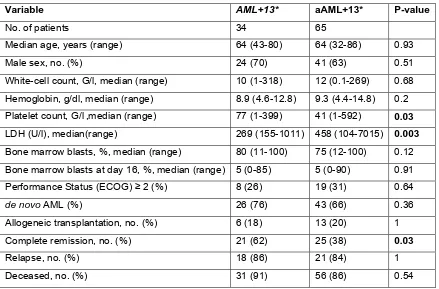
Clinical data were available for 34 patients with isolated +13 and 850 patients with other cytogenetic findings that also fall into the same risk group. Patients with +13 were significantly older and had higher blast counts at diagnosis. Moreover, relapse-free survival and overall survival were inferior for the AML +13 group compared with the other intermediate-risk patients.
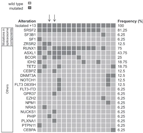
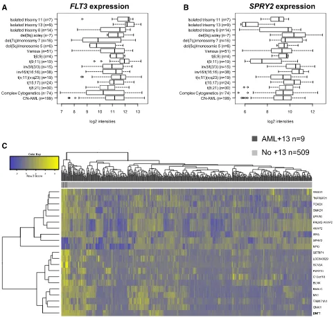
Exome sequencing of paired diagnostic and remission samples from two patients with +13 identified leukemia-specific mutations in 36 genes, including RUNX1, ASXL1, BCOR, ZRSR2, NUP188 and CEBPZ. Next, targeted amplicon sequencing was performed on 16 AML +13 samples, revealing high frequencies of mutations in RUNX1 (n=12, 75%) and the spliceosome complex (SRSF2: 81%, SF3B1: 6%, SF1: 6% and ZRSR2:13%). Moreover, novel mutations in CEBPZ were identified. The frequency of SRSF2 mutations in AML +13 is the highest to be so far reported in any AML subgroup, pointing towards a joint contribution to cell transformation. Similarly, gene expression analysis identified genes that were significantly deregulated in AML +13, including FLT3 (upregulation) and SPRY2 (downregulation).
Contribution to this project as co-author:
19
Paper II: ZBTB7A mutations in t(8;21) positive AML
Hartmann L, Dutta S, Opatz S, Vosberg S, Reiter K, et al. ZBTB7A Mutations in
Acute Myeloid Leukemia with t(8;21) Translocation, Nat Commun. 2016
The t(8;21) translocation is one of the most frequent chromosomal abnormalities in AML and leads to the fusion gene RUNX1/RUNX1T1. However, in vivo models indicate the requisite of additional lesions for leukemogenesis as RUNX1/RUNX1T1 alone is not able to induce leukemia. Exome sequencing of matched diagnostic and remission samples of two patients with t(8;21) rearrangement identified leukemia-specific ZBTB7A mutations in both patients. ZBTB7A is a transcriptional repressor and plays a role in normal hematopoiesis. Previous studies indicated that ZBTB7A has both proto-oncogenic and tumor suppressor properties in a tissue-dependent fashion.
The aim of this study were to (I) assess the mutation frequency of ZBTB7A mutations in a large cohort of AML patients with t(8;21) translocation, (II) functionally characterize ZBTB7A mutations and (III) evaluate the clinical impact of ZBTB7A mutations and expression.
Using targeted amplicon sequencing, ZBTB7A mutations were identified in 13/56 (23%) of screened RUNX1/RUNXT1 positive AML patients. Importantly, ZBTB7A mutations were not detected in 50 CN-AML patients. Two mutational hotspots (R402 and A175fs) were identified and further characterized on a functional level. The R402 mutations affect the zinc finger structure of ZBTB7A while the A175fs mutation leads to complete loss of the zinc finger domain. DNA pull-down assays and luciferase-based transcription reporter assays indicated that the analyzed ZBTB7A mutations lead to loss-of-function. Retroviral expression of wild-type ZBTB7A in a RUNX1/RUNXT1 positive cell line as well as lineage negative murine bone marrow cells (co-expressing RUNX1/RUNX1T1) inhibited cell growth, whereas this anti-proliferative effect was lost or weakened upon expression of ZBTB7A mutants.
20
4. Conclusion and outlook
The two studies presented in this thesis provided novel insides into the biology of acute myeloid leukemia:
-Isolated trisomy 13 is a rare cytogenetic finding but associated with inferior clinical outcome. Consequently, patients with this cytogenetic aberration should be stratified into the group of adverse risk.
-For the first time we have shown that trisomy 13 is associated with a high frequency of SRSF2 mutations (13 of 16 patients, 81%). SRSF2 is a splicing factor and part of the spliceosome. It was shown that the common SRSF2 P59H mutation leads to deregulated splicing because of altered RNA-binding affinities (Zhang et al, 2015). How this effect contributes to leukemogenesis and how mutated SRSF2 and trisomy 13 may collaborate remains to be investigated.
-ZBTB7A mutations are a novel finding in AML. Just recently, another group also identified ZBTB7A mutations in 3/20 patients with t(8;21) translocation (Lavallée et al, 2016), independently confirming our data. Given the high frequency of these mutations, it is worth analyzing ZBTB7A mutations in a larger patient cohort to gain reliable information about the prognostic relevance of ZBTB7A mutations. This information can help to refine risk-stratification for t(8;21) positive patients.
-Our data indicates a specific association of ZBTB7A mutations and RUNX1/RUNX1T1 suggesting oncogenic collaboration, however, the underlying mechanism remains elusive.
-ZBTB7A has been reported to act either as a tumor suppressor or oncogene, in a tissue-dependent fashion. The presented study indicates that ZBTB7A functions as a tumor suppressor in AML.
21
restricted in ZBTB7A mutated AML by treatment with glycolysis inhibitors such as 2-Deoxy-D-glucose (2-DG). For solid tumors, mouse transplantation assays already indicated that 2-DG treatment leads to reduced growth of ZBTB7A-knock down cells (Liu et al 2014). Importantly, clinical trials confirmed that the administration of 2-DG alone or combined with other anticancer therapies, such as chemotherapy and radiotherapy was safe and well tolerated by patients with solid tumors (Dwarakanath et al, 2009; Raez et al, 2013). It is therefore worthwhile investigating whether similar effects can also be observed in AML.
In 2013, the cancer genome atlas (TCGA) consortium published a series of 200 AML cases that were comprehensively characterized for gene mutations by either whole genome sequencing (n=50) or exome sequencing (n=150). The cohort comprised adult AML patients representing the major cytomorphologic and cytogenetic subtypes, including 7 patients that were RUNX1/RUNX1T1 positive and a single patient with isolated trisomy 13. A total of 2315 somatic single nucleotide variants (SNV) and 270 small insertions or deletions (INDEL) in coding regions were identified. However, no ZBTB7A mutations and only a single SRSF2 mutation were reported in this patient cohort (the SRSF2 mutation was not found in the patient with isolated trisomy 13). This highlights that the genetic landscape of AML is still not fully understood and that focused analyses of cytogenetic subgroups is important for the discovery of novel mutations that might play an important role in leukemogenesis and provide the basis for tailored therapies that overcome the poor clinical outcome of patients with AML.
22
5. References
Akashi, K., D. Traver, T. Miyamoto and I. L. Weissman (2000). "A clonogenic common myeloid progenitor that gives rise to all myeloid lineages." Nature 404(6774): 193-197.
Aplan, P. D. (2006). "Causes of oncogenic chromosomal translocation." Trends Genet 22(1): 46-55.
ar-Rushdi, A., K. Nishikura, J. Erikson, R. Watt, G. Rovera and C. M. Croce (1983). "Differential expression of the translocated and the untranslocated c-myc oncogene in Burkitt lymphoma." Science 222(4622): 390-393.
Bakhoum, S. F. and D. A. Compton (2012). "Chromosomal instability and cancer: a complex relationship with therapeutic potential." J Clin Invest 122(4): 1138-1143. Becker, H., K. Maharry, K. Mrozek, S. Volinia, A. K. Eisfeld, M. D. Radmacher, J.
Kohlschmidt, K. H. Metzeler, S. Schwind, S. P. Whitman, J. H. Mendler, Y. Z. Wu, D. Nicolet, P. Paschka, B. L. Powell, T. H. Carter, M. Wetzler, J. E. Kolitz, A. J. Carroll, M. R. Baer, M. A. Caligiuri, R. M. Stone, G. Marcucci and C. D. Bloomfield (2014). "Prognostic gene mutations and distinct gene- and microRNA-expression signatures in acute myeloid leukemia with a sole trisomy 8." Leukemia 28(8): 1754-1758.
Beghini, A., P. Peterlongo, C. B. Ripamonti, L. Larizza, R. Cairoli, E. Morra and C. Mecucci (2000). "C-kit mutations in core binding factor leukemias." Blood 95(2): 726-727.
Bennett, J. M., D. Catovsky, M. T. Daniel, G. Flandrin, D. A. Galton, H. R. Gralnick and C. Sultan (1976). "Proposals for the classification of the acute leukaemias. French-American-British (FAB) co-operative group." Br J Haematol 33(4): 451-458.
Bochtler, T., S. Frohling and A. Kramer (2015). "Role of chromosomal aberrations in clonal diversity and progression of acute myeloid leukemia." Leukemia 29(6): 1243-1252.
Bonnet, D. and J. E. Dick (1997). "Human acute myeloid leukemia is organized as a hierarchy that originates from a primitive hematopoietic cell." Nat Med 3(7): 730-737.
Bos, J. L., D. Toksoz, C. J. Marshall, M. Verlaan-de Vries, G. H. Veeneman, A. J. van der Eb, J. H. van Boom, J. W. Janssen and A. C. Steenvoorden (1985). "Amino-acid substitutions at codon 13 of the N-ras oncogene in human acute myeloid leukaemia." Nature 315(6022): 726-730.
23
Care, R. S., P. J. Valk, A. C. Goodeve, F. M. Abu-Duhier, W. M. Geertsma-Kleinekoort, G. A. Wilson, M. A. Gari, I. R. Peake, B. Lowenberg and J. T. Reilly (2003). "Incidence and prognosis of c-KIT and FLT3 mutations in core binding factor (CBF) acute myeloid leukaemias." Br J Haematol 121(5): 775-777.
de The, H., C. Lavau, A. Marchio, C. Chomienne, L. Degos and A. Dejean (1991). "The PML-RAR alpha fusion mRNA generated by the t(15;17) translocation in acute promyelocytic leukemia encodes a functionally altered RAR." Cell 66(4): 675-684. Döhner, H., E. H. Estey, S. Amadori, F. R. Appelbaum, T. Buchner, A. K. Burnett, H.
Dombret, P. Fenaux, D. Grimwade, R. A. Larson, F. Lo-Coco, T. Naoe, D. Niederwieser, G. J. Ossenkoppele, M. A. Sanz, J. Sierra, M. S. Tallman, B. Lowenberg and C. D. Bloomfield (2010). "Diagnosis and management of acute myeloid leukemia in adults: recommendations from an international expert panel, on behalf of the European LeukemiaNet." Blood 115(3): 453-474.
Döhner, H., D. J. Weisdorf and C. D. Bloomfield (2015). "Acute Myeloid Leukemia." N Engl J Med 373(12): 1136-1152.
Dolnik, A., J. C. Engelmann, M. Scharfenberger-Schmeer, J. Mauch, S. Kelkenberg-Schade, B. Haldemann, T. Fries, J. Kronke, M. W. Kuhn, P. Paschka, S. Kayser, S. Wolf, V. I. Gaidzik, R. F. Schlenk, F. G. Rucker, H. Dohner, C. Lottaz, K. Dohner and L. Bullinger (2012). "Commonly altered genomic regions in acute myeloid leukemia are enriched for somatic mutations involved in chromatin remodeling and splicing." Blood 120(18): e83-92.
Dwarakanath, B. S., D. Singh, A. K. Banerji, R. Sarin, N. K. Venkataramana, R. Jalali, P. N. Vishwanath, B. K. Mohanti, R. P. Tripathi, V. K. Kalia and V. Jain (2009). "Clinical studies for improving radiotherapy with 2-deoxy-D-glucose: present status and future prospects." J Cancer Res Ther 5 Suppl 1: S21-26.
Erickson, P., J. Gao, K. S. Chang, T. Look, E. Whisenant, S. Raimondi, R. Lasher, J. Trujillo, J. Rowley and H. Drabkin (1992). "Identification of breakpoints in t(8;21) acute myelogenous leukemia and isolation of a fusion transcript, AML1/ETO, with similarity to Drosophila segmentation gene, runt." Blood 80(7): 1825-1831.
Estey, E. and H. Dohner (2006). "Acute myeloid leukaemia." Lancet 368(9550): 1894-1907.
Falini, B., C. Mecucci, E. Tiacci, M. Alcalay, R. Rosati, L. Pasqualucci, R. La Starza, D. Diverio, E. Colombo, A. Santucci, B. Bigerna, R. Pacini, A. Pucciarini, A. Liso, M. Vignetti, P. Fazi, N. Meani, V. Pettirossi, G. Saglio, F. Mandelli, F. Lo-Coco, P. G. Pelicci and M. F. Martelli (2005). "Cytoplasmic nucleophosmin in acute myelogenous leukemia with a normal karyotype." N Engl J Med 352(3): 254-266. Fiedler, K. and Brunner, C (2012). "Mechanisms Controlling Hematopoiesis, Hematology
24
Figueroa, M. E., O. Abdel-Wahab, C. Lu, P. S. Ward, J. Patel, A. Shih, Y. Li, N. Bhagwat, A. Vasanthakumar, H. F. Fernandez, M. S. Tallman, Z. Sun, K. Wolniak, J. K. Peeters, W. Liu, S. E. Choe, V. R. Fantin, E. Paietta, B. Lowenberg, J. D. Licht, L. A. Godley, R. Delwel, P. J. Valk, C. B. Thompson, R. L. Levine and A. Melnick (2010). "Leukemic IDH1 and IDH2 mutations result in a hypermethylation phenotype, disrupt TET2 function, and impair hematopoietic differentiation." Cancer Cell 18(6): 553-567.
Freireich, E. J., P. H. Wiernik and D. P. Steensma (2014). "The leukemias: a half-century of discovery." J Clin Oncol 32(31): 3463-3469.
Goyama, S. and J. C. Mulloy (2011). "Molecular pathogenesis of core binding factor leukemia: current knowledge and future prospects." Int J Hematol 94(2): 126-133.
Greif, P. A., A. Dufour, N. P. Konstandin, B. Ksienzyk, E. Zellmeier, B. Tizazu, J. Sturm, T. Benthaus, T. Herold, M. Yaghmaie, P. Dorge, K. P. Hopfner, A. Hauser, A. Graf, S. Krebs, H. Blum, P. M. Kakadia, S. Schneider, E. Hoster, F. Schneider, M. Stanulla, J. Braess, M. C. Sauerland, W. E. Berdel, T. Buchner, B. J. Woermann, W. Hiddemann, K. Spiekermann and S. K. Bohlander (2012). "GATA2 zinc finger 1 mutations associated with biallelic CEBPA mutations define a unique genetic entity of acute myeloid leukemia." Blood 120(2): 395-403.
Grimwade, D., R. K. Hills, A. V. Moorman, H. Walker, S. Chatters, A. H. Goldstone, K. Wheatley, C. J. Harrison and A. K. Burnett (2010). "Refinement of cytogenetic classification in acute myeloid leukemia: determination of prognostic significance of rare recurring chromosomal abnormalities among 5876 younger adult patients treated in the United Kingdom Medical Research Council trials." Blood 116(3): 354-365.
Grimwade, D., A. Ivey and B. J. Huntly (2016). "Molecular landscape of acute myeloid leukemia in younger adults and its clinical relevance." Blood 127(1): 29-41.
Gröschel, S., M. A. Sanders, R. Hoogenboezem, E. de Wit, B. A. Bouwman, C. Erpelinck, V. H. van der Velden, M. Havermans, R. Avellino, K. van Lom, E. J. Rombouts, M. van Duin, K. Dohner, H. B. Beverloo, J. E. Bradner, H. Dohner, B. Lowenberg, P. J. Valk, E. M. Bindels, W. de Laat and R. Delwel (2014). "A single oncogenic enhancer rearrangement causes concomitant EVI1 and GATA2 deregulation in leukemia." Cell 157(2): 369-381.
25
Haferlach, C., F. Dicker, H. Herholz, S. Schnittger, W. Kern and T. Haferlach (2008). "Mutations of the TP53 gene in acute myeloid leukemia are strongly associated with a complex aberrant karyotype." Leukemia 22(8): 1539-1541.
Hermans, A., N. Heisterkamp, M. von Linden, S. van Baal, D. Meijer, D. van der Plas, L. M. Wiedemann, J. Groffen, D. Bootsma and G. Grosveld (1987). "Unique fusion of bcr and c-abl genes in Philadelphia chromosome positive acute lymphoblastic leukemia." Cell 51(1): 33-40.
Horton, S. J. and B. J. Huntly (2012). "Recent advances in acute myeloid leukemia stem cell biology." Haematologica 97(7): 966-974.
Janssen, A., M. van der Burg, K. Szuhai, G. J. Kops and R. H. Medema (2011). "Chromosome segregation errors as a cause of DNA damage and structural chromosome aberrations." Science 333(6051): 1895-1898.
Juliusson, G., P. Antunovic, A. Derolf, S. Lehmann, L. Mollgard, D. Stockelberg, U. Tidefelt, A. Wahlin and M. Hoglund (2009). "Age and acute myeloid leukemia: real world data on decision to treat and outcomes from the Swedish Acute Leukemia Registry." Blood 113(18): 4179-4187.
Kampen, K. R. (2012). "The discovery and early understanding of leukemia." Leuk Res 36(1): 6-13.
Kondo, M., I. L. Weissman and K. Akashi (1997). "Identification of clonogenic common lymphoid progenitors in mouse bone marrow." Cell 91(5): 661-672.
Larsson, C. A., G. Cote and A. Quintas-Cardama (2013). "The changing mutational landscape of acute myeloid leukemia and myelodysplastic syndrome." Mol Cancer Res 11(8): 815-827.
Lavallee, V. P., S. Lemieux, G. Boucher, P. Gendron, I. Boivin, R. N. Armstrong, G. Sauvageau and J. Hebert (2016). "RNA-sequencing analysis of core binding factor AML identifies recurrent ZBTB7A mutations and defines RUNX1-CBFA2T3 fusion signature." Blood 127(20): 2498-2501.
Ley, T. J., L. Ding, M. J. Walter, M. D. McLellan, T. Lamprecht, D. E. Larson, C. Kandoth, J. E. Payton, J. Baty, J. Welch, C. C. Harris, C. F. Lichti, R. R. Townsend, R. S. Fulton, D. J. Dooling, D. C. Koboldt, H. Schmidt, Q. Zhang, J. R. Osborne, L. Lin, M. O'Laughlin, J. F. McMichael, K. D. Delehaunty, S. D. McGrath, L. A. Fulton, V. J. Magrini, T. L. Vickery, J. Hundal, L. L. Cook, J. J. Conyers, G. W. Swift, J. P. Reed, P. A. Alldredge, T. Wylie, J. Walker, J. Kalicki, M. A. Watson, S. Heath, W. D. Shannon, N. Varghese, R. Nagarajan, P. Westervelt, M. H. Tomasson, D. C. Link, T. A. Graubert, J. F. DiPersio, E. R. Mardis and R. K. Wilson (2010). "DNMT3A mutations in acute myeloid leukemia." N Engl J Med 363(25): 2424-2433.
26
Miner, L. Fulton, V. Magrini, T. Wylie, J. Glasscock, J. Conyers, N. Sander, X. Shi, J. R. Osborne, P. Minx, D. Gordon, A. Chinwalla, Y. Zhao, R. E. Ries, J. E. Payton, P. Westervelt, M. H. Tomasson, M. Watson, J. Baty, J. Ivanovich, S. Heath, W. D. Shannon, R. Nagarajan, M. J. Walter, D. C. Link, T. A. Graubert, J. F. DiPersio and R. K. Wilson (2008). "DNA sequencing of a cytogenetically normal acute myeloid leukaemia genome." Nature 456(7218): 66-72.
Liu, X. S., J. E. Haines, E. K. Mehanna, M. D. Genet, I. Ben-Sahra, J. M. Asara, B. D. Manning and Z. M. Yuan (2014). "ZBTB7A acts as a tumor suppressor through the transcriptional repression of glycolysis." Genes Dev 28(17): 1917-1928.
Liu, X. S., Z. Liu, C. Gerarduzzi, D. E. Choi, S. Ganapathy, P. P. Pandolfi and Z. M. Yuan (2015). "Somatic human ZBTB7A zinc finger mutations promote cancer progression." Oncogene.
Meyer, S. C. and R. L. Levine (2014). "Translational implications of somatic genomics in acute myeloid leukaemia." Lancet Oncol 15(9): e382-394.
Metzeler, K. H., T. Herold, M. Rothenberg-Thurley, S. Amler, M. C. Sauerland, D. Goerlich, S. Schneider, N. P. Konstandin, A. Dufour, K. Braundl, B. Ksienzyk, E. Zellmeier, L. Hartmann, P. A. Greif, M. Fiegl, M. Subklewe, S. K. Bohlander, U. Krug, A. Faldum, W. E. Berdel, B. Wormann, T. Buchner, W. Hiddemann, J. Braess and K. Spiekermann (2016). "Spectrum and prognostic relevance of driver gene mutations in acute myeloid leukemia." Blood (in press).
Micol, J. B., N. Duployez, N. Boissel, A. Petit, S. Geffroy, O. Nibourel, C. Lacombe, H. Lapillonne, P. Etancelin, M. Figeac, A. Renneville, S. Castaigne, G. Leverger, N. Ifrah, H. Dombret, C. Preudhomme, O. Abdel-Wahab and E. Jourdan (2014). "Frequent ASXL2 mutations in acute myeloid leukemia patients with t(8;21)/RUNX1-RUNX1T1 chromosomal translocations." Blood 124(9): 1445-1449.
Okuda, T., J. van Deursen, S. W. Hiebert, G. Grosveld and J. R. Downing (1996). "AML1, the target of multiple chromosomal translocations in human leukemia, is essential for normal fetal liver hematopoiesis." Cell 84(2): 321-330.
Papaemmanuil, E., M. Gerstung, L. Bullinger, V. I. Gaidzik, P. Paschka, N. D. Roberts, N. E. Potter, M. Heuser, F. Thol, N. Bolli, G. Gundem, P. Van Loo, I. Martincorena, P. Ganly, L. Mudie, S. McLaren, S. O'Meara, K. Raine, D. R. Jones, J. W. Teague, A. P. Butler, M. F. Greaves, A. Ganser, K. Dohner, R. F. Schlenk, H. Dohner and P. J. Campbell (2016). "Genomic Classification and Prognosis in Acute Myeloid Leukemia." N Engl J Med 374(23): 2209-2221.
Pouzolles, M., L. Oburoglu, N. Taylor and V. S. Zimmermann (2016). "Hematopoietic stem cell lineage specification." Curr Opin Hematol 23(4): 311-317.
dose-27
escalation trial of 2-deoxy-D-glucose alone or combined with docetaxel in patients with advanced solid tumors." Cancer Chemother Pharmacol 71(2): 523-530. Rhoades, K. L., C. J. Hetherington, N. Harakawa, D. A. Yergeau, L. Zhou, L. Q. Liu, M.
T. Little, D. G. Tenen and D. E. Zhang (2000). "Analysis of the role of AML1-ETO in leukemogenesis, using an inducible transgenic mouse model." Blood 96(6): 2108-2115.
Rowley, J. D. (1973). "Identificaton of a translocation with quinacrine fluorescence in a patient with acute leukemia." Ann Genet 16(2): 109-112.
Russler-Germain, D. A., D. H. Spencer, M. A. Young, T. L. Lamprecht, C. A. Miller, R. Fulton, M. R. Meyer, P. Erdmann-Gilmore, R. R. Townsend, R. K. Wilson and T. J. Ley (2014). "The R882H DNMT3A mutation associated with AML dominantly inhibits wild-type DNMT3A by blocking its ability to form active tetramers." Cancer Cell 25(4): 442-454.
Solh, M., S. Yohe, D. Weisdorf and C. Ustun (2014). "Core-binding factor acute myeloid leukemia: Heterogeneity, monitoring, and therapy." Am J Hematol 89(12): 1121-1131.
Speck, N. A. and D. G. Gilliland (2002). "Core-binding factors in haematopoiesis and leukaemia." Nat Rev Cancer 2(7): 502-513.
Tan, B. T., C. Y. Park, L. E. Ailles and I. L. Weissman (2006). "The cancer stem cell hypothesis: a work in progress." Lab Invest 86(12): 1203-1207.
Tanaka, T., K. Tanaka, S. Ogawa, M. Kurokawa, K. Mitani, J. Nishida, Y. Shibata, Y. Yazaki and H. Hirai (1995). "An acute myeloid leukemia gene, AML1, regulates hemopoietic myeloid cell differentiation and transcriptional activation antagonistically by two alternative spliced forms." Embo j 14(2): 341-350.
Tenen, D. G. (2003). "Disruption of differentiation in human cancer: AML shows the way." Nat Rev Cancer 3(2): 89-101.
The Cancer Genome Atlas (2013). "Genomic and epigenomic landscapes of adult de novo acute myeloid leukemia." N Engl J Med 368(22): 2059-2074.
Thiede, C. (2012). "Impact of mutational analysis in acute myeloid leukemia." Hematology Education: the education program for the annual congress of the European Hematology Association; 6:33-40
Vardiman, J. W., J. Thiele, D. A. Arber, R. D. Brunning, M. J. Borowitz, A. Porwit, N. L. Harris, M. M. Le Beau, E. Hellstrom-Lindberg, A. Tefferi and C. D. Bloomfield (2009). "The 2008 revision of the World Health Organization (WHO) classification of myeloid neoplasms and acute leukemia: rationale and important changes." Blood 114(5): 937-951.
28
Welch, J. S. and D. C. Link (2011). "Genomics of AML: clinical applications of next-generation sequencing." Hematology Am Soc Hematol Educ Program 2011: 30-35.
Westendorf, J. J., C. M. Yamamoto, N. Lenny, J. R. Downing, M. E. Selsted and S. W. Hiebert (1998). "The t(8;21) fusion product, AML-1-ETO, associates with C/EBP-alpha, inhibits C/EBP-alpha-dependent transcription, and blocks granulocytic differentiation." Mol Cell Biol 18(1): 322-333.
Wiemels, J. L., Z. Xiao, P. A. Buffler, A. T. Maia, X. Ma, B. M. Dicks, M. T. Smith, L. Zhang, J. Feusner, J. Wiencke, K. Pritchard-Jones, H. Kempski and M. Greaves (2002). "In utero origin of t(8;21) AML1-ETO translocations in childhood acute myeloid leukemia." Blood 99(10): 3801-3805.
Wilson, N. K., S. D. Foster, X. Wang, K. Knezevic, J. Schutte, P. Kaimakis, P. M. Chilarska, S. Kinston, W. H. Ouwehand, E. Dzierzak, J. E. Pimanda, M. F. de Bruijn and B. Gottgens (2010). "Combinatorial transcriptional control in blood stem/progenitor cells: genome-wide analysis of ten major transcriptional regulators." Cell Stem Cell 7(4): 532-544.
Yuan, Y., L. Zhou, T. Miyamoto, H. Iwasaki, N. Harakawa, C. J. Hetherington, S. A. Burel, E. Lagasse, I. L. Weissman, K. Akashi and D. E. Zhang (2001). "AML1-ETO expression is directly involved in the development of acute myeloid leukemia in the presence of additional mutations." Proc Natl Acad Sci U S A 98(18): 10398-10403.
29
6. Acknowledgements
30
7. Curriculum vitae – Luise Hartmann
1987 Born April 6th 1987 in Hannover (Germany) 2003 Mittlere Reife (Secondary school graduation)
Johann-Winklhofer Realschule Landsberg am Lech
2005 Fachhochschulreife (Technical college entrance qualification) Staatliche Fachoberschule Landsberg am Lech
2005-2010 University of Applied Sciences Weihenstephan-Triesdorf Course: Biotechnology, degree: Diplom-Ingenieur (FH) 2010-2012 Technical University of Munich (TUM)
Course: Biology, degree: Master of Science (passed with high distinction)
Since 2013 Doctoral student
German Cancer Consortium (DKTK), partner site Munich Medizinischen Klinik und Poliklinik III - Großhadern Ludwig-Maximilians-Universität München
Supervisors: Prof. Dr. med. Karsten Spiekermann and Dr. med. Philipp Greif
Publications
Metzeler, K. H., T. Herold, M. Rothenberg-Thurley, S. Amler, M. C. Sauerland, D. Goerlich, S. Schneider, N. P. Konstandin, A. Dufour, K. Braundl, B. Ksienzyk, E. Zellmeier, L. Hartmann, P. A. Greif, M. Fiegl, M. Subklewe, S. K.
Bohlander, U. Krug, A. Faldum, W. E. Berdel, B. Wormann, T. Buchner, W. Hiddemann, J. Braess and K. Spiekermann (2016). "Spectrum and prognostic relevance of driver gene mutations in acute myeloid leukemia." Blood (in press).
Hartmann, L., S. Dutta, S. Opatz, S. Vosberg, K. Reiter, G. Leubolt, K. H. Metzeler,
T. Herold, S. A. Bamopoulos, K. Braundl, E. Zellmeier, B. Ksienzyk, N. P. Konstandin, S. Schneider, K. P. Hopfner, A. Graf, S. Krebs, H. Blum, J. M. Middeke, F. Stolzel, C. Thiede, S. Wolf, S. K. Bohlander, C. Preiss, L. Chen-Wichmann, C. Chen-Wichmann, M. C. Sauerland, T. Buchner, W. E. Berdel, B. J. Wormann, J. Braess, W. Hiddemann, K. Spiekermann and P. A. Greif (2016). "ZBTB7A mutations in acute myeloid leukaemia with t(8;21) translocation." Nat Commun 7: 11733.
Vosberg, S., T. Herold, L. Hartmann, M. Neumann, S. Opatz, K. H. Metzeler, S.
31
Herold, T., K. H. Metzeler, S. Vosberg, L. Hartmann, C. Rollig, F. Stolzel, S.
Schneider, M. Hubmann, E. Zellmeier, B. Ksienzyk, V. Jurinovic, Z. Pasalic, P. M. Kakadia, A. Dufour, A. Graf, S. Krebs, H. Blum, M. C. Sauerland, T. Buchner, W. E. Berdel, B. J. Woermann, M. Bornhauser, G. Ehninger, U. Mansmann, W. Hiddemann, S. K. Bohlander, K. Spiekermann and P. A. Greif (2014). "Isolated trisomy 13 defines a homogeneous AML subgroup with high frequency of mutations in spliceosome genes and poor prognosis." Blood 124(8): 1304-1311.
Hartmann, L., C. F. Stephenson, S. R. Verkamp, K. R. Johnson, B. Burnworth, K.
Hammock, L. E. Brodersen, M. E. de Baca, D. A. Wells, M. R. Loken and B. K. Zehentner (2014). "Detection of clonal evolution in hematopoietic malignancies by combining comparative genomic hybridization and single nucleotide polymorphism arrays." Clin Chem 60(12): 1558-1568.
Zehentner, B. K., L. Hartmann, K. R. Johnson, C. F. Stephenson, D. B. Chapman,
M. E. de Baca, D. A. Wells, M. R. Loken, B. Tirtorahardjo, S. R. Gunn and L. Lim (2012). "Array-based karyotyping in plasma cell neoplasia after plasma cell enrichment increases detection of genomic aberrations." Am J Clin Pathol 138(4): 579-589.
Hartmann, L., J. S. Biggerstaff, D. B. Chapman, J. M. Scott, K. R. Johnson, K. M.
Ghirardelli, W. K. Fritschle, D. L. Martinez, R. K. Bennington, M. E. de Baca, D. A. Wells, M. R. Loken and B. K. Zehentner (2011). "Detection of genomic abnormalities in multiple myeloma: the application of FISH analysis in combination with various plasma cell enrichment techniques." Am J Clin Pathol 136(5): 712-720.
Awards
2014 ASH Abstract Achievement Award
Abstract #17 ‘Genetic Evolution of Cytogenetically Normal Acute Myeloid Leukemia (CN-AML) during Therapy and Relapse: An Exome Sequencing Study of 47 Cases’, selected for oral presentation.
2015 ASH Abstract Achievement Award
Abstract #690 ‘Mutations of Genes Linked to Epigenetic Regulation Are Frequently Gained in Relapsed Cytogenetically Normal Acute Myeloid Leukemia’, selected for oral presentation.
2016 EHA Travel Grant
32
Appendix:
Regular Article
MYELOID NEOPLASIA
Isolated trisomy 13 de
fi
nes a homogeneous AML subgroup with
high frequency of mutations in spliceosome genes and
poor prognosis
Tobias Herold,1-4Klaus H. Metzeler,1-4Sebastian Vosberg,1-4Luise Hartmann,1-4Christoph R ¨ollig,3-5Friedrich St ¨olzel,5
Stephanie Schneider,1Max Hubmann,1,2Evelyn Zellmeier,1Bianka Ksienzyk,1Vindi Jurinovic,6Zlatana Pasalic,1
Purvi M. Kakadia,7Annika Dufour,1Alexander Graf,8Stefan Krebs,8Helmut Blum,8Maria Cristina Sauerland,9
Thomas B ¨uchner,10Wolfgang E. Berdel,10Bernhard J. Woermann,11Martin Bornh ¨auser,3-5Gerhard Ehninger,3-5
Ulrich Mansmann,3,4,6Wolfgang Hiddemann,1-4Stefan K. Bohlander,12Karsten Spiekermann,1-4and Philipp A. Greif1-4 1Department of Internal Medicine 3, University Hospital Grosshadern, Ludwig-Maximilians-Universit ¨at, Munich, Germany;2Clinical Cooperative Group
Leukemia, Helmholtz Zentrum M ¨unchen, German Research Center for Environmental Health, Munich, Germany;3German Cancer Consortium (DKTK),
Heidelberg, Germany;4German Cancer Research Center (DKFZ), Heidelberg, Germany;5Medizinische Klinik und Poliklinik I, Universit ¨atsklinikum Dresden,
Dresden, Germany;6Institute for Medical Informatics, Biometry and Epidemiology, Ludwig-Maximilians-Universit ¨at, Munich, Germany;7Center for Human Genetics, Philipps University, Marburg, Germany;8Laboratory for Functional Genome Analysis (LAFUGA), Gene Center, Ludwig-Maximilians-Universit ¨at, Munich, Germany;9Institute of Biostatistics and Clinical Research, and10Department of Medicine A - Hematology, Oncology and Pneumology, University of
M ¨unster, M ¨unster, Germany;11German Society of Hematology and Oncology, Berlin, Germany; and12Department of Molecular Medicine and Pathology,
The University of Auckland, Auckland, New Zealand
Key Points
• AML patients with isolated trisomy 13 have a very poor clinical outcome
• Isolated trisomy 13 in AML is associated with a high frequency of
mutations in SRSF2(81%)
and RUNX1(75%)
In acute myeloid leukemia (AML), isolated trisomy 13 (AML113) is a rare chromosomal abnormality whose prognostic relevance is poorly characterized. We analyzed the clinical course of 34 AML113 patients enrolled in the German AMLCG-1999 and SAL trials and performed exome sequencing, targeted candidate gene sequencing and gene expression profiling. Relapse-free (RFS) and overall survival (OS) of AML113 patients were inferior compared to other ELN Intermediate-II patients (n5855) (median RFS, 7.8 vs 14.1 months,
P5.006; median OS 9.3 vs. 14.8 months,P5.004). Besides the known high frequency of
RUNX1mutations (75%), we identified mutations in spliceosome components in 88%, including SRSF2codon 95 mutations in 81%. Recurring mutations were detected in
ASXL1(44%) andBCOR(25%). Two patients carried mutations inCEBPZ, suggesting that
CEBPZis a novel recurrently mutated gene in AML. Gene expression analysis revealed a homogeneous expression profile including upregulation of FOXO1and FLT3and downregulation ofSPRY2. This is the most comprehensive clinical and biological characterization of AML113 to date, and reveals a striking clustering of lesions in a few genes, defining AML113 as a genetically homogeneous subgroup with alterations in a few critical cellular pathways. Clinicaltrials.gov identifiers: AMLCG-1999: NCT00266136; AML96: NCT00180115; AML2003: NCT00180102; and AML601: NCT00893373 (Blood. 2014;124(8):1304-1311)
Introduction
Acquired isolated trisomy 13 (113) is a rare cytogenetic alteration in acute myeloid leukemia (AML). In a retrospective study of 22 856 AML patients from the Mayo Clinic, its incidence was 0.7%.1 So far, the prognostic relevance of AML113 has not been extensively studied, but assumed to be unfavorable based on small or heterogeneous patient cohorts.2-4However, according to the European LeukemiaNet (ELN) classification, AML113 is currently classified in the Intermediate-II genetic group.5AML113 is frequently associated with FAB M0 morphology and shows a high frequency (80% to 100%) ofRUNX1 mutations.6,7Overexpression ofFLT3(located in band q12 on chromosome 13) due to a gene dosage effect was proposed as
a potential mechanism of leukemogenesis in AML113.6,7 The possibility that AML113 might be a marker for treatment response to lenalidomide has recently been raised.8
Constitutional aneuploidy is linked to increased cancer risk.9For example, Down syndrome (trisomy 21) predisposes to megakaryoblastic leukemia with a high frequency of acquired GATA1 mutations.10 Trisomy 13 (Patau syndrome) is a severe congenital disorder with cerebral, cardiac, and renal malformations.11An association of Patau syndrome and solid neoplasms including neuroblastoma and nephroblastoma was reported.12In the literature, we found a single case report of Patau syndrome with congenital myeloid leukemia.13
Submitted December 1, 2013; accepted May 28, 2014. Prepublished online as
Blood First Edition paper, June 12, 2014; DOI 10.1182/blood-2013-12-540716.
Presented in abstract form at the 55th annual meeting of the American Society of Hematology, New Orleans, LA, December 7-10, 2013.
The online version of this article contains a data supplement.
The publication costs of this article were defrayed in part by page charge payment. Therefore, and solely to indicate this fact, this article is hereby marked “advertisement” in accordance with 18 USC section 1734.
©2014 by The American Society of Hematology
1304 BLOOD, 21 AUGUST 2014xVOLUME 124, NUMBER 8
For personal use only.
on May 16, 2016. by guest
www.bloodjournal.org
Considering that the vast majority of infants with Patau syndrome die before 1 year of age,11 it remains unclear whether constitutional trisomy 13 predisposes to myeloid neoplasia.
We set out to characterize the clinical course of AML113 patients and to elucidate the underlying spectrum of molecular genetic changes by exome sequencing, targeted sequencing, and gene expression profiling.
Materials and methods
Patients
In this analysis, a subgroup of patients enrolled in the German AML Cooperative Group (AMLCG) (NCT00266136) multicenter AMLCG-1999 trial, and the AML96, AML2003, and AML601trials of the Study Alliance Leukemia (SAL) was studied (for details, see supplemental Figure 1A-B on theBloodWeb site).14-17All patients received intensive induction chemo-therapy as described elsewhere.14-17The AMLCG and SAL clinical trials were approved by the local institutional review boards of all participating centers and informed consent was obtained from all patients in accordance with the Declaration of Helsinki.
Exome sequencing
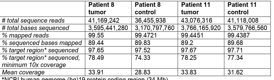
To perform exome sequencing, genomic DNA of available paired diagnostic and remission samples was extracted from archived bone marrow (BM) samples and fragmented for library preparation as described previously.18,19Protein-coding regions were enriched using the SureSelect Human All Exon V4 Kit (Agilent), followed by multiplexed 80 bp paired-end sequencing on an Illumina Genome Analyzer IIx. In total, at least 3.2 Gb of raw sequence data were generated per sample (mean 3.5 Gb; quality metrics are summarized in supplemental Table 1). Raw sequence reads werefiltered by Illumina’s chastityfilter and mapped to the NCBI human hg19 RefSeq reference genome using BWA mapper with default parameters.20Insufficiently mapped sequence reads (cutoff Q13, according to 95% confidence of correct mapping) and polymerase chain reaction (PCR) duplicate reads were removed using SAMtools21; realignment of mapped reads was performed using the Genome Analysis Toolkit to reduce false-positive single nucleotide variant calls.22Candidates for somatically acquired mutations were detected using VarScan with the following parameters: coverage$103, variant allele frequency$20%, variant base calling quality$Q13, and variant reads$3.23Positions with evidence for a variant in the corresponding remission sample or annotated polymorphism (as listed in dbSNP v135) were excluded.
Targeted amplicon sequencing
A selection of genes identified by exome sequencing (n59) and a panel of genes recurringly mutated in AML (n 5 42) were studied by targeted amplicon sequencing (Haloplex; Agilent) in all AMLCG AML113 patients with available material (16 of 23). The resulting libraries were sequenced in a single run on a MiSeq instrument. Sequence data were aligned to the human reference genome (version hg19) using BWA.20Single nucleotide variants and short insertions or deletions were called using VarScan 2 and Pindel, respectively.24,25
In addition, Sanger sequencing of genomic DNA was performed for additional validation of selected mutations. Primer sequences and PCR conditions (forSRSF2) are shown in supplemental Tables 2 and 3). PCR products were purified using NucleoFast 96 PCR Clean-up Kit (Macherey Nagel, D¨uren, Germany) and bi-directional sequencing was performed on an ABI 3500xL Genetic Analyzer using the BigDye Terminator v1.1 Cycle Sequencing Kit (Applied Biosystems, Foster City, CA). Sequences were aligned and compared with the reference sequences (NCBI accession numbers: NC_000002.11 [CEBPZ], NG_027868.1 [ASXL1], and NG_032905.1 [SRSF2]) using the Sequencher software (Gene Codes Corporation, Ann Arbor, MI)
Gene expression analysis
To further characterize the AML113 subgroup, we compared gene ex-pression profiles of 9 patients with AML113 to 509 AML patients with various genetic abnormalities (except for numerical alterations affecting chromosome 13). The gene expression data set was published previously and is publicly available through the Gene Expression Omnibus Web site (GSE37642).26Eight of 9 patients were also included in the genetic analysis. Details of sample preparation, hybridization, and image acquisition were described previously.26 For probe-to-probe set summarization, we used custom chip definitionfiles based on GeneAnnot version 2.0 (available at http://www.xlab.unimo.it/GA_CDF/) as reported before.18Only the 17 389 probe sets present on both the Affymetrix HG-U133A and B chips, and the HG-U133 plus 2.0 chips were included in the analysis. To eliminate the batch effect resulting from the use of different chip designs, we applied an empirical Bayesian method as described previously.27
Gene set enrichment analysis (GSEA) was performed with GSEA software (MIT) using the“c5_all”collection consisting of 1454 gene sets derived from the controlled vocabulary of the Gene Ontology project.28
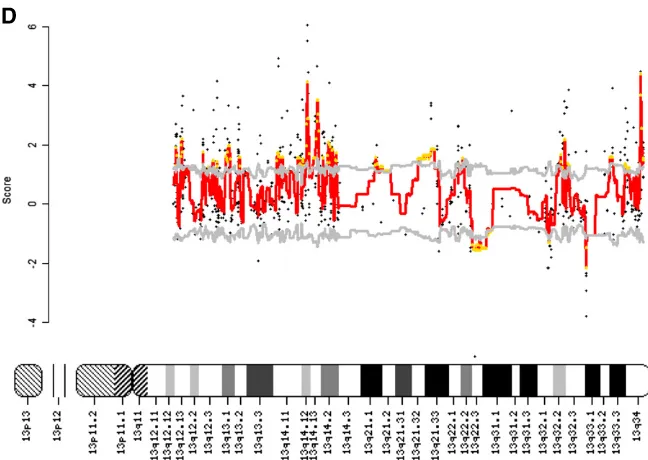
The Linear Models for Microarray Data package was used to compute differentially regulated probe sets. Differential regional gene expression on chromosome 13 was analyzed using MACAT (MicroArray Chromosome Analysis Tool) as described previously.29,30
Table 1. Patient characteristics
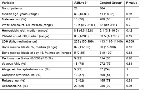
Variable AML113* Control Group* P
No. of patients 34 850
Median age, years(range) 64 (43-80) 59 (17-84) .004
Male sex, no.(%) 24 (70) 465 (55) .08
WBC count, G/l, median(range) 10 (1-318) 11 (0.1-365) .64
Hemoglobin, g/dl, median(range) 8.9 (4.6-12.8) 9.2 (2.9-17.2) .2 Platelet count, G/l, median(range) 77 (1-399) 54 (1-1760) .23
LDH (U/l), median(range) 269 (155-1011) 414 (115-11140) .009
BM blasts, %, median(range) 80 (11-100) 68 (11-100) .02
BM blasts at day 16, %, median(range) 5 (0-85) 9 (0-100) .78
Performance status (ECOG)$2(%) 8 (26) 263 (34) .44
de novo AML(%) 26 (76) 646 (76) 1.0
Allogeneic transplantation, no.(%) 6 (18) 180 (21) .83
CR, no.(%) 21 (62) 471 (55) .49
Relapse, no.(%) 18 (86) 327 (69) .14
Deceased, no.(%) 31 (91) 644 (76) .04
SignificantPvalues are indicated in bold.
*All patients were enrolled in the AMLCG-99 or SAL trials and received intensive induction treatment. All patients are classified as ELN Intermediate-II; AML113: patients with isolated tri- or tetrasomy 13, additional aberrations of the sex chromosomes are allowed.
BLOOD, 21 AUGUST 2014xVOLUME 124, NUMBER 8 SRSF2 MUTATIONS IN AML113 1305
For personal use only.
on May 16, 2016. by guest
www.bloodjournal.org
Statistical analyses
All statistical analyses were performed using the R 2.12.2 and 3.0.1 software31and routines from the biostatistics software repository Biocon-ductor, and SPSS version 21.0 (SPSS Inc., Chicago, IL). Two-sided Fisher’s exact test was used to compare categorical variables, while Wilcoxon Mann-WhitneyUtest was applied for continuous variables. Adjustment for multiple hypothesis testing was performed using the Benjamini-Hochberg pro-cedure.32Complete remission (CR) was defined as hematologic recovery with at least 1000 neutrophils permL and at least 100 000 platelets permL, and
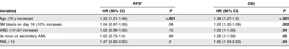
,5% BM blasts in at least one measurement.33Relapse-free survival (RFS) was defined as time from the date of CR until relapse, or death. Overall survival (OS) was defined as time from study entry until death from any cause. Patients alive without an event were censored at the time of their last follow-up. The prognostic impact of AML113 was evaluated according to the Kaplan-Meier method and the log-rank test. To adjust for other potential prognostic variables, we derived multivariate Cox models for RFS and OS. The following variables were included in the models, based on their role as potential confounders and availability of data: age (as a continuous parameter), sex, BM blasts at initial diagnosis and on day 16, Eastern
Cooperative Oncology Group (ECOG) performance status, white blood cell (WBC) count, platelet count, hemoglobin, serum lactate dehydrogenase (LDH) level, de novo vs secondary AML, and presence of AML113. No variable selection technique was applied, and all variables were retained in the
final models.P#.05 was considered significant.
Results
Isolated trisomy 13 is associated with poor prognosis We evaluated the cytogenetic reports of 6836 AML patients with available follow up data treated within the multicenter AMLCG-1999 and SAL trials for aneuploidy of chromosome 13. A total of 264 patients (3.9%) lacked sufficient cytogenetic data. Additional copies of chromosome 13 were reported in 99 of 6572 patients (incidence, 1.5%). Our analyses focused on patients with isolated trisomy (n533) or tetrasomy 13 (n51) (incidence, 0.5%). Patients with additional
Figure 1. RFS and OS in AML patients.(A-B) AMLCG
cohort. (C-D) Combined AMLCG and SAL cohort. Kaplan–Meier estimates of RFS and OS are signifi-cantly reduced for the AML113 subgroup within the ELN Intermediate-II genetic group.
Table 2. Multivariate analysis
Variable‡
RFS* OS†
HR (95% CI) P HR (95% CI) P
Age(10 y increase) 1.33 (1.21-1.46) <.001 1.38 (1.27-1.5) <.001
BM blasts on day 16 (10% increase) 1.04 (0.97-1.09) .08 1.02 (1.02-1.09) .002 WBC (10 G/l increase) 1.02 (0.99-1.05) .15 1.02 (1-1.05) .04 de novo vs secondary AML 1.02 (0.75-1.4) .89 1.26 (1-1.59) .05
AML113 1.47 (0.82-2.62) .2 1.65 (1.03-2.63) .04
Significant P values are indicated in bold.
*n5378, number of events5275 (114 patients excluded due to missing covariables). †n5549, number of events5410 (335 patients excluded due to missing covariables).
‡Only variables withP#.05 in either model are shown. The following variables were included in both models: sex, age (continuous variable), BM blasts at initial diagnosis and day 16, ECOG performance status, WBC count, platelet count, hemoglobin, serum LDH level, de novo vs secondary AML, and AML113 status.
1306 HEROLD et al BLOOD, 21 AUGUST 2014xVOLUME 124, NUMBER 8
For personal use only.
on May 16, 2016. by guest
www.bloodjournal.org
![Bis(cucurbit[6]uril) bis(hexane 1,6 diyldipyridinium) tetrabromide tridecahydrate](data:image/gif;base64,R0lGODlhAQABAIAAAP///wAAACH5BAEAAAAALAAAAAABAAEAAAICRAEAOw==)