HETEROCHROMATIN I N DROSOPHILA MELANOGASTER
KIRSTEN H. WALEN
Naval Biological Laboratory, School of Public Health and Department of Genetics Uniuersity of California, Berkeley
Received November 21. 1963
OMATIC crossing over, was first demonstrated by STERN (1936) in Drosophila srneZarzogaster, and has since been shown to occur in fungi. From the observa- tion of single and twin mosaic spots in the hypoderm, STERN concluded that the exchanges occur at the four-strand stage, and that the chromatids segregate equationally proximal to the point of exchange and reductionally distal to the point of exchange. H e also observed that somatic crossing over occurred most frequently adjacent to the centromere. KAPLAN (1953) and BROSSEAU (1957) showed that the preferential location for somatic crossing over is the proximal heterochromatic region. BROSSEAU also concluded that somatic crossing over early in embryogeny occurs only in the proximal heterochromatin of the X chromo- somes, but that later occurrences involve both hetero- and euchromatic exchanges, but with a higher frequency in the proximal heterochromatic region.
The preferential location of the exchange in the proximal heterochromatic region led to the following two hypotheses to explain the observed higher somatic instability of ring X chromosomes as compared to rod X chromosomes (BROWN, WALEN and
BROSSEAU
1962) : ( 1 ) since somatic crossing over largely takes place in heterochromatin then the greater amount of proximal heterochromatin, usually present in ring chromosomes as compared to rod chromosomes, increases the probability of somatic exchange. (2) The inherent shape of ring chromosomes may, for mechanical reasons, cause more stress and, therefore, more opportunities for breaks resulting in increased frequency of somatic crossing over. The present study was undertaken in a n attempt to discriminate between these two hypotheses.MATERIALS A N D M E T H O D S
The following four groups of Drosophila melanogaster stocks were used in this study: (1) T h e original stocks consisted of: (a) normal rods, either the wild-type stock Samarkand or variously marked with y ac and sn3; (b) the ring chrommomes Xc2 and Xc y , with or without sn3; (c) a
rod chromosome derived from a ring of approximately normal size as viewed i n oogonial smears and with a reversed gene order, i.e., I n ( l ) E n H from DR. ALOHA HANNAH-ALAVA (presumably similar i n structure to I n ( 1 ) E n of NOVITSKI (1954), although probably not identical i n regard
to heterochromatin); (d) YSIn(1)En B y Y L (NOVITSKI 1954) henceforth called XY y B.
(2) A rod X chromosome derived by crossing over between In s( l )sc S ' L, S , s c 8 R , s c S 1 + 8 ~ ~ (Muller-5) and XY y B . Crossovers between y+ and B were selected as diagrammed:
906 K. H. WALEN
Three independent crossover products were selected, since the distal heterochromatin may have been partially or wholly replaced by YS heterochromatin. These chromosomes will henceforth be called XYLy+y-1,2 and 3. These chromosomes were kept in attached-X (yf:=/Y) cultures for the duration of these experiments.
( 3 ) Ring XY and rod XY chromosomes presumed to contain different amounts of hetero- chromatin were synthesized by means of X-ray treatments of males with the attached XY Y B
chromosome. Suspected altered XY y B chromosomes were given code numbers and kept in stocks either heterozygous with a balancer chromosome (delta 49) or as males in attached-X cultures (yf:=/Y) depending on the fertility of the males. The morphology of the chromo- somes was determined cytologically by the aceto-orcein squash method on oogonial cells from heterozygotes of the various supected altered XY y B chromosomes. The ring derivatives were given the general brief designation, XYcy-1,2,3, etc., to 6. Unlike the ring derivatives, the rod derivatives gave normal results on crossover tests with In(l)EnF1 sn3, and were selected at random on the presumption that the radiation may have produced deficiencies in the heterochromatin in some of them. The six treated XY y B rods were labeled with the superscript D to indicate linear derivatives, e.g., XYD y-1,2,3, etc. to 6. Cytological examination did not reveal which of the chromosomes had more or less heterochromatin, but it did permit classification into either ring or rod chromosomes. This classification was found to fit two frequencies of gynandromorph production. Ring chromosomes produced many more gynandromorphs than did the rods. Sterile males resulted from ring chromosomes Nos. 5 and 6 and rod chromosomes Nos. 2, 5 and 6.
(4) The following three stocks were obtained from DR. D. L. LINDSLEY, Oak Ridge National Laboratory: y f YS y cu U
.
YL, YS y sn3.
YL, and Ins(l)scl12 s G R , y sc4 car m & sea. The first has Y heterochromatin intercalated between y+ and y in contrast to X heterochromatin in chro- mosomes XYLy+y-1,2 and 3. The last chromosome is deficient for a large block of centric hetero- chromatin.Data were obtained for many of the experimental series from two separate series of abdomens collected from different culture bottles. Temperature conditions, age of flies at mating, observa- tions of mosaic spots and mounting procedures of abdomens were all identical to those previously reported (WALEN 1961).
RESULTS
The two hypotheses mentioned in the introduction regarding the higher fre- quency of somatic crossing over for ring/ring and ring/rod as compared to normal combinations will be designated heterochromatin and ring-shape. The former hypothesis assumed that the amount of heterochromatin influences the frequency of somatic crossing over, whereas the latter hypothesis assumed that the particular shape of ring chromosomes is the responsible factor. The results expected can be outlined as follows.
exchange event, as opposed to a stimulatory effect on the natural frequency of somatic crossing over in the proximal heterochromatic region. This assumption can be checked by comparing the phenotypes of mosaic classes expected a priori
to those observed.
Data on frequency of crossing over for the various types of chromosomes will therefore be presented in the following order: ( 1 ) A rod chromosome deficient in heterochromatin; (2) A rod chromosome with an excess of heterochromatin located terminally (XY y B ) ; (3) Rod chromosomes with Y-derived heterochro- matic right arms and additional heterochromatin intercalated between the marker loci; (4) Ring chromosomes derived by X-ray treatment of rods with extra termi- nal heterochromatin (XY y B ) ; ( 5 ) Rod chromosomes with extra terminal hetero- chromatin (XY y B ) which had been treated with X rays and therefore perhaps containing altered amounts of heterochromatin; ( 6 ) A rod chromosome with a Y-derived right arm and additional heterochromatin intercalated between the marker loci in heterozygous combination with a normal ring chromosome.
According to the ring-shape hypothesis, the chromosomes of all classes except (4) should show about the same frequency of somatic crossing over, but should differ from class (4). According to the heterochromatin hypothesis, class ( 1 )
should show a low, classes (2) and ( 3 ) a high frequency of crossing over. Classes
(4)
and( 5 )
should have an upper limit about that of class (2), but class (4) certainly, and class ( 5 ) probably, would have considerable variation in fre- quency. The chromosomes of class (4) having sustained two breaks in the terminal heterochromatin, one in the right and one in the left arm to yield the rings, must therefore, be deficient as compared to the original chromosome in some of the heterochromatin. The chromosomes of class ( 5 ) , however, need not necessarily have sustained any damage from the treatment. Class (6) was used in an experiment in which it was attempted to distinguish between single and multiple exchange events in a single heterochromatic region.Next, the major classes and nomenclature of mosaic spots in this study will be briefly considered. Figure 1 is a schematic representation of exchange between two normal rod X chromosomes with the marker genes y and sn3 in the repulsion phase. When the exchange occurs to the right of sn3 (that is, in centric hetero- chromatin) the segregating chromatids may give rise to two cells homozygous for the marker genes
y
and sn3 respectively. Ifboth
cells give rise to hypoderm tissue,Exchange: ( 1 ) y - z twin spots
FIGURE 1 .-Normal rod chromosomes with
sns)
.
908 K. H. WALEN
there will be observed two mosaic spots, one with yellow hypoderm and bristle(s) ;
the other with wild-type coloration and singed bristle (s) j these spots will lie close to each other and will often be contiguous. STERN referred to these mosaic spots as twin mosaic spots in contrast to single mosaic spots of either phenotype. Single mosaic spots can be due either to the production of hypoderm by only one
of the crossover derivatives o r to the loss of one of the X chromosomes. As out- lined in Figure 1 both twin and single mosaic spots will generally be composed of cells with an XX constitution. However, in special cases of chromosome rearrangements, somatic crossing over may lead to a n XO constitution of the spot-forming cells. Such a genetic constitution is an imperative result in the case of simple chromosome elimination. Mosaic spots composed of cells with the
XO constitution and which occur in the sexually dimorphic region of the female abdomen will exhibit the dark male coloration of the hypoderm. These “male XO”
spots may give important information as to the underlying mosaic producing mechanism. Finally, it is customary to distinguish between single and multiple bristle spots. This offers a convenient evaluation of the time (early us. late) dur- ing development when the event took place which led to the spot formation.
A rod X chromosome deficient in centric heterochromatin: The first hetero- zygote tested was sc4 scs, y+/+sn3 (deficient rod/normal rod). The control for this and other experiments was y+/+sn3 (normal rod/normal rod). Both twin and single phenotype spots are expected. The results are presented in Table 1.
No twin spots were observed in the experimental series and the total percent mosaicism (6 percent) was much lower than the average value (average of per- cent mosaicism of rows 3 and 4, Table 1) obtained for two normal chromosomes
( 5 3 percent). A second experiment was performed to rule out the possibility that heterozygosity for the sc4 scs inversion was responsible for the reduced frequency of somatic exchange. In this experiment the heterozygote Z n ( l ) E n H , +sn3/sc4 sc8,
TABLE 1
Mosaicism following somatic crossing ouer of different rod chromosomes with deleted proximal, normal, and extra terminal heterochromatin
Xlosaic spots
Twin Singles Multiples Number of
Genotypes Pcrcent y ~ s n Y sn Y sn abdomens
a.
100
6 0 3 2 1 0
sc4 SCS, y
+
+
sn315 0 10 1 0 4 100
sc4 scs, y
+
EnH,+
s ~ 3h.
Y t 64 9 13 20 12 10 100
+
sn3 43 5 8 18 8 4 100 (control)C.
xy,
Y+
538 147 92 149 61 89 100y+ was used. These chromosomes are isosequential in their euchromatic portion and similar to sc4 scs/nonnal rod in the amount of heterochromatin. In this case (Table 1, column 3, rows 1 and 2) only a slight increase (10 us. 3) in single yellow-bristle spots was observed, which could be due to better pairing of the chromosomes. The total percent mosaicism in this case was 15 percent, again much lower than the average control value.
A rod chromosome with an excess of heterochromatin located terminally ( X Y y B) : Individuals heterozygous for the standard
XY
y B chromosome and a normal rod carrying sn3 were examined for mosaic spots. Figure 2 shows possible heterochromatic exchanges and the expected phenotypes of the resulting mosaic spots. It should be noted that several pairing patterns are indicated. The same normal rod X chromosomes as in the previous experiment served as the control. The frequencies of the different spot classes of the two independent abdomen readings are in good agreement (Table 1 ).
Furthermore, when these data are compared with the two readings of the normal rod X chromosomes (rows 3 and 4), a tenfold increase in total percent mosaicism is observed. Since only twin spots can be considered as a definitive measure of somatic crossing over, it is of con- siderable importance that these values also are approximately ten times higher for the series with the terminally located extra heterochromatin than for the control.a)
( 1)
---
y+ sn3YS sn+ y YL
y+ sn3 (1)
YL Y sn+ YS
J
;
=
-
- --
-Exchange: (1) y-SJ twin spots (XX constitution)
b)
-x ,:I y _
Exchange: (1) dicentric-elimination, Y-SJ twin
C) spots (XO constitution)
v+ ,"3 (1)
YS sn+ y (2)
Exchanges: (1) attached X,no spots
d)
(1) & (2) y-=twin spots (XX constitution)
Pairing of heterochromatic regions within one single chromosome.
Chromosome: XYJ -2 (XO) spots
910 K. H. WALEN
Rod chromosomes with Y L appended to the right a r m and additional hetero- chromatin intercalated between the marker loci: The previous series of abdomen readings showed that additional heterochromatin located terminally had an effect on the frequency of somatic crossing over. Next, rod chromosomes with inter- calated heterochromatin were observed for mosaic-spot production. The derivation and genetic constitution of these chromosomes (XYLy+y-l, 2 and 3 ) were men- tioned above. These chromosomes contain
X
heterochromatin between y + andsn+ y , and have
YL
attached to the right of the centromere. Figure 3 is a schematic representation of the possible heterochromatic exchanges between this chromo- some and the standard XY y sn3 B chromosome. According to this scheme, (3a), no twin spots are expected, but the contrary would be true if the sn3 marker were located on the chromosome with intercalated heterochromatin (Figure 3b, c).
The sn3 marker was, therefore, crossed into two of these chromosomes. The data on these two series of abdomen readings are listed in Table 2. As predicted, the occurrence of twin spots ( y - s n ) in the one series (a) is negligible in contrast to the expected high frequency in the other series (b).
Total percent mosaicism forYS t y (2)
Exchanges: (1) attached X, no spots
(1) & (2) y-E twin spots (XX constitution) (3) y spots
(1) & (2) & (3) I_” spots d)
Pairing of heterochromatic regions w i t h i n one single chromosome. Chromosome: XY$’f y-E (XO) a n d y ( X X ) spots
XY$+ % 3 y - y ( X X ) spots XYy +-I_” (XO) spots
FIGURE 3.-Rod with extra heterochromatin intercalated in the euchromatic part and YL attached to the right arm i n combination with a rod with extra heterochromatin terminally
TABLE 2
Mosaicism following somatic crossing over between rods with extra heterochromatin and normal X Y rod chromosomes*
91
Mosaic spots
Twin y - y sn Singles Multiples Number of
Genotypes Percent y-sn sn-ysn y sn y s n y sn y s n abdomens
a.
362 1 1 68 19 37 16 8 31 50
XYL, y +
+
y-l XY , sn3y394 0 4 63 21 47 28 3 31 50
XYL, y+
+
y-2 XY , sn3yXYL, y+
+
y-3XY
.
s n $ y 630 4 6 98 26 6 2 4 6 17 56 50b.
561 61 2 242 118 2 81 54 2 100
XYL, y+ sn3 y-1 x y , + Y XYL, y+ sn3 y-3
X Y , + r 626 71 4 274 119 19 97 47 1 100
C.
710 49 27 256 85 97 105 48 43 100
y + Y S y + .YL Ys y sn3. YL
* Control values, Table 1, y ac
+/+
4- sn8, 64 percent and 43 percent mosaicism.the chromosomes with extra heterochromatin is somewhat higher in the series in which twin spots are observed. Furthermore, as in the previous experiment, with extra heterochromatin located terminally, the results of these series (a and b) show, on the average, a tenfold increase in mosaicism as compared to the normal control chromosomes.
Another fact borne out by the results of the present experiment is that the ex- change events occur in the extra heterochromatin. The latter conclusion is based on the prediction of the lack of twin spots in series (a), and that the occurrence of both yellow and singed spots in this series involve exchanges in the intercalated heterochromatin (Figure 3a). Moreover, since the production of singed spots involves double crossing over, these spots are a priori expected to appear with a lower frequency than yellow spots, which is fully in agreement with the data
(Table 2a).
To study the possibility of a specific difference between intercalated X and Y heterochromatin behavior in somatic crossing over, females of the genetic consti- tution y+
Ys
y+
YL/Ys y sn3YL
(detailed above) were examined. The inter- calated heterochromatin is Ys rather than centric X heterochromatin present in the chromosomes of the previous experimental series. Unfortunately no cyto- logical data are available for a consideration of the relative length of the X and Ys intercalated pieces of heterochromatin. The cytogenetic information on Muller-5 (see MATERIALS (2) ),
however, gives an approximate length of the X-912 K. H. WALEN
of Ys in accordance with COOPER’S scale f o r X and Y heterochromatin, Ys is the longer piece of heterochromatin.
The expected phenotypes of the mosaic spots in this Ys intercalated series arise as diagrammed in Figure 3a. From Table 2c it can be seen that a n average of seven spots per abdomen was observed in this series, which is an average increase of two spots per abdomen over the previous series (Table 2a, b) with intercalated
X heterochromatin. If this increase in mosaicism is viewed in the light of the spot frequency for normal rod chromosomes (one spot for every two abdomens), it represents a significant increase. As mentioned above, the same spot classes and no twin spots are expected for the two series ( a ) and (c), whereas i n series (b), twin spots are expected which could result in an increase in mosaicism (Figure 3a and b). The data from chromosome 1 and 2 in series (a) are in agreement with each other, but not with the data of the third chromosome, which are con- siderably different (approximately four versus six spots per abdomen). The ob- servations on the third chromosomes in series ( a ) will be evaluated separately in the discussion. I t is concluded that total percent mosaicism is significantly higher in the YS intercalated series than the X heterochromatin intercalated series. The possible reasons for this difference in behavior of the two “types” of heterochromatin will be considered in the discussion.
Finally, it is necessary to elaborate on an apparent discrepancy between results listed in Table 2 f o r series (a) and (c) and the predictions outlined in Figure 3a. The figure does not indicate the appearance of twin or other dual spots (e.g., other combinations of spots than the usual y-sn twin spots; y sn-y and y sn-sn), but these spot categories are listed (c) at percentages as high as 49 and 27, respec- tively. It is believed that some of these mosaic spots are actually two separate, independently produced spots which happen to lie close to each other, and that others may be due to two independent exchanges in the same cell lineage.
somatic crossing over and fertility versus sterility would be expected if more tension and stress is assumed in a smaller ring than in a larger ring. Thus, the male-fertile chromosomes, because they are larger than the male-sterile chromo- somes, should show a lower frequency of somatic crossing over than the male- sterile ring chromosomes. Finally, it follows that in ring/rod combinations in which the ring is kept constant and the rod is variable in respect to content of heterochromatin, the ring-shape hypothesis predicts about the same frequencies of somatic crossing over, while different frequencies are expected according to the heterochromatin hypothesis.
The six derived ring chromosomes were studied for mosaicism in females heterozygous for a sn3 rod chromosome. Of these ring chromosomes, Numbers 1, 2, 3 and
4
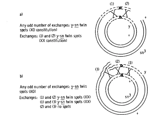
are male fertile, while 5 and 6 are male sterile. Figure4
is a sche- matic representation of possible exchanges between the heterochromatic regions of a usualx"
ring chromosome and a normal rod chromosome. Considering the number of exchanges, the following spot phenotypes are expected: (1) single (odd number) exchange results in twin spots (y-sn) composed of XO cells; (2) double (even number) exchanges on both sides of the centromere lead to twin spots with cells of an XX constitution. The results of duplicate series of the ex- periments are given in Table 3. Mosaic spots that could only have arisen througha)
Any odd number of exchanges: y-E twin spots (XO constitution)
Exchanges: (1) and (2) y - z twin spots
(XX constitution)
Any odd number of exchanges: y - 5 ~ twin
Exchanges: (1) and (2) y-% twin spots (XX) (1) and (3) y-E twin spots (XX)
(2) and (3) no spots
spots (XO)
914 K. H. WALEN
TABLE 3
Mosaicism following somatic crossing over or loss, with ring chromosomes containing different amounts of heterochromatin
Percent
due to somatic Twin Singles Xlultiples Sumher of Genotypes Percent crossing OYW' p s n 1. sn y sr1 abdomens
XYC, y
+
-1 645 192 16 36 220 4 53 50+
sns 671 223 23 94 461 6 88 100XYC, y
+
-2 510 188 6 43 158 1 47 50+
sn3 4.08 177 7 83 289 2 27 100XYC, y
+
-3 562 198 9 43 188 2 44 50+
sn3 614 246 13 52 194 3 45 50~
XYC, y
+
-4 622 186 9 37 217 5 44 50+
v n 3 614 224 14 42 186 7 58 50XYC. y
+
-5 228 112 2 26 152 1 47 50+
snJ 21 3 88 6 19 164 0 24 50XYC, y
+
-6 260 114 7 23 169 2 59 50+
sns 245 92 8 17 165 2 53 50Controls XC. Y
+
+
sn3484 26 1 17 110 296 12 49 100
46 7 223 31 89 262 7 78 100
Xc2,
+
sns 514 205t 21 343 87 58 5 100Y + 478 193t 13 343 82 32 8 100
885 603t 43 467 248 97 32 100
* Twin spots plus twice the number of yellow spots.
t Twin spots plus twice the number of singed spots.
somatic crossing over, in contrast to spots due to simple loss of the ring chromo- some, have been estimated (Table 3, column 2) in view of the following facts. One genetic marker is on the ring, the other on the rod, so that simple loss of the ring will result in mosaic spots showing the marker of the rod, whereas the marker of the ring would appear only after somatic crossing over. This latter component of the total mosaicism has been doubled since both types of spots are expected after somatic crossing over, and to this estimate the number of twin spots has been added (BROWN et al., 1962). It is clear from these results that the percentage of spots due to somatic crossing over fall into one of two categories, either 100 percent or 200 percent. The former category reflects the frequencies of somatic crossing over for the male-sterile chromosomes and the latter, those of the male- fertile chromosomes.
915
ing over, but the same ring chromosome together with the rod having extra heterochromatin raises the frequency approximately three times.
X-ray treated rod chromosomes ( X Y y B) with presumably altered quantities
of extra terminal heterochromatin: Finally, it was reasoned that X-ray treatment of rod X chromosomes with extra heterochromatin terminally attached (XU y B )
could produce rods with different amounts of heterochromatin which in turn would result in varying frequencies of somatic crossing over. Moreover, a n upper limit frequency near that of the normal XY y
+/+
sn3 series (Table 1, series b) was expected. The percent mosaicism from the different X-ray treated rods in combination with a normal rod with the marker sn3 are listed in Table 4. As controls to this experiment the results of the two previously discussed series (Table l b and c) are also listed in Table 4. I t can be seen in this table that the predictions are realized: that is, none of the frequencies of mosaicism for the X-ray treated chromosomes are greater than the control series (last row) and at least for two of these chromosomes( 5
and 6 ) the percent mosaicism is signifi- cantly reduced. In view of the evidences presented in this study in support of the quantitative significance of heterochromatin in somatic crossing over, it seems reasonable to assume that at least chromosomes5
and 6 in Table4
are deficient for heterochromatin. Further support for such a conclusion comes from the fact that both of these chromosomes were sterile in males which indicate a deficiency for fertility factors carried on Ys and/orYL.
TABLE 4
Mosaicism following somatic crossing over befiween rods with different amounts of heterochromatin and control chromosomes
Mosaic spots
Twin Singles Multiples Number of Genotypes Percent y-sn Y sn Y sn abdomens
XyD, Y
+
-1 464 92 32 46 15 48 50+
sns 310 60 34 36 10 15 50XYD, y
+
-2 578 103 39 52 26 69 50+
sn3 7% 1 42 54 71 4 0 6 6 50XYD, Y
+
-3 316 56 19 39 14 30+
sn3 426 68 43 45 22 3550 50
XYD, y
+
-4 6% 100 43 84 32 64 50+
sns 448 70 42 52 27 33 50XYD, Y
+
-5 160 10 13 32 2 23 50+
sn3 196 15 20 23 1 39 50XyD, Y
+
-6 160 12 8 23 2 35 50+
sn3 172 13 9 25 7 32 50Controls
Y + 64 9 13 20 12 10 100
+
sns 43 5 8 18 8 4 100xy,
Y+
538 147 92 149 61 89 100916 K. H. WALEN
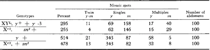
A rod chromosome with Y L appended to the right a r m and additional hetero- chromatin intercalated between the marker loci in combination with a normal ring chromosome: The experiments described above suggested a rather precise and cumulative effect of heterochromatin on somatic crossing over. It was, there- fore, assumed that the positive correlation observed between the amount of heterochromatin and the frequency of somatic crossing over signified more oppor- tunities for exchange. Furthermore, since male spots (spots composed of XO cells) i n the sexually dimorphic region of the abdomen (Figure 4) were only observed occasionally for the ring/rod combinations, it was hypothesized that many of the mosaic spots recorded in Table 3 were of an XX constitution instead of XO. This meant that more than one exchange must have occurred in the heterochromatic regions of both ring and rod chromosomes. To test this hypothesis an experiment was performed for which certain predictions could be made regarding relative spot-class frequencies. Simple elimination of the ring chromosome for this par- ticular ring/rod combination will result in no spots, since the rod chromosome carries both a y and a y+ locus (Figure 5 ) . A single exchange between the ring and the rod will lead to the formation of a dicentric chromosome which may sub- sequently be lost, followed by normal segregation of the two remaining noncross- over chromatids. In this case singed spots with a n XO constitution showing male characteristics in the dimorphic region, and no yellow spots are expected. Double crossovers at ( 1 ) and (2) and ( 1 ) and ( 3 ) will also result in singed spots, but only simultaneous crossovers at (1) and ( 3 ) and (4) will give rise to yellow spots. These latter spots presumably consist of normal, diploid tissue. If singed spots owing to single exchanges are the primary source of mosaic spots in this experi-
Single exchanges will result inJnspots with an XO constitution Exchanges: (1) and (2) s i spots
(1) and (3) spots
(l), (3) and (4)y spots
FIGURE 5.-Rod with extra heterochromatin intercalated i n the euchromatic part and Y L
TABLE 5
Mosaicism following somatic crossing ouer between a rod with heterochromatin intercalated into the euchromatic part and a usual ring chromosome
Mosaic spots
Twin Singles Multiples Number of Genotypes Percent y-sn Y sn Y sn abdomens
XYL, y +
+
y -3 295 1 1 69 158 1 7 41) 100Xc2, sn3
+
255 4 62 145 15 29 100Y + 514 21 343 87 58 5 100
xc2,
+
sn3 478 13 343 82 32 8 100ment, then no correlation between sn and y spots is expected. However, if multiple exchanges are the underlying process for the formation of yellow and singed spots and if double exchanges occur with equal frequency as triple exchanges (see Figure 5 ) , then singed spots are expected twice as frequently as yellow spot? Again duplicate series of individuals were collected from two separate culture bottles and all slides bearing mounted abdomens were coded and “read” along with those of other experiments. All spots appearing in the dimorphic region of the abdomen were critically examined for any indication of maleness (XO tissue), but none of the typical morphological male spot characteristics was observed. The results of this experiment are listed in Table 5 , together with the data from a normal ring/rod combination from Table 3, as the control. Both single and multi- ple yellow bristle spots are recorded for the experimental series, and the frequen- cies of the respective spot classes in the two independent series of abdomens are quite similar. Comparing the numbers in the experimental series with those in the control, it is evident that the relative frequencies of y and sn spots (single and multiple spots) are reversed. The rather high frequency of yellow spots in the control series is mostly due to simple elimination of the ring chromosome, where- as singed spots are only due to somatic crossing over, as already discussed above for this particular ring/rod combination. It is important to note that in the experi- mental series which involved a rod with extra heterochromatin, the frequency of singed spots is doubled. It was also reasoned earlier (see Figure 5 ) that if multiple exchanges occurred preferentially between chromosomes with extra heterochro- matin then singed spots should be twice as frequent as yellow spots in this par- ticular experiment. According to the values in Table 5 , the ratio of single y to single sn spots is 2.2 and 2.3, and for multiple bristle spots 2.3 and 1.9, respec- tively, for the two series of abdomens. These results will be considered in the discussion.
DISCUSSION
91 8 K. H. WALEN TABLE 6
Summary of results on somatic crossing ouer
Chroniosonies
rod/rod (control) rod/rod
rod/rod rods/rods
ring/rod (control) ring/rod
rings/rods
Heterochromatin content
normal/normal deficiency/normal duplication/normal duplication variable/normal normal/normal
normal/duplication deficiency variable/normal
Frequency of mosaicism' (percent)
53
10 532 172-491 220 603 101-204
* Due to somatic crossing over only. The percentages are averages taken from Tables 1 , 2, 3 and 4.
present in ring chromosomes. HINTON (1957) suggested that some element in the heterochromatic region of the ring chromosomes and the ring fragments made for greater somatic instability of ring chromosomes as compared to rod chromo- somes. KAPLAN (1953)
,
studying somatic crossing over in the second chromosome, found that most of the mosaics resulted from crossing over close to the centromere, and that crossovers could occur simultaneously in the heterochromatin of the left and the right arms. Since ring chromosomes usually contain heterochromatin on both sides of the centromere, the conditions for crossing over between two rings may, therefore, be similar to those observed for the second chromosome. Accord- ingly, when the marker genes are in the repulsion phase, simultaneous crossing over in the heterochromatic regions of paired ring chromosomes would result in twin spots composed of XX tissues, whereas twin spots with an XO constitution would be the result of single exchanges. The same two types of twin spots, that is, spots composed of XO or XX tissues, are expected for ring/rod combinations following single or double exchanges (Figure 4). STERN (1 936) originally dis- cussed the fate of dicentric chromosomes resulting from single crossing over between a ring and a rod chromosome. From a consideration of sister-strand crossing over as the cause for simple ring elimination (double dicentric) it is believed that dicentric chromosomes generally do not survive in the sense of giving rise to mosaic spots. This has been indicated for the figures of the present study. Sister-strand crossing over as the basic cause for ring-chromosome insta- bility was proposed by MCCLINTOCK (1938) and was discussed for gynandro- morphs of Drosophila melanogaster by BROWN and HANNAH (1952). Suffice it to say for the present that sister-strand crossing over has recently been demon- strated in some animal and plant cells (TAYLOR 1958; PRESCOTT and BENDER 1963; PEACOCK 1963; WALEN 1963).919
twin spots, namely, that they really constitute XX tissue originating by means of double crossing over, will be evaluated in view of some of the data of STERN
(1936) and from the present study as well.
As outlined in Figure 4, twin spots with an XX constitution would occur for
ring/rod combinations with the marker genes in repulsion if one of the crossovers involved the short heterochromatic arm of the rod chromosome. Since the present data do not furnish evidence of the participation of XR, the very short right arm of the X chromosome, in somatic crossing over, it seems justified to consider COOPER’S (1959) results on meiotic exchanges. He came to the conclu- sion regarding meiotic crossing over that “No portion of the heterochromatin half of X, including the right arm of X, can be eliminated as a possible site of exchange with Y at this time
.
. .”
and that the right arm“.
. .
is breakable at many points along its length.. . .”
It is, of course, questionable whether these conclusions apply also to mitotic exchanges. Yet, such may be the case, especially in view of the results obtained by STERN (1936) with the Theta duplication and the present results from the experiment involving combinations of the usual X“ chromosome with a rod having X-heterochromatin intercalated in the euchro- matic left arm (Table5 ) .
The frequency of mosaic spots obtained by STERN (1936) cannot be compared with the present data, since different methods of observation were employed in the two studies, but it is noteworthy that STERN in the experiments involving the Theta duplication observed twin ( y - s n ) and yellow spots. Both types of mosaic spots were explained by him as having arisen through exchange between the homologous left end of the normal X chromosome (euchromatic exchange) and the Theta duplication, resulting in cells with a constitution of 3X and 1X 4- frag- ment. Spots of the same phenotypes, and presumably of XX origin, may, however, appear after exchange between XR and the heterochromatic part of the Theta arm.
Less direct evidence in favor of the participation of the right arm of the X in somatic crossing over is found in the last experiment in the present study (Table
5 and Figure 5) . This particular rod chromosome consisted of YL attached to
XR (XRYL)
.
The results indicated that yellow mosaic spots were due to three simultaneous exchanges, with one of these exchanges involving the heterochro- matic arm on the right side of the centromere. A similar participation of the combination XRYL in the somatic crossing over process may be deduced from the approximately three times higher frequency of somatic crossing over for a ring chromosome and a rod with heterochromatin terminally attached than for normal ring/rod combinations (Table 3, last series).920 K. H. WALEN
increased frequency of exchange in the heterochromatic region on the left side of the centromere, without itself participating physically in the process. An argu- ment against such a “catalytic action” of the added heterochromatin, however, stems from an evaluation of the different pairing patterns in Figure 2, and the resulting mosaic spots if the exchanges occur equally freely between XX and XY heterochromatin. Evidence for “nonhomologous” exchanges of heterochromatin (i.e. between X and Y heterochromatin) in somatic crossing over comes from STERN (1936) and from a consideration of the spot classes expected a priori and those actually observed in the third experiment of this study (Table 2, Series a and b; Figure 3). In this experiment certain spot phenotypes could only have arisen as a result of exchanges between X- and Y-heterochromatin. Accepting these arguments in favor of fairly frequent X and Y heterochromatic exchanges, it is evident from Figure 2 that twin spots do result from all the possible ex- changes in this case. On the other hand, none of the spots showed male charac- teristics in the dimorphic area although they were predicted for some of the exchange patterns. The absence of such spots may again be due to the general smallness of mosaic spots which result from somatic crossing over (HANNAH
1953; STERN 1956).
From these considerations, it is concluded that the frequency of somatic cross- ing over is to a great extent dependent on the amount of heterochromatin avail- able for physical exchange. This means the addition of yet another factor to the list of those which have already been shown to influence somatic crossing over: the presence o r absence of Minutes (STERN 1936; KAPLAN 1953; DEMARINIS
1959) ; a multigenic system (WEAVER 1960) ; and environmental factors (STERN and RENTSCHLER 1936; KAPLAN 1953; BROSSEAU 1957).
In the third experiment of this study, which involved a rod with intercalated
X heterochromatin between the marker loci, it was observed that the third experi- mental X chromosome (Table 2a) was involved in a higher degree of mosaicism than the other two chromosomes. This is believed to be due to a greater amount of intercalated heterochromatin in this X chromosome. The procedure by which these three X chromosomes were derived involved a meiotic crossing over of a
type that could result in the extension of the original piece of intercalated X heterochromatin by the addition of Ys heterochromatin. Such an interpretation also explains the significantly higher percent mosaicism for the Ys intercalated series as compared to the other two X-heterochromatin intercalated series (Table 2, Series c and a, chromosome 1 and 2 ) . The short arm of Y is, as discussed above, longer than the piece of centric heterochromatin transposed to the left end in the Muller-5 chromosome. A logical extrapolation from these arguments is that somatic crossing over is positively correlated with the amount of heterochromatin present at any one particular location in the chromosome. Such a conclusion is supported by the observed differences in percent mosaicism in the two experi- ments i n which X-ray derived chromosomes were analyzed for mosaicism, These considerations also indicate that there are no “type” specific effects of X versus Y heterochromatin on the frequency of somatic crossing over.
92 1
into only two groups in regard to the frequency of somatic crossing over (200 percent-100 percent, Table 3).
If
the grouping is other than spurious it could mean preferential breakages ofYL
and Ys for the reunion of the broken ends to form a ring chromosome.Finally, some considerations of single and double exchanges in somatic cross- ing over merit mention. As already discussed, KAPLAN (1953) observed double crossing over across the centromere. This observation may be indicative of lack of interference across the centromere, a phenomenon known for meiotic cross- ing over (MORGAN, BRIDGES and STURTEVANT 1925; STERN 1933). Other types of double exchanges in somatic crossing over have already been considered by STERN (1936)
,
although he explained the majority of mosaic spots as having arisen through single exchange events. Most of the mosaic spots observed in the present study can be explained by single exchange events also. There are, how- ever, some mosaic classes from some of the present experiments in which two heterochromatic regions are separated by almost the entire euchromatic arm, which can best be interpreted as resulting from double crossovers (Table 2a). For these particular genotypes, such a conclusion seems quite reasonable, con- sidering the distance between the two exchanges. Multiple exchanges in the same heterochromatic region, however, must also be taken into consideration as some of the figures indicate. In Figure 5, for instance, crossovers ( 3 ) and (4) are shown close to each other in heterochromatin. It is, however, possible that cross- over (3) lies anywhere in the adjacent euchromatin up to the sn. locus. If this is so, then, it is possible that a crossover (5) could occur i n the euchromatic region between y and sn, since the length of this region is one third of the total eucromatic length. Such a crossover together with either crossovers (2) or (3) will give rise to singed spots. Considering all the spots possible from both hetero- and euchromatic double exchanges, the ratio of yellow to singed spots becomes1:4 and not 1:2 as previously discussed for double and triple heterochromatic exchanges. The experimental data (Table 5, see text also) were strikingly close to a 1:2 ratio. Less direct evidences for the localization of crossover ( 3 ) in the heterochromatin comes from an overall consideration of all the experiments in the present study, especially the fact that a deficiency for heterochromatin almost completely suppresses crossing over (Table 1 ) . I t is therefore believed that par- ticularly the last experiment in this study gives evidences for a type of multiple crossing over in relatively short heterochromatic regions of the X chromosomes.
I wish to thank PROFESSORS SPENCER W. BROWN and CURT STERN as well as DRS. ALOHA HANNAH-ALAVA and WALTER A. NELSON-REES for invaluable criticisms, comments and sugges- tions offered during this investigation.
SUMMARY
922 K. H. W A L E N
crossing over is positively correlated with the amount of heterochromatin present in the chromosomes. The positive correlation is due to the participation of the extra heterochromatin in exchange events. There is no specificity of X versus
Y heterochromatin in somatic crossing over, since exchanges occur between X
and X as well as between X and Y heterochromatin. Some of the mosaic spots observed for ring/ring and ring/rod combinations are most probably due to simultaneous crossing over on opposite sides of the centromere. The majority of
mosaic spots observed for normal rod chromosomes are due to single exchange events, and the number of exchanges increases with added amount of heterochro- matin; certain mosaic spots especially in the last series of experiments, involving a usual ring chromosome and a rod with extra heterochromatin intercalated between the marker loci and YL attached to the right side of the centromere, favored the interpretation of multiple exchanges within one relatively short heterochromatic region. Heterozygous inversions do not suppress somatic cross- ing over measurably.
L I T E R A T U R E C I T E D
BAKER, W. K., 1958 BROSSEAU, G. E., JR., 1957
Crossing over in heterochromatin. Am. Naturalist 92: 59-60.
The environmental modification of somatic crossing-over in Dro- sophila melanogaster with special reference to developmental phase. J. Exptl. Zool. 136: 567-593.
BROWN, S. W., and A. HANNAH, 1952
BROWN, S. W., K. H. WALEN, and G. E. BROSSEAU, 1962 COOPER, K. W., 1959
An induced maternal effect on the stability of the ring-X- chromosome of Drosophila melanogaster. Proc. Natl. Acad. Sci. U.S. 38: 687-693.
Somatic crossing-over and elimination of ring X chromosomes of Drosophila melanogaster. Genetics 47: 1573-1579.
Cytogenetic analysis of major heterochromatic elements (especially Xh and Y) in Drosophila melanogaster, and the theory of “heterochromatin.” Chromosoma Frequency of somatic crossing-over in various parts of the body of Non-autonomy of yellow in gynandromorphs of Drosophila melanogaster.
An analyses o f rod derivatives of an unstable ring chromosome of Dro- The influence of Minutes upon somatic crossing-over in Drosophila 10: 535-588.
DEMARINIS, F., 1959 HANNAH, A., 1953 HINTON, C. W., 1957
KAPLAN, W. D., 1953
Minute-n Drosophila. (Abstr.) Genetics 44: 505-506. J. Exptl. 2001. 123: 523-560.
sophila melanogaster. Genetics 42 : 55-65. melanogaster. Genetics 38: 630451.
The production of homozygous deficient tissues with mutant character- istics by means of the aberrant mitotic behavior of ring-shape chromosomes. Genetics 23: 315-376.
The genetics of Drosophila. Biblio. MCCLINTOCK, B., 1938
MORGAN, T. H., C. B. BRIDGES, and A. H. STURTEVANT, 1925 NOVITSKI, E., 1954
PEACOCK, W. J., 1963
Proc. Natl. Acad. Sci. U S . 49: 793-801. PRESCOTT, D. M., and M. A. BENDER, 1963
of labeled DNA in two types of mammalian cells in uitro. Exptl. Cell Res. 29: 430-442. Genet. 2: 1-98.
The compound X chromosomes in Drosophila. Genetics 39: 127-140. Chromosome duplication and structure as determined by autoradiography.
Faktorenkoppelung und Faktorenaustausch. Hdbch. Vererbgswiss. 1 :H (Lief. 19).
-
1936 Somatic crossing-over and segregation in Drosophila melamgaster. Ge- netics 21: 625-730. - 1956 The genetic control of developmental competence and morphogenetic tissue interactions i n genetic mosaics. Wilhelm Roux’ Arch. Entwicklungs- mech. 149: 1-25.The effect of temperature on the frequency of somatic crossing-over i n Drosophila melanogaster. Proc. Natl. Acad. Sci. U.S. 22 : 451453.
Sister chromatid exchanges in tritium-labeled chromosomes. Genetics 43 :
513-529.
Studies of cell lethality of a small deficiency in Drosophila melanogaster.
Genetics 46: 93-103. - 1963 The pattern of DNA synthesis in the chromosomes of the marsupial Potorous tridactylis. (Abstr.) Proc. 11th Inter. Congr. Genetics 1: 106.
Somatic crossing-over and its genetic control i n Drosophila. Genetics 45: 345357,
STERN, C., 1933
STERN, C., and V. RENTSCHLER, 1936 TAYLOR, H. J., 1958
WALEN, IC. H., 1961