O R I G I N A L R E S E A R C H
Photothermal transforming agent and
chemotherapeutic co-loaded electrospun
nano
fi
bers for tumor treatment
This article was published in the following Dove Press journal: International Journal of Nanomedicine
Jiulong Zhao1,* Yangbei Zhu1,* Changqing Ye2,* Ying Chen2 Shige Wang2 Duowu Zou3 Zhaoshen Li1
1Department of Gastroenterology,
Changhai Hospital, Second Military Medical University, Shanghai 200433,
People’s Republic of China;2College of
Science, University of Shanghai for Science and Technology, Shanghai
200093, People’s Republic of China;
3Department of Gastroenterology, Ruijin
Hospital, School of Medicine, Shanghai Jiaotong University, Shanghai 200025,
People’s Republic of China
*These authors contributed equally to this work
Background: Photothermal and chemotherapy treatment has been frequently studied for
cancer therapy; however, chemotherapy is equally toxic to both normal and cancer cells. The clinical application value of most kinds of photothermal transforming agents remains limited, due to their poor degradation and minimal accumulation in tumors.
Materials and methods:We reported the synthesis of photothermal transforming agents
(MoS2) and chemotherapeutic (doxorubicin, DOX) co-loaded electrospun nanofibers using blend electrospinning for the treatment of postoperative tumor recurrence.
Results: Under the irradiation of an 808 nm laser, the as-prepared chitosan/polyvinyl
alcohol/MoS2/DOX nanofibers showed an admirable photothermal conversion capability with a photothermal conversion efficiency of 23.2%. These composite nanofibers are in vitro and in vivo biocompatible. In addition, they could control the sustained release of DOX and the generated heat can sensitize the chemotherapeutic efficacy of DOX via enhancing its release rate. Their chemo-/photothermal combined therapy efficiency was systematically studied in vitro and in vivo. Instead of circulating with the body fluid, MoS2was trapped by the nanofibrous matrix in the tumor and so its tumor-killing ability was not compromised, thus rendering this composite nanofiber a promising alternative for future clinical translation within biomedical applicationfields.
Conclusion:Chitosan/polyvinyl alcohol/MoS2/DOX nanofibers showed an excellent
photo-thermal conversion capability with a photophoto-thermal conversion efficiency of 23.2% and can completely inhibit the postoperative tumor reoccurrence.
Keywords:electrospinning, chitosan, chemotherapy, photothermal tumor therapy, tumor
Introduction
Surgery, radiotherapy, and chemotherapy have played dominant roles in current cancer therapy; unfortunately, these cancer therapies, especially radiotherapy and chemotherapy, are equally toxic to both normal and cancer cells.1,2As a common chemotherapeutic drug, doxorubicin (DOX) has been frequently applied in tumor chemotherapy. However, apart from the high toxicity of DOX, its high in vivo permeability has caused an extremely low utilization ratio.3,444In order to solve the above problems, different kinds of drug carriers have been designed to simulta-neously deliver DOX into the tumor to enhance drug usage and safety.
Another solution to realize safe and efficient cancer therapy is to explore minimally invasive therapy methods like photothermal therapy (PTT),5 photody-namic therapy,6,7microwave ablation,8magnetic-hyperthermia regression,9,10gene Correspondence: Zhaoshen Li
Department of Gastroenterology, Changhai Hospital, Second Military Medical University, No. 168 Changhai Road, Yangpu District, Shanghai 200433,
People’s Republic of China
Tel +86 213 116 1353 Email li.zhaoshen@hotmail.com
Duowu Zou
Department of Gastroenterology, Ruijin Hospital, School of Medicine, Shanghai Jiaotong University, No. 197 Ruijin Second Road, Huangpu District, Shanghai 200025,
People’s Republic of China
Tel +86 213 116 1355 Email zdw_pi@163.com
International Journal of Nanomedicine
Dove
press
open access to scientific and medical research
Open Access Full Text Article
International Journal of Nanomedicine downloaded from https://www.dovepress.com/ by 118.70.13.36 on 22-Aug-2020
therapy,11,12 and high-intensity focused ultrasound ablation.13,14 Among them, PTT has been progressively unveiled in recent years for highly specific cancer therapy. The success of tumor PTT depends on the rational design of a biocompatible and multifunctional photothermal transforming agent (PTA) which can efficiently absorb and transform the near-infrared (NIR) laser into heat and burn out the tumor tissue.15,16 In the past few decades, interest in the PTA design was continually aroused, and different kinds of inorganic PTAs such as noble metal,17 transition metal sulfide,18,19 transition metal carbide,1,20 and organic PTAs including graphene oxide,21 reduced graphene oxide,22 and conductive polymers23–26 have been continuously proposed. However, the clinical appli-cation value of most kinds of PTAs, especially inorganic PTAs, remains limited, probably owing to their poor degradation and minimal accumulation in tumors.
Bearing the aforementioned issues of chemotherapeu-tics and PTAs in mind, researchers have proposed various kinds of intelligent platforms including injectable hydro-gel, oleosol, and polymeric implants for safely localized tumor treatments.9,27 Electrospun polymer nanofibers, owing to their unique properties including high surface-to-volume ratio, tunable porosity, and ease of surface functionalization, have attracted high attention in a number offields, such asfiltration, textile manufacturing, catalysts, and pharmaceutics.28–30As a therapeutic carrier, nanofiber can improve the drug-loading capacity and effectively control drug release. Moreover, the nanofibrous matrix may exert a discernible impact on the safety of guest materials while not compromising their function.27,31,32
In a previous study, MoS2nanosheets and DOX-loaded poly(lactic-co-glycolic) acid (PLGA) nanofibers have been successfully prepared via directly electrospinning the mix-ture solution of PLGA, MoS2, and DOX, for the efficient postoperative treatment of tumor in NIR I and NIR II biowindows.27However, due to the strong hydrophobicity of PLGA, PLGA/MoS2/DOX nanofibers severely shrank after being implanted into the tumor site. As the N-deace-tylated product of chitin, chitosan (CS) is the second-most abundant natural polysaccharide. CS has many unique properties like biocompatibility, biodegradability, antimi-crobial activity, etc.33 Nevertheless, due to the strong molecular interaction, CS has poor spinnability and is difficult to form into uniform nanofibers. In this study, we reported the design of an electrospun CS nanofi ber-based carrier, using polyvinyl alcohol (PVA) as an adju-vant to enhance the spinnability of CS, to concurrently
deliver PTA (MoS2nanosheets) and DOX for highly effi -cient multimodal postoperative tumor treatment. Using typical blending electrospinning, MoS2 nanosheets and DOX were directly incorporated within the formed CS/ PVA (CP) nanofibers. The multimodal postoperative tumor treatment efficiency of the as-prepared CP/MoS2/DOX nanofibers was evaluated in vitro and in vivo. With tunable physicochemical properties such as the diameter and guest material-loading amount, the biocompatible nanofibers can control the release of DOX with a NIR laser-responsive manner and so retain MoS2nanosheets in tumors, thereby assuring highly efficient and safe postoperative tumor therapy.
Materials and methods
Electrospinning and crosslinking
The powder of PVA was dissolved in acetic acid aqueous solution (70 wt.%) to form a solution with concentration of 10 wt.% by magnetic stirring for 8 h at 80 °C. The powder of CS was dissolved in acetic acid aqueous solution (70 wt.%) at a concentration of 7 wt.%. The electrospinning solution was prepared by mixing the above two solutions as a CS/PVA solution (volume ratio 1:2). After this, MoS2 nanosheets that were produced based on our previous report34 were blended with CS/PVA solution for electro-spinning. CP, CP/MoS2, and CP/MoS2/DOX nanofibers were fabricated via electrospinning under ambient condi-tions. Based on a previous study,27 the concentration of MoS2nanosheets and DOX in the electrospinning solution was selected as 6 mg/mL and 10 mg/mL, respectively. The applied voltage was 20 kV, the collecting distance was 20 cm, and the solution feeding rate was 0.3 mL/h by home-made syringe pump. When the electrospinning process was finished, the nanofibrous mats were taken off the aluminum foil and dried in a vacuum oven.
The electrospun CP, CP/MoS2, and CP/MoS2/DOX nanofibers were crosslinked via a GA crosslinking method. The crosslinking solution was made up of 0.66 mL of GA, 15 mL of acetone, and 0.016 mL of concentrated hydro-chloric acid. The nanofibers were immersed in the cross-linking solution for 1 h, and then taken out and washed with PBS three times to remove the unbound GA, acetone, and hydrochloric acid. After that, the nanofibrous mat was dried in a vacuum oven for 4 h, and underwent a subsequent heating treatment at 60 °C. The surface morphology of nanofibers was characterized usingfield-emission scanning transmission electron microscopy (FEI Magellan 400).
International Journal of Nanomedicine downloaded from https://www.dovepress.com/ by 118.70.13.36 on 22-Aug-2020
In vitro photothermal performance of
CP/MoS
2nano
fi
bers
The in vitro photothermal performance of CP (control) and the CP/MoS2nanofibrous mat was systematically studied by altering the laser power density (0.3 W/cm2, 0.5 W/ cm2, and 1 W/cm2) and irradiation time (0–5 min). The temperature increase (△T) of irradiated nanofibers was monitored with a FLIR™E60 infrared camera. The△T values of the CP/MoS2nanofibrous mat during 5 cycles of laser on/off irradiation (power density 1 W/cm2, laser on 5 min, laser off 5 min in each circle) were recorded to explore the photothermal stability. The photothermal con-version efficiency (η) of the CP/MoS2 nanofibers was quantified according to Korgel’s report using equa-tion(1):35
η¼hs Tð maxTsurrÞ Qin;surr
I 110ðAð ÞλÞ (1)
In this equation, S stands for the surface area of the nanofibrous mat, and hSwas calculated by measuring the natural cooling rate of the nanofibrous mat after switching off the light source. Tmax represents the maximum tem-perature of the nanofibrous mat under NIR laser irradia-tion. Tsurr represents the temperature of the surroundings.
Qin,surris the lost heat of nanofiber to the surroundings. I represents the laser power (mW). A(λ) represents the absorbance of CP/MoS2 nanofibers at a wavelength of 808 nm. More details to calculate η can be found in Supplementary materials.
Drug loading and in vitro drug release
To calculate the drug content of the CP/MoS2/DOX nanofi -brous mat, the absorbance of the crosslinking solution was recorded using an ultraviolet–visible (UV–Vis) spectrophot-ometer (Lambda 25; Perkin Elmer Inc., Waltham, MA, USA) and the concentration of DOX was calculated accord-ing to the standard curve. To study the in vitro drug release, 5 mg CP/MoS2/DOX nanofibrous mat was put in a glass vial that contained 5 mL PBS with a pH of 7.4 (duplicate the normal physiological environment) or 5 mL sodium acetate–acetic acid buffer solution with a pH of 5.4 (dupli-cate the in vivo acidic circumstance of tumor). Then, the sample was incubated in a 37 °C or 54 °C vapor-bathing vibrator and vibrated at a speed of 90 rpm. At predeter-mined time points, 1.5 mL solution containing the released DOX was taken from each glass vial. After that, 1.5 mL corresponding fresh buffer solution was replenished. Theabsorbance of the released solution at 490 nm was recorded using a UV–Vis spectrophotometer (Lambda 25; Perkin Elmer) and the concentration of DOX was calculated according to the standard curve (pH 5.4 and 7.4,Figure S1).
Cell culture and in vitro biocompatibility
For cell culture, DMEM was added with 10% FBS, penicillin (100 units/mL), and streptomycin (100μg/mL). Mousefi bro-blast (L929) and human colorectal carcinoma (HT29) cells were cultured in a humidified incubator that wasfilled with 5% CO2 at 37 °C. To study the in vitro biocompatibility, pristine CP and CP/MoS2nanofibers were deposited on a coverslip with diameter of 1 cm and then put in a 24-well plate andfixed with a homemade stainless ring. Cells directly cultured in cell culture plate were set as control. Before cell seeding, the nanofibrous mat was sterilized with 75% ethanol for 2 h. The L929 cell density was set at 5×10⁴ cells/well when seeding the cells. After 24 h, 48 h, and 72 h of incuba-tion, the viability of cells was studied using a cell counting kit-8 (CCK-8) and Dead/Live staining. For the cell morphol-ogy observation, the nanofibrous mat with cells was rinsed with PBS 3 times andfixed with 2.5 wt.% glutaraldehyde (4 ° C, 2 h). Then, cells were dehydrated with ethanol and air dried. The cell morphology was observed usingfi eld-emis-sion scanning transmiseld-emis-sion electron microscopy (FEI Magellan 400).In vitro hemocompatibility
All animal experiments were performed under the guidance of Changhai Hospital, Second Military Medical University (Shanghai, People’s Republic of China) and in accordance with the policies of the National Ministry of Health. Balb/c nude mice and Kunming (KM) mice were bought from Shanghai Slac Laboratory Animal Centre (Shanghai, People’s Republic of China) and fed in Changhai Hospital, Second Military Medical University in accordance with the policies ethically and legally approved by the National Ministry of Health. The in vitro hemocompatibility of CP and CP/MoS2 nanofibers was evaluated using mouse red blood cells (mRBCs). For this study, 0.9 mL mRBCs dis-persed in PBS were mixed with 3.6 mL distilled water (served as positive control), 3.6 mL PBS (served as negative control), or 3.6 mL PBS containing 5 mg CP or CP/MoS2 nanofibers. The above specimens were cultured at 37 °C for 2 h. After the incubation, the above mRBCs were centrifuged (10,000 rpm, 60 s) and the absorbance at 541 nm of super-natant was recorded using the Lambda 25 UV–Vis spectro-photometer. The absorbance value was recorded as Dt
International Journal of Nanomedicine downloaded from https://www.dovepress.com/ by 118.70.13.36 on 22-Aug-2020
(absorbance of supernatant from mRBCs treated with nano-fibrous mat), Dpc (absorbance of supernatant from mRBCs treated with water), and Dnc(absorbance of supernatant from mRBCs treated with saline). The hemolytic percentage (HP) was quantified according to equation (2):13
HPð%Þ ¼ ðDtDncÞ
ðDpcDncÞ
100% (1)
In vivo hemocompatibility
The in vivo hemocompatibility of the nanofibrous mat was assessed by performing serum biochemistry and a routine blood parameters test. The routine blood parameters test was carried out using a Sysmex XS-800i automated hema-tology analyzer, and the serum biochemistry was per-formed using a DxC 800 automatic biochemical analyzer. The blood samples of these tests were obtained from KM mice that were hypodermically embedded with 5 mg CP/ MoS2 nanofibrous mat. Healthy KM mice were set as control. The time points of blood collection were day 7 and day 28 after nanofiber embedding. At each time point, 3 mice were anesthetized for heart puncturing to collect the blood. The analyzed parameters are listed in Supplemental materials.
Biodistribution of Mo ions and
histocompatibility
The biodistribution of Mo ions was analyzed by selecting KM mice as a model. In brief, KM mice with subcutaneous CP/MoS2nanofibers (5 mg) embedded were euthanized after being fed for 7 days and 28 days. Major organs (heart, liver, spleen, lung, and kidney) were collected and digested using aqua regia to form a clear solution. The concentration of Mo ions in the above solution was quantified using an Agilent 700 Series ICP-OES. The in vivo histocompatibility of CP/ MoS2nanofibers was evaluated with standard H&E staining. To this end, KM mice were hypodermically embedded with 5 mg CP (control) and CP/MoS2nanofibrous mat. Mice were euthanized after being fed for 7 days and 28 days to collect their major organs. The pictures of H&E staining were recorded using a Leica DM IL LED inverted phase contrast microscope. The body weight of KM mice was also weighed every 2 days during the feeding period.
In vitro tumor therapeutic ef
fi
ciency
HT29 cells (5×10⁴cells/well) were seeded into a cell culture plate (24-well) and incubated in the cell incubator overnight. Then, 5 mg CP, CP/DOX, CP/MoS2, or CP/MoS2/DOXnanofibrous mat (sterilized with 75% alcohol for about 2 h and washed with PBS 3 times) was placed in the individual hole of a 24-well cell culture plate. After that, cells were irradiated with 808 nm for 5 min. The Dead/Live kit and CCK-8 were used to determine the metabolic activities of HT29 cells according to the instruction book. The absor-bance collection was done using a microplate reader (MK3; Thermo Fisher Scientific, Waltham, MA, USA). The Dead/ Live stained cell images were collected using the Leica DM IL LED inverted phase contrast microscope. The phase con-trast microscopic andfluorescent photograph of HT 29 cells treated with CP/MoS2/DOX nanofibers were recorded using an Olympus BX43fluorescence microscope.
Postoperative tumor recurrence
inhibition study
Approximately 107HT29 cells were hypodermically injected into the back of Balb/c nude mice. A human colorectal carcinoma nodule with diameter of about 1 cm was formed after 3 weeks of feeding. Then, the tumor nodule was surgi-cally excised. The postoperative tumor recurrence inhibition efficiencies of CP, CP/MoS2, and CP/MoS2/DOX nanofi -brous mats were assessed as follows: 5 mg of CP, CP/ MoS2, or CP/MoS2/DOX nanofibrous mats were embedded into tumor after surgery (group I, without any further treat-ment; group II, CP; group III, CP/MoS2; group IV, CP/MoS2/ DOX; n=6). After sewing the wound, scars of mice were irradiated with the NIR laser for a period of 5 min (1 W/cm2). The thermal images and temperature of laser-irradiated mice were recorded using the FLIR™ E60 camera. The tumor recurrence was evaluated by measuring the tumor volume and appearance was also recorded to assess the tumor post-operative treatment efficiency.
Statistical analysis
Statistical analysis was performed using one-way
ANOVA. The significance level was 0.05, and the data were indicated for p<0.05, p<0.01, and p<0.001. The sample size was set as 3 (n=3) unless specified.
Results and discussions
Nano
fi
ber preparation and
characterization
In this study, the doped MoS2nanosheet with diameter of ~20
nm (Figure S2) was prepared according to our previous
study.34MoS2nanosheets and DOX molecules were directly
International Journal of Nanomedicine downloaded from https://www.dovepress.com/ by 118.70.13.36 on 22-Aug-2020
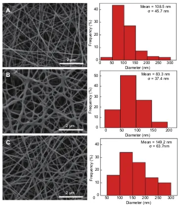
dissolved in the electrospinning solution for the fabrication of composite nanofibers using blend electrospinning. During the electrospinning, the mixed solution was surface charged by the electricfield, leading to a mutual charge repulsion force among polymer chains to counterbalance the surface tension. When the electricfield intensity increased to a critical value, the repulsive electrical forces among polymer chains over-came the surface tension of polymers, making the polymers eject from the syringe needle and travel to the collecting plate. The solvent evaporated during the traveling process and the polymerfibers were formed randomly on the collecting plate. The surface morphologies of the resultant CP, CP/MoS2, and CP/MoS2/DOX nanofibers were observed using scanning electron microscopy (Figures 1 and 2). It was found that pristine CP nanofibers exhibit a typical 3-dimensional mor-phology with a smooth surface and diameter of 108.5±45.7 nm (Figure 1A). After adding MoS2, the 3-dimensional mor-phology of CP nanofibers was not changed, but the diameter decreased to 83.3±37.4 nm (Figure 1B). When MoS2and DOX were added, the diameter of CP/MoS2/DOX nanofibers increased to a size of 149.2±63.7 nm (Figure 1C). The change of nanofiber diameter may be attributed to the variation of electrospinning solution properties after adding MoS2 and DOX.
Due to the hydrophilic nature of PVA, the composite CP nanofibers were dissolved upon contacting water. Therefore, in order to facilitate their biomedical application, GA was selected as a crosslinker to render the nanofibers with water stability. After crosslinking, the morphology of nanofibers did not change significantly; however, the diameter of the crosslinked CP, CP/MoS2, and CP/MoS2/DOX nanofibers increased to 117.4±42.4 nm (Figure 2A), 89.1±34.8 nm (Figure 2B), and 166.9±59.1 nm (Figure 2C), respectively. The increase of diameter is likely ascribed to swelling of the nanofibers during the crosslinking.
Photothermal performance
As a representative member of 2-dimensional transition metal sulfides, the MoS2 nanosheet has been recognized as a typical biocompatible PTA. It is no wonder that doping of the MoS2 nanosheet will render the CP/MoS2 nanofibers with high photothermal conversion efficiency. To prove this hypothesis, the photothermal performance of CP/MoS2 nanofibers was then performed with irradiation from the 808 nm NIR laser. The UV–Vis–NIR spectrum of CP/MoS2nanofibers was firstly studied, which found that CP/MoS2nanofibers showed an apparent light absorption in the test wavelength range (Figure S3A). Apparently,
pure DOX has no classical light absorbance in the NIR region (Figure S3B); therefore, it can be inferred that adding DOX will not influence the light absorbance of CP/MoS2/DOX nanofibers. It was verified that the photo-thermal transformation outcome of CP/MoS2 nanofibers was strongly related to the NIR laser power density and irradiation time (Figure 3). Under the laser power densities of 0.3, 0.5, and 1.0 W/cm2, the △T values of CP/MoS2 containing saline were determined as 13.2 °C, 21.5 °C, and 30.7 °C, respectively, after 5 min of laser irradiation (Figure 3A,B). However, without MoS2, pure CP nanofi -ber showed no meaningful photothermal performance (Figure S4A,B), indicating that the photothermal conver-sion efficiency was solely originated from MoS2 nanosheets. The corresponding infrared thermal images of CP/MoS2 nanofibers under NIR irradiation were recorded using an infrared camera (FLIR™E60) to further intuitively evaluate the photothermal performance. As shown in Figure 3B, the high-temperature area swiftly extended and reached whole coverage of the cell culture hole, further significantly suggesting the admirable in vitro photothermal transforming capability of CP/MoS2nanofi -bers. The photothermal conversion efficiency of CP/MoS2 nanofibers was determined as 23.2% (Figure 3C, 1 W/cm2, 5 min). Besides, CP/MoS2 nanofibers were proved to be photothermally stable and the△T values did not show any apparent retrogress during the 5 cycles of continuous laser irradiation (△T=34.3 °C (cycle 1), 34.6 °C (cycle 2), 34.2 °C (cycle 3), 34.8 °C (cycle 4), and 34.7 °C (cycle 5), power density 1 W/cm2;Figure 3D). CP/MoS2nanofi -bers with such an apparent photothermal efficiency and stability are anticipated to act as an encouraging PTA to find universal applications for PTT cancer treatment.
DOX loading and release from CP/MoS
2/
DOX nano
fi
bers
As a drug reservoir, the CP/MoS2nanofibers can load and control the release of DOX. The concentration of DOX released to the crosslinking solution was calculated to be 0.74 mg/mL. Therefore, the drug content of the CP/MoS2/DOX nanofibrous mat was computed as 3.68%. The absorbance of the crosslinking solution was recorded using a UV–Vis spec-trophotometer (Lambda 25; Perkin Elmer) to calculate the DOX concentration according to the standard curve. It was found that the DOX release was characterized with a pH/ temperature dual-modal responsive and sustainable manner (Figure 4). The initial release rate was slightly fast, likely
International Journal of Nanomedicine downloaded from https://www.dovepress.com/ by 118.70.13.36 on 22-Aug-2020
owing to the hydrophilic surface of CP nanofibers. The cumu-lative release amount reached a plateau after 6 h at all of the tested pHs and temperatures. In addition, the release rate can be promoted by the increasing temperature and decreasing pH, likely due to the increased DOX solubility in mild acidic conditions and high temperature. For example, the total release amount was 11.0±1.7% under physiological conditions (pH 7.4, T=37 °C), but it sharply increased to 65.8±2.5% under tumor PTT conditions (pH 5.4, T=54 °C). Such a pH and temperature dual-modal responsive drug release profile ren-ders the nanofibers with the ability to augment the tumor therapy, since the generated heat under NIR laser irradiation not only can kill tumor cells by hyperthermia, but can also sensitize the chemotherapeutic efficacy of DOX via enhancing its release rate. Moreover, this drug loading is anticipated to be
universally extended to the loading of many categories of other drugs with a high loading capacity, providing the drug was soluble in electrospinning solution.
In vitro cytocompatibility
We then studied the cytocompatibility, which is the basic prerequisite for safe biomedical application of the nanofi -brous mat, using CCK-8 and Dead/Live staining (Figure 5
and Figure S5). It was found that the viability of L929 cells on CP and CP/MoS2nanofibers was higher than 95% (CP: 98.4%; CP/MoS2: 98.1%), and showed no detectable difference when compared with healthy L929 cells (p>0.05) after 24 h of culture (Figure S5A). The cell viability of healthy cells cultured on CP and CP/MoS2 nanofibers was then qualitatively evaluated by performing
0 0
2 μm
2 μm
2 μm
10 20 30 40
50 100 150 Diameter (nm)
A
B
C
Frequency (%)
200 250 300 Mean = 108.5 nm
σ = 45.7 nm
00 10 20 30 40
50 100 150 Diameter (nm)
Frequency (%)
200 250 300 Mean = 149.2 nm
σ = 63.7nm 0
0 10 20 30 40 50
50 100 150
Diameter (nm)
Frequency (%)
200 Mean = 83.3 nm
σ = 37.4 nm
Figure 1Characterization of nanofibers. (A) Scanning electron microscopy pictures and diameter distribution of (A) chitosan/polyvinyl alcohol (CP), (B) CP/MoS2, and (C)
CP/MoS2/doxorubicin nanofibers.
International Journal of Nanomedicine downloaded from https://www.dovepress.com/ by 118.70.13.36 on 22-Aug-2020
Dead/Live staining, which can stain live and dead cells green and red, respectively. In accordance with the CCK-8, almost all cells turned green during the Dead/Live stain-ing, confirming the insignificant cytotoxicity of the com-posite nanofibers (Figure S5B–D).
To further evaluate the biocompatibility of the nanofi -brous carrier, the longer-term proliferation level of L929 cells cultured on nanofibers was monitored by performing CCK-8 and cell morphology observation. L929 cells pro-liferated smoothly on both the CP and CP/MoS2 nanofi -bers, and their proliferation viability was comparable with cells cultured onto the cell culture plate at different time points (Figure 5). The morphology of L929 cells cultured onto CP and CP/MoS2 nanofibrous mats for 24 h, 48 h, and 72 h was observed by scanning electron microscopy
(Figure 5C). Qualitatively corroborating with the CCK-8
assay results (Figure 5A), it was found that both CP and CP/MoS2nanofibers can support the migration, adhesion, and proliferation of L929 cells onto thefiber surface, and a typical three-dimensional cell-matrix structure was formed at all of the tested time points, further suggesting the excellent biocompatibility of the CP and CP/MoS2 nanofibers.
In vitro hemocompatibility
Red blood cells play a dominant role in the transportation of oxygen and nutrients. Therefore, the hemocompatibility of CP/MoS2 nanofibers necessarily needs to be verified. The hemocompatibility assessment was initially performed by performing in vitro hemolysis, where the mRBCs were
0
2 μm
2 μm
2 μm
10 20 30 40
50 100 150 Diameter (nm)
A
B
C
Frequency (%)
200 250 300 Mean = 117.4 nm
σ = 42.4 nm
00 10 20 30 40
50 100 150 Diameter (nm)
Frequency (%)
200 250 300 Mean = 166.9 nm
σ = 59.1nm 0
0 10 20 30 40 50 60
50 100 150
Diameter (nm)
Frequency (%)
200 Mean = 89.1 nm
σ = 34.8 nm
Figure 2Characterization of crosslinked nanofibers. (A) Scanning electron microscopy pictures and diameter distribution of GA-crosslinked (A) chitosan/polyvinyl alcohol
(CP), (B) CP/MoS2, and (C) CP/MoS2/doxorubicin nanofibers.
International Journal of Nanomedicine downloaded from https://www.dovepress.com/ by 118.70.13.36 on 22-Aug-2020
individually treated with saline (negative control), water (posi-tive control), CP, and CP/MoS2 nanofibers. The structural integrity of mRBCs can be completely preserved and the hemolysis ratio was defined as 0 in saline, while the structural integrity of mRBCs can be totally destroyed in water and the hemolysis ratio was defined as 100%. As shown inFigure 5B, the hemolysis ratio of CP and CP/MoS2nanofibers was cal-culated as 1.8±0.4% and 2.8±0.8% after 2 h of incubation, respectively. In accord with the hemolysis ratio, mRBCs can be readily separated from the solution upon incubation with saline, CP, and CP/MoS2nanofibers, further indicating that the impact of CP and CP/MoS2nanofibers on the structural integ-rity of mRBCs is negligible.3636
In vivo biocompatibility
Before moving to the in vivo tumor therapy study, the bio-compatibility of CP/MoS2was further explored using healthy KM mice. After embedding the subcutaneous nanofibers, all of the KM mice gained weight progressively and showed negligible difference compared with healthy KM mice, pre-liminarily confirming that the CP/MoS2 nanofibers do not cause obvious side effects on the physical condition of KM mice (Figure 6A). To prove that the CP/MoS2 nanofibrous
carrier can ensure highly efficient tumor therapy by retaining the great mass of MoS2nanosheets in tumor sites, the in vivo release of MoS2nanosheets from the polymeric matrix was analyzed by studying the biodistribution of Mo ions in major organs of KM mice using ICP, at 7 days and 28 days after subcutaneous nanofiber embedding (Figure 6C). It was dis-covered that the Mo ion accumulation in the heart, liver, spleen, lung, and kidney was limited. For example, the content of Mo in the spleen was about 4.7±1.0μg/g and 3.5±0.3μg/g after 7 days and 28 days of feeding; this content only accounted for about 0.2% of the total dose of MoS2in the composite nanofibrous mats, indicating that the vast majority of MoS2nanosheets were entrapped within tumor and thereby their potential health threats to living body can be eliminated. It is worth noting that the laser irradiation will not lead to obvious influence on the biodistribution of Mo ions, because MoS2 nanosheets34 and CP/MoS2 nanofibers have been proved photothermal stable under 5 cycles of laser irradiation. The in vivo compatibility of CP/MoS2nanofibers was further studied by performing in vivo hemocompatibility using the serum biochemistry and routine blood test on day 7 and day 28 after subcutaneous embedding. Similar to healthy mice, blood routine parameters and serum
0
0 0
0 1.4 1.2
Heating period
Cooling period 1.0 0.8 0.6 0.4 0.2 0.0
100 200 300 400 500 600300 250 200 150 100 50
600 1200
Time (s) Time (s)
T
ime (s)
-Ln(θ)
1800 2400 3000
50
A
B
D
C
0 s 30 s 60 s 180 s 300 s
60.0
25.0
100 150 Time (S)
T
emperature (°C)
T
emperature (°C)
200
Control
Control
0.3 W/cm2 0.3 W/cm
2
0.5 W/cm2
0.5 W/cm
2
1 W/cm2
1 W/cm
2
250
On On On On On
Off Off Off Off Off
300
Power density = 1 W/cm2 0
20 25 30 35 40 45 50
30 40 50 60 70 80 5
10 15 20 25 30 35
λex = 808 nm R = 0.997
ηT = 23.2 %
τS = 225.7 s
∆T
(°C)
Figure 3In vitro photothermal performance of nanofibers. (A) Power density-related temperature changes (△T) of CP/MoS2nanofibers during 300 s of irradiation by 808
nm near-infrared laser. (B) Thermal images of CP/MoS2nanofibrous mat at different time points corresponding to (A). (C) Steady state heating curve and heat transfer time
constant of CP/MoS2nanofibers (power density 1 W/cm
2
, 5 min; R=0.997,τs=225.7 s). (D) 808 nm laser-induced recycling photothermal ability of CP/MoS2nanofibers.
Abbreviation:CP, chitosan/polyvinyl alcohol.
International Journal of Nanomedicine downloaded from https://www.dovepress.com/ by 118.70.13.36 on 22-Aug-2020
biochemistry parameters of experimental animalsfluctuate in the normal range (Figure 6BandFigure S7), implying
healthy physiological functions of the main organs and blood of CP/MoS2 nanofiber-treated mice. Then, the above KM mice were euthanized to harvest their major organs for standard H&E staining (Figure 6D). Consistent with the body weight and in vivo hemocompatibility assay, the main organs of the tested KM mice showed no obvious pathological abnormity after 7 days and 28 days of feed-ing. The in vitro and in vivo analysis data confirm the excellent hemocompatibility of the as-synthesized compo-site nanofibers, providing more evidence for the feasibility and practicality of safe in vivo applications of CP/MoS2 nanofibers. Together with the above proved photothermal conversation capacity, such CP/MoS2/DOX nanofibers are anticipated to have different applications for highly effi -cient and secure tumor treatments.
In vitro tumor therapeutic ef
fi
ciency
After proving the excellent photothermal efficiency, photo-thermal stability, controlled drug release, and biocompatible0 5 10 15 20 25
0 10 20 30 40 50 60 70
pH = 5.4, T = 54°C
pH = 7.4, T = 54°C
pH = 5.4, T = 37°C
pH = 7.4, T = 37°C
Cumulative release (%)
Time (h)
Figure 4Doxorubicin release from CP/MoS2nanofibrous mat at different pH with
(T=54 °C) or without (T=37 °C) near-infrared irradiation (1 W/cm2).
Abbreviation:CP, chitosan/polyvinyl alcohol.
24 h Control CP CP/MoS2
0.0 0
CP
CP
CP/MoS2
CP/MoS
2
1 2 3 4 5 6 7
0.2 0.4 0.6 0.8 1.0 1.2 1.4 1.6 1.8
24 h
A
B
C
OD
HP
(%)
48 h
48 h 72 h
72 h (1) (2) (3) (4)
5 μm
5 μm
5 μm
5 μm
5 μm
5 μm
Figure 5In vitro cytocompatibility and hemocompatibility assays of crosslinked nanofibers. (A) Proliferation viability of L929 cells on cell culture plate, CP, and CP/MoS2
nanofibers after 24 h, 48 h, and 72 h of culture. (B) Hemolytic percentage (HP) of mRBCs treated with CP and CP/MoS2nanofibers. Inset images are the centrifuged mRBCs
after 2 h of incubation with (1) water, (2) saline, (3) CP, and (4) CP/MoS2nanofibers. (C) Scanning electron microscopy micrographs of L929 cells cultured on CP and CP/
MoS2nanofibers after 24 h, 48 h, and 72 h culture.
Abbreviations:CP, chitosan/polyvinyl alcohol; OD, optical density; HP, hemolytic percentage; mRBC, mouse red blood cell.
International Journal of Nanomedicine downloaded from https://www.dovepress.com/ by 118.70.13.36 on 22-Aug-2020
nature of the electrospun CP/MoS2nanofibers, we then sys-tematically studied the combined chemotherapy and PTT efficiency in vitro using HT29 cells and in vivo using the HT29 tumor-bearing nude mice. As shown in Figure S6, obvious red color and red fluorescence can be detected from the phase contrast microscopic andfluorescent photo-graph of HT29 cells treated with CP/MoS2/DOX nanofibers
(Figure S6C,D). However, no visible and fluorescent red
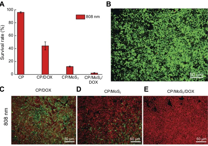
signal was observed from HT29 cells treated with saline (Figure S6A,B), inferring that DOX release from CP/MoS2/ DOX nanofibers can be efficiently internalized by HT29 cells. The viability of HT29 cells cultured onto CP/DOX nanofibers decreased to 46.2±6.2% after 24 h of incubation (Figure 7A). After being exposed under the 808 nm laser for 5 min, the viability of HT29 cells onto CP/MoS2nanofibers significantly decreased to 12.7±0.8% (Figure 7A), resulting from the photothermal effect of the doped MoS2nanosheets. Under the coexistence of the photothermal effect of MoS2 and the chemotherapeutic efficiency of DOX, CP/MoS2/ DOX nanofibers can completely suppress the malignant pro-liferation of tumor cells, and the viability of HT29 cells
cultured therein for 24 h sharply decreased to 2.1±0.8% after being irradiated with the 808 nm laser for 5 min. To further prove the in vitro photothermal and chemotherapy tumor therapy capacity of CP/MoS2nanofibers, HT29 cells after the above treatments were further stained using the Dead/Live kit. Without DOX and MoS2, pure CP nanofibers showed no anti-tumor efficiency; therefore, almost all of the cells cultured with CP nanofibers were stained green. In accordance with the CCK-8 assay, a section of HT29 cells were stained red after single photothermal or chemotherapy, and the red-stained cell portion of CP/DOX was smaller than that of CP/MoS2+NIR. Oppositely, nearly all of HT29 cells were killed and stained red (Figure 7B–E) when adopting the combined therapy, confirming the combined tumor photo-thermal and chemotherapy efficiency of CP/MoS2/DOX nanofibers.
Postoperative tumor recurrence
inhibition study
Encouraged by the obvious in vitro tumor recurrence inhi-bition efficiency, CP/MoS2/DOX nanofibers were 100 μm
100 μm
100 μm
100 μm
100 μm
100 μm
100 μm
100 μm
100 μm
100 μm
100 μm
100 μm
100 μm
100 μm
100 μm
0 0.0 0.0 1.0 1.5 2.0
3 6
Relative body weight
9 12
Control
Day 7 Day 28 Control
Control
Day 7
Day 28
CP/MoS2
CP/MoS2 Control
Heart Liver Spleen Lung Kidney
Heart
A
B
C
D
Liver Spleen Lung KidneyTB(μ mol/L) ALT (U/L) AST (U/L) UREA (mg/dL) Crea
CP/MoS2
15 18 Time (days)
21 24 27 300.0 0
1 2 3 4 5
6 Day 7
Day 28
0.5 1.0 20
[Mo
2+
] μg/g
40 60 80 100 120
Figure 6In vivo biocompatibility assays of crosslinked nanofibers. (A) Body weight evolution curves of healthy KM mice and KM mice with subcutaneous CP/MoS2nanofiber
embedding. (B) Blood biochemistry parameter. (C) Biodistribution of Mo element. (D) H&E staining images of KM mice with subcutaneous CP/MoS2nanofiber embedding.
Abbreviations:ALT, alanine aminotransferase; AST, aspartate aminotransferase; CP, chitosan/polyvinyl alcohol; KM, Kunming; TB, total bilirubin.
International Journal of Nanomedicine downloaded from https://www.dovepress.com/ by 118.70.13.36 on 22-Aug-2020
hypodermically embedded within the tumor site that was excised to see their in vivo tumor postoperative recurrence inhibition ability. The in vivo photothermal conversion performance of CP/MoS2/DOX nanofibers was firstly monitored, which revealed that the in vitro photothermal conversion ability of CP/MoS2nanofibers was well inher-ited after subcutaneous embedding within the tumor site. Upon 30 s of laser irradiation (1 W/cm2), a swift tumor temperature augmentation of 8.1 °C was found, and the maximum△T can reach 31.8 °C during 5 min of contin-uous laser irradiation (Figure 8A,B). Moreover, the intro-duction of DOX exerted no apparent influence on the in vivo photothermal conversion of CP/MoS2 nanofibers
(Figure 8A,B). However, the maximum △T values of
untreated mice and mice treated with pristine CP nanofi -bers were merely 6.5 °C and 7.7 °C (Figure 8Aand Figure
S2). The in vivo infrared thermal images further proved the remarkable photothermal transform capacity of CP/ MoS2/DOX nanofibers (Figure 8B). CP/MoS2/DOX
nano-fibers with excellent in vivo photothermal conversion and controllable DOX release induce a satisfactory therapeutic result. Due to the photothermal and chemotherapy effi cien-cies of CP/MoS2/DOX, the postoperative tumor recurrence of mice with CP/MoS2/DOX embedding was totally pro-hibited and the scar was completely healed after 28 days of feeding (Figure 8C,D). In sharp contrast, owing to the
incomplete clearance of tumor cells, untreated mice and mice treated with pristine CP nanofibers endured a natural tumor recurrence and the tumor volume expanded to 12.3 ±2.3 cm3 and 10.3±2.4 cm3 after 28 days of feeding, respectively (Figure 8C). Notably, although the photother-mal conversion capability (η=23.2%) of CP/MoS2nanofi -bers is lower than previously reported MoS2-containing nanofibers,27 the NIR-induced hyperthermia of CP/MoS2 nanofibers can still efficiently restrain the tumor recur-rence in this study. It is no wonder that total inhibition of tumor recurrence can also be realized under a lower power density (lower than 1 W/cm2) since a combined photother-mal and chemotherapy capacity of CP/MoS2/DOX nanofi -bers exists.
Conclusion
In summary, the primary aim of this nanofiber design is to deliver a sufficient amount of PTAs and chemothera-peutics to tumors. To this end, smooth and uniform MoS2 and DOX co-loaded composite nanofibers were fabricated
using blend electrospinning. After loading MoS2
nanosheets, the as-prepared CP/MoS2/DOX nanofibers showed an excellent photothermal conversion capability (η=23.2%). In addition, the composite nanofibers could control the sustained release of DOX. It was revealed that the designed composite nanofibers possess excellent in
0 20 40 60 80 100
CP/MoS2
CP/MoS2
CP/MoS2/
DOX
CP/MoS2/DOX
CP/DOX
CP/DOX CP
808 nm
808 nm
C
D
E
A
B
Survival rate (%)
50 μm
50 μm 50 μm
50 μm
Figure 7In vitro therapeutic efficacy of crosslinked nanofibers. (A) In vitro photothermal therapy and chemotherapy of CP, CP/DOX, and CP/MoS2and CP/MoS2/DOX
nanofibers. (B–E) Dead/Live staining of HT29 cells treated with (B) control, (C) CP/DOX+NIR, (D) CP/MoS2+NIR, and (E) CP/MoS2/DOX+NIR.
Abbreviations:CP, chitosan/polyvinyl alcohol; DOX, doxorubicin; NIR, near infrared.
International Journal of Nanomedicine downloaded from https://www.dovepress.com/ by 118.70.13.36 on 22-Aug-2020
vitro and in vivo biocompatibility. Moreover, the gener-ated heat under NIR laser irradiation can sensitize the chemotherapeutic efficacy of DOX via controlling its release rate. The combined chemo-/photo thermal therapy efficiency of the composite nanofibers was systematically studied in vitro using HT29 cells and in vivo using HT29 tumor-bearing nude mice. The results prove that the post-operative tumor reoccurrence was completely prohibited owing to the combined tumor photothermal and che-motherapy efficiency of CP/MoS2/DOX nanofibers. Instead of circulating with the body fluid, MoS2 was confined to the tumor site by the nanofiber matrix while the tumor-killing ability was not compromised, therefore rendering the concept to design composite nanofibers a promising clinical translation in biomedical application fields.
Acknowledgments
The authors are grateful for thefinancial support from the National Natural Science Foundation of China (Grant Nos 51702214, 81470800, and 81670485), and Shanghai
Sailing Program (17YF1412600) supported by the
Shanghai Committee of Science and Technology.
Disclosure
The authors report no conflicts of interest in this work.
References
1. Lin H, Gao S, Dai C, Chen Y, Shi J. A two-dimensional biodegradable niobium carbide (MXene) for photothermal tumor eradication in NIR-I and NIR-II biowindows.J Am Chem Soc.2017;139(45):16235–16247. doi:10.1021/jacs.7b07818
2. Wang S, Zhao J, Yang H, et al. Bottom-up synthesis of WS2 nanosheets with synchronous surface modification for imaging guided tumor regression. Acta Biomater. 2017;58:442–454. doi:10.1016/j. actbio.2017.06.014
3. Zhitnyak I, Bychkov I, Sukhorukova IV, et al. Effect of BN nanopar-ticles loaded with doxorubicin on tumor cells with multiple drug resistance. ACS Appl Mater Interfaces. 2017;9(38). doi:10.1021/ acsami.7b08713
4. Ma Y, Liang X, Tong S, Bao G, Ren Q, Dai Z. Gold nanoshell nanomicelles for potential magnetic resonance imaging, light-triggered drug release, and photothermal therapy. Adv Funct Mater. 2013;23 (7):815–822. doi:10.1002/adfm.v23.7
5. Yang H, Zhao J, Wu C, Ye C, Zou D, Wang S. Facile synthesis of colloidal stable MoS2nanoparticles for combined tumor therapy.Chem Eng J.2018;351:548–558. doi:10.1016/j.cej.2018.06.100
6. Lan G, Ni K, Xu R, et al. Nanoscale metal-organic layers for deeply penetrating X-ray-induced photodynamic therapy. Angew Chem Int Edit.2017;56(40):12102–12106. doi:10.1002/anie.201704828 7. Rong P, Kai Y, Avinash S, et al. Photosensitizer loaded nano-graphene
for multimodality imaging guided tumor photodynamic therapy. Theranostics.2014;4(3):229–239.
CP/MoS
2
CP/MoS2
CP/MoS2
CP/MoS2
CP/MoS
2
/DOX
CP/MoS2/DOX
CP/MoS2/DOX
CP/MoS2/DOX
Control
Control
Day 1
Day 28
0 s 30 s 60 s 300 s
0 s 30 s 60 s 300 s
0 s 30 s 60 s 300 s
CP
CP 0
0 0 2 4 6 8 10 12 14
1 4 8 12 Time (days)
Relative tumor volume
∆T
(°C)
16 20 24 28 0
5 10 15 20 25 30 35
50 100 150 Time (S)
Control
Control
200 250 300
°C 65.0
30.0
FLIR FLIR 30.0 FLIR 30.0 FLIR 30.0 65.0 65.0 65.0 °C °C °C
°C 65.0
30.0
FLIR FLIR 30.0 FLIR 30.0 FLIR 30.0 65.0 65.0 65.0 °C °C °C
°C 65.0
30.0
FLIR FLIR 30.0 FLIR 30.0 FLIR 30.0 65.0 65.0 65.0 °C °C °C
C
D
A
B
Figure 8In vivo therapeutic efficacy of crosslinked nanofibers. (A) In vivo photothermal performance of CP/MoS2nanofibrous mat (control: mice without treatment). (B) In
vivo thermal images of mice receiving different treatments as noted. (C) Postoperative tumor volume of mice receiving different treatments as noted. (D) Representative
photographs of HT29 tumor-bearing mice corresponding to (C).
Abbreviations:△T, temperature increase; CP, chitosan/polyvinyl alcohol; DOX, doxorubicin.
International Journal of Nanomedicine downloaded from https://www.dovepress.com/ by 118.70.13.36 on 22-Aug-2020
8. Ma L, Zhou Y, Zhu Y, et al. 3D printed personalized titanium plates improve clinical outcome in microwave ablation of bone tumors around the knee.Sci Rep.2017;7(1):7626.
9. Chen Y, Jiang L, Wang R, et al. Injectable smart phase-transfor-mation implants for highly efficient in vivo magnetic-hyperther-mia regression of tumors.Adv Mater.2014;26(44):7468–7473. 10. Sonvico F, Mornet S, Vasseur S, et al. Folate-conjugated iron oxide
nanoparticles for solid tumor targeting as potential specific magnetic hyperthermia mediators: synthesis, physicochemical characterization, and in vitro experiments. Bioconjugate Chem. 2005;16(5):1181– 1188. doi:10.1021/bc050050z
11. Li C, Hu J, Li W, Song G, Shen J. Combined bortezomib-based chemotherapy and p53 gene therapy using hollow mesoporous silica nanospheres for p53 mutant non-small cell lung cancer treatment. Biomater Sci. 2016;5(1):77–88. doi:10.1039/ c6bm00449k
12. Shen J, Sheng X, Chang Z, et al. Iron metabolism regulates p53 signaling through direct heme-p53 interaction and modulation of p53 localization, stability, and function.Cell Rep. 2014;7(1):180– 193. doi:10.1016/j.celrep.2014.02.042
13. Wang S, Zhao J, Hu F, et al. Phase-changeable and bubble-releasing implants for highly efficient HIFU-responsive tumor surgery and chemotherapy.J Mat Chem B.2016;4(46):7368–7378. doi:10.1039/ C6TB01861K
14. Chen Y, Chen H, Sun Y, et al. Multifunctional mesoporous composite nanocapsules for highly efficient MRI-guided high-intensity focused ultrasound cancer surgery.Angew Chem.2011;123(52):12713–12717. doi:10.1002/ange.201106180
15. Yang B, Chen Y, Shi J. Material chemistry of two-dimensional inorganic nanosheets in cancer theranostics.Chem.2018;4(6):1284– 1313. doi:10.1016/j.chempr.2018.02.012
16. Wu C, Wang S, Zhao J, et al. Biodegradable Fe(III)@WS2-PVP nanocapsules for redoxReaction and TME-enhanced nanocatalytic, photothermal,and chemotherapy. Adv Funct Mater.
2019;201901722. doi:10.1002/adfm.201901722
17. Tang S, Chen M, Zheng N. Sub-10-nm Pd nanosheets with renal clearance for efficient near-infrared photothermal cancer therapy. Small. 2014;10(15):3139–3144. doi:10.1002/smll.2013 03631
18. Chen L, Zhou X, Nie W, et al. Marriage of albumin-gadolinium complexes and MoS2 nanoflakes as cancer theranostics for dual-modality magnetic resonance/photoacoustic imaging and photother-mal therapy.ACS Appl Mater Interfaces.2017;9(21):17786–17798. doi:10.1021/acsami.7b04488
19. Chhowalla M, Liu Z, Zhang H. Two-dimensional transition metal dichalcogenide (TMD) nanosheets.Chem Soc Rev.2015;44(9):2584– 2586. doi:10.1039/c5cs90037a
20. Lin H, Wang YW, Gao SS, Chen Y, Shi JL. Theranostic 2D tantalum carbide (MXene). Adv Mater. 2018;30(4):1703284. doi:10.1002/ adma.v30.4
21. Gulzar A, Xu J, Yang D, et al. Nano-graphene oxide-UCNP-Ce6 covalently constructed nanocomposites for NIR-mediated bioimaging and PTT/PDT combinatorial therapy.Dalton T.2018;47(11):3931– 3939. doi:10.1039/C7DT04141A
22. Shao L, Zhang R, Lu J, Zhao C, Deng X, Wu Y. Mesoporous silica coated polydopamine functionalized reduced graphene oxide for synergistic targeted chemo-photothermal therapy.ACS Appl Mater Inter.2017;9(2):1226–1236. doi:10.1021/acsami.6b11209
23. Liu H, Li W, Cao Y, Guo Y, Kang Y. Theranostic nanoplatform based on polypyrrole nanoparticles for photoacoustic imaging and photothermal therapy.J Nanopart Res.2018;20(3):57. doi:10.1007/s11051-018-4157-y 24. Zhang D, Wu M, Zeng Y, et al. Chlorin e6 conjugated poly(dopa-mine) nanospheres as PDT/PTT dual-modal therapeutic agents for enhanced cancer therapy. ACS Appl Mater Interfaces. 2015;7 (15):8176–8187. doi:10.1021/acsami.5b01027
25. Wang J, Guo Y, Hu J, et al. Development of multifunctional polydopa-mine nanoparticles as a theranostic nanoplatform against cancer cells. Langmuir.2018;34:9516–9524. doi:10.1021/acs.langmuir.8b01769 26. Li W, Wang X, Wang J, et al. Enhanced photoacoustic and
photo-thermal effect of functionalized polypyrrole nanoparticles for near-infrared theranostic treatment of tumor. Biomacromolecules.
2019;20:401–411. doi:10.1021/acs.biomac.8b01453
27. Ye C, Zhao J, Zheng Y, et al. Preparation of poly(lactic-co-glycolic acid)-based composite microfibers for postoperative treatment of tumor in NIR I and NIR II biowindows. Macromol Biosci.
2018;1800206. doi10.1002/mabi.201800206
28. Wang S, Hu F, Li J, et al. Design of electrospun nanofibrous mats for osteogenic differentiation of mesenchymal stem cells. Nanomed-Nanotechnol.2018;14:2505–2520. doi:10.1016/j.nano.2016.12.024 29. Chen M, Li YF, Besenbacher F. Electrospun nanofibers-mediated
on-demand drug release. Adv Healthc Mater. 2014;3(11):1721–1732. doi:10.1002/adhm.201400166
30. Yang YY, Liu ZP, Yu DG, Wang K, Liu P, Chen X. Colon-specific pulsatile drug release provided by electrospun shellac nanocoating on hydrophilic amorphous composites.Int J Nanomed.2018;13:2395– 2404. doi:10.2147/IJN.S154849
31. Wang K, Liu XK, Chen XH, Yu DG, Yang YY, Liu P. Electrospun hydrophilic janus nanocomposites for the rapid onset of therapeutic action of helicid.ACS Appl Mater Interfaces.2018;10(3):2859–2867. doi:10.1021/acsami.7b17663
32. Liu X, Shao W, Luo M, Bian J, Yu DG. Electrospun blank nanocoat-ing for improved sustained release profiles from medicated gliadin nanofibers.Nanomaterials.2018;8(4):184. doi:10.3390/nano8040184 33. Liao H, Qi R, Shen M, et al. Improved cellular response on multi-walled carbon nanotube-incorporated electrospun polyvinyl alcohol/ chitosan nanofibrous scaffolds.Colloid Surface-B.2011;84(2):528– 535. doi:10.1016/j.colsurfb.2011.02.010
34. Zhao J, Zhou C, Mao L, et al. Bottom-up synthesis of ultra-small molybdenum disulfide-polyvinylpyrrolidone nanosheets for imaging-guided tumor regression. Oncotarget. 2017;8(63):106707–106720. doi:10.18632/oncotarget.22477
35. Tian Q, Hu J, Zhu Y, et al. Sub-10 nm Fe3O4@ Cu2-xS core-shell nanoparticles for dual-modal imaging and photothermal therapy.J Am Chem Soc.2013;135(23):8571–8577. doi:10.1021/ja4013497 36. Guo B, Zhao J, Wu C, et al. One-pot synthesis of polypyrrole
nanoparticles with tunable photothermalconversion and drug loading capacity. Colloid Surface-B. 2019;177:346–355. doi:10.1016/j. colsurfb.2019.02.016
International Journal of Nanomedicine downloaded from https://www.dovepress.com/ by 118.70.13.36 on 22-Aug-2020
Supplementary materials
Calculation of the photothermal conversion efficiency and supplementaryfigures.
Materials
Chitosan (medium molecular weight, degree of deacetyla-tion>90%), glutaraldehyde (50% aqueous solution), acetic acid, polyvinyl alcohol (high molecular weight, 88% hydrolyzed), PBS, cell counting kit-8 (CCK-8), and Dead/Live kit were purchased from Sigma Chemicals (Perth, Australia). DOX was commercially purchased from Beijing Huafeng Pharmaceutical Co., Ltd. (People’s Republic of China). Mouse fibroblast (L929) and human colorectal carcinoma (HT29) cell lines were commercially purchased from the Institute of Biochemistry and Cell
Biology (Chinese Academy of Science, Shanghai,
People’s Republic of China). DMEM, FBS, penicillin, and streptomycin were products of Gibco (Thermo Fisher Scientific, Waltham, MA, USA). Water with resistivity higher than 18.2 MΩ.cm was purified using a Milli-Q Plus 185 water purification system (EMD Millipore, Billerica, MA, USA).
Calculation of the photothermal
conversion ef
fi
ciency
The formulas related to the calculation of efficiency are listed as follows:
(1) The product of heat transfer coefficient (h) and the surface area of the container (S) is:
hS¼mCH2O
τs
wheremandCH2Oare the mass and specific heat capacity of the CP/MoS2nanofibers, andτsis the time constant.
(2) τscan be calculated by the following formula:
t¼ τslnð TTsurr
TmaxTsurrÞ
wheret(s) is the time of the cooling process,T(°C) is the real-time temperature of t, Tmax (°C) is the highest tem-perature of the sample, andTsurr(°C) is the temperature of the surroundings.
(3) The baseline energy input without a sample (QDis) independently measured at about 808 nm was 17.12 mW. (4) The absorbance of the sample (A(λ)) at the excitation wavelength of 808 nm was measured as about 4.1.
The studied routine blood test and
biochemistry test parameters
The analyzed routine blood parameters include red blood cells (RBC), white blood cells (WBC), hemoglobin (HGB), hematocrit (HCT), mean corpuscular volume (MCV), mean corpuscular hemoglobin (MCH), mean cor-puscular hemoglobin concentration (MCHC), platelet (PLT), and red cell distribution width (RDW). The studied biochemistry parameters include aspartate aminotransfer-ase (AST), alanine aminotransferaminotransfer-ase (ALT), total bilirubin (TBIL), and carbamide (CAR).
A
B
0 0 10 20
Concentration (μg/mL) Concentration (μg/mL)
Absorbtance (a.u.) Absorbtance (a.u.) y = 0.0156 x - 0.0044 R2 = 0.9989
y = 0.0112 x + 0.0065 R2 = 0.9994
30 40 50 5 10 15 20 25
0.0
0.0 0.1 0.2 0.3 0.4 0.5 0.6
0.1 0.2 0.3 0.4
Figure S1The standard curve of doxorubicin at (A) pH 5.4 and (B) pH 7.4.
International Journal of Nanomedicine downloaded from https://www.dovepress.com/ by 118.70.13.36 on 22-Aug-2020
Figure S2Transmission electron microsopy image of MoS2nanosheets.
200 500 600 700 800 900 1000
CP/MoS2
1100 300
Wavelength (nm) Wavelength (nm)
400 500 600 700 800 900 0.0
0 1 2 3 4 5
0.1 0.2 0.3 0.4
25 ppm 0.5
Absorbtance
Absorbtance (a.u.)
A
B
Figure S3Ultraviolet–visible–near-infrared spectrum of(A)CP/MoS2nanofibers and(B)DOX. Abbreviation:CP, chitosan/polyvinyl alcohol.
0 s 30 s 60 s 300 s
°C 65.0
30.0
65.0
30.0
65.0
30.0
65.0
30.0
FLIR FLIR FLIR FLIR
°C °C °C
0
∆T
(°C)
0 2 4 6 8 10
50 100 150 Time (S)
200 250 300
CP
A
B
Figure S4(A) In vivo photothermal performance of CP nanofibrous mat. (B) In vivo thermal images of mice corresponding to (A). Abbreviations:△T, temperature increase; CP, chitosan/polyvinyl alcohol.
International Journal of Nanomedicine downloaded from https://www.dovepress.com/ by 118.70.13.36 on 22-Aug-2020
CP/MoS2 Control
Cell viability (%)
0 20 40 60 80 100
CP Sample
A
B
D
C
50 μm
50 μm 50 μm
Figure S5(A) The viability of L929 cells after incubation with saline, CP, and CP/MoS2nanofibers. (B–D) Dead/Live phase contrast images of L929 cells after treatment with
(B) saline, (C) CP, and (D) CP/MoS2nanofibers for 24 h.
Abbreviation:CP, chitosan/polyvinyl alcohol.
A
B
C
D
100 μm
Figure S6Phase contrast microscopic (A, C) andfluorescent (B, D) photographs of HT29 cells treated with saline (A, B) and CP/MoS2/DOX nanofibers (C, D). Abbreviation:CP, chitosan/polyvinyl alcohol.
International Journal of Nanomedicine downloaded from https://www.dovepress.com/ by 118.70.13.36 on 22-Aug-2020
International Journal of Nanomedicine
Dove
press
Publish your work in this journal
The International Journal of Nanomedicine is an international, peer-reviewed journal focusing on the application of nanotechnology in diagnostics, therapeutics, and drug delivery systems throughout the biomedical field. This journal is indexed on PubMed Central, MedLine, CAS, SciSearch®, Current Contents®/Clinical Medicine,
Journal Citation Reports/Science Edition, EMBase, Scopus and the Elsevier Bibliographic databases. The manuscript management system is completely online and includes a very quick and fair peer-review system, which is all easy to use. Visit http://www.dovepress.com/ testimonials.php to read real quotes from published authors.
Submit your manuscript here:https://www.dovepress.com/international-journal-of-nanomedicine-journal CP/MoS2
Control
CP/MoS2
Control
CP/MoS2 CP/MoS2
Control Control
CP/MoS2
Control CP/MoS2
Control
CP/MoS2
Control
CP/MoS2
Control
CP/MoS2
Control Day 7
Day 7
Day 7 0
0
0 4 8 12 10 20 30 40 50 60
0
0
0 0
2 4 6 8
4 8 12 16
0 50 100 150 200 250 300 350
3 6 9 12 15 18
0 200 400 600 800 1000 1200 1400
10 20 30 40 50
40 80
HB (g/L)
MCV (FL)
RBC (10
12
/L)
WBC (10
9 /L)
RDW (%)
MCH (pg) MCHC (g/L)
HCT
(%)
PL
T
(10
9 /L)
120 160
Day 28
Day 28
Day 28
Day 7
Day 7
Day 7
Day 28
Day 28
Day 28 Day 7 Day 28 Day 7
Day 7
Day 28
Day 28
Figure S7Changes of hemoglobin (HB), hematocrit (HCT), platelet (PLT), mean corpuscular volume (MCV), mean corpuscular hemoglobin (MCH), mean corpuscular hemoglobin concentration (MCHC), red blood cell count (RBC), red cell distribution width (RDW), and white blood cell count (WBC) of healthy Kunming mice (control)
and mice treated with CP/MoS2nanofibrous mat for 7 and 28 days (mean±SD, n=3).
Abbreviation:CP, chitosan/polyvinyl alcohol.
International Journal of Nanomedicine downloaded from https://www.dovepress.com/ by 118.70.13.36 on 22-Aug-2020