Ali R1, 2, Chauhan V2, Farooq S1, Khan A2, Farooq U2* 1
The Himalaya Drug Company, Dehradun, U.K. 2
Faculty of Biotechnology, Shoolini University of Biotechnology and Management Sciences, Solan, Himachal Pradesh, India. *Corresponding author’s E-mail:[email protected]
Accepted on: 04-12-2013; Finalized on: 31-03-2014.
ABSTRACT
Ocimum sanctum (Tulsi) is known to be an important medicinal plant from ancient period in India. Ursolic acid (UA), Oleanolic acid
(OA) and Betulinic acid (BA), the derivatives of triterpenoid saponins, acts as a bioactive compounds and have been isolated from many medicinal plants and reported to possess a potent anti-bacterial activity against the pathogenic bacteria. The present study was aimed to undergo the phytochemical screening and to investigate the antibacterial efficacy of the leaf extract of Ocimum
sanctum in various solvents like acetone, methanol, chloroform, diethyl ether, dimethyl sulfoxide and aqueous extracts. The
Minimum Inhibition Concentration (MIC) of UA, OA and BA against pathogenic bacteria was also determined. The methanolic and aqueous extracts of crude leaf powder were found to be most effective against the pathogenic bacterial strains selected for the study. Of the three triterpenoids, UA was most effective followed by OA and BA against most of the pathogens. Furthermore, the concentration of UA and OA in leaf powder of Ocimum sanctum was also determined by HPLC. The total concentration of UA and OA was 0.352% in leaf of Tulsi.
Keywords: Betulinic acid, Minimum Inhibitory Concentration (MIC), Ocimum sanctum, Oleanolic acid, Ursolic acid.
INTRODUCTION
icroorganisms have developed resistance to many antibiotics and this has created an immense clinical problem in treatment of infectious diseases. The resistance against drugs has increased due to indiscriminate use of commercial antimicrobial drugs commonly used for the treatment of infectious diseases. Methicillin-resistant Staphylococcus
aureus (MRSA) and Vancomycin resistant Enterococcus
sps (VRE) are one of the most common nosocomial pathogens throughout the world and markedly increases the morbidity and mortality in hospitalized patients.1-3 Therefore, to combat multi drug resistance it is necessary to discover new antimicrobial compounds. This situation forced scientist to search for new anti-microbial substances from various sources such as medicinal plants.4 Plants are well known for medical importance and have been used as a good source of many effective drugs around the globe.5 Ocimum sanctum belongs to the family Lamiaceae and is used as an important component for the ayurvedic treatment of various diseases and also possesses several pharmacological properties such as antifertility, anticancer, antidiabetic, antifungal and antimicrobial.6 The medicinal value of plants lies in some chemical substances that produce a definite physiological action on the human body. The most important of these bioactive compounds of plants are alkaloids, tannins and phenolic compounds.7 Ursolic acid (UA, 3β-hydroxy-urs-12-en-28-oic acid), Oleanolic acid (OA, 3β-3-hydroxyolean-12-en-28-oic acid) and Betulinic acid (3β-hydroxylup-20-(29)-en-28-oic acid) the three bioactive compounds, are the derivatives of triterpenoid saponins,
and possess anti-bacterial activity against many pathogenic bacterial strains.8-11
Keeping these facts in mind, the present work was undertaken to screen the leaf extract of Ocimum sanctum for the presence of various phytochemical constituents and to determine its antibacterial efficacy against human pathogenic bacteria and estimation of Ursolic and Oleanolic acid in its leaves.
MATERIALS AND METHODS
Collection of Plant Materials
Ocimum sanctum leaves were collected in the month of
May & June from semiarid, unshaded land of Dehradun (Uttranchal) and Solan (Himachal Pradesh), India. Leaves suitable for extraction were plucked from plants and were washed under running tap water followed by sterilized distilled water. Leaves were air-dried, powdered and were subjected for the extraction.
Aqueous Extraction
Air-dried powder of Ocimum sanctum leaves (10 gm) were boiled in 400 ml distilled water till one fourth of the extract initially taken, was left behind after evaporation. The solution was then filtered using muslin cloth. Filtrate was centrifuged at 5000 rpm for 15 minutes. The supernatant was again filtered using Whatman filter paper no. 1 under aseptic conditions and the filtrate was collected in fresh sterilized bottles and stored at 4°C until used.
In-vitro Analysis of Antibacterial Activity of Ocimum Sanctum Against Pathogenic
Bacteria and Quantification of Ursolic Acid and Oleanolic Acid
M
Organic Solvent Extraction
Air-dried powder (10 gm) was thoroughly mixed with 100 ml organic solvent viz., acetone, chloroform, di-ethyl ether, hexane and methanol. The mixture was placed at room temperature for 24 h on shaker with 150 rpm. Solution was then filtered through muslin cloth and then re-filtered through Whatman filter paper no. 1. The filtrate thus obtained was concentrated by complete evaporation of solvent at room temperature to yield the pure extract. Stock solutions of various organic crude extracts were prepared by mixing well the appropriate amounts of dried extracts and suitable solvent to give rise to a final concentration of 100 mg/ml. Each solution was stored at 4°C in sterilized bottles until further use. Phytochemical Screening
The phytochemical screening of methanol and aqueous extracts of Ocimum sanctum was performed using standard procedures.11, 12 The qualitative analysis for alkaloids, tannins, saponin, steroid, glycosides, flavonoids, terpenoids, amino acids and carbohydrates were carried out for both the extracts.
Collection of bacterial strain
The bacterial strains, S. aureus (MTCC-737), P. aeruginosa (MTCC-429), S. mutans (MTCC-890), E. coli (MTCC-1303),
B. subtilis (MTCC-121), K. pneumoniae (MTCC-109), Vibrio fischeri (MTCC-1738), S. pneumoniae (MTCC-655), Aeromonas veronii (MTCC-3249), Methicillin-resistant S. aureus (MTCC-84) and Vancomycin-resistant enterococci
(VRE-912) were obtained from MTCC, IMTECH, Chandigarh, India. These strains were maintained in nutrient agar slants at 37°C. Each of the bacteria was reactivated prior to susceptibility testing by transferring them into a separate test tube containing nutrient broth and incubated overnight at 37°C.
Bacterial Susceptibility Assay
In vitro antibacterial activities of all extracts were
determined by standard agar well diffusion assay.13 Petri dishes (size 100 mm diameter) containing 18 ml of cool and molten Mueller Hinton Agar (MHA) (at 40°C) were seeded with 100 µl inoculums of bacterial strain (inoculums size was adjusted so as to deliver a final inoculum of approximately 1.0 x 10⁸ CFU/ml). Wells of 6 mm diameter were cut into solidified agar media with the help of sterilized cork borer. An aliquot of 100 µl of each extract was poured in the respective well and the plates were incubated at 37°C overnight. Organic solvents, in which extracts were prepared, were used as negative control while ciprofloxacin (10µg/ml) was used as a control. The experiment was performed in triplicate under aseptic conditions. The antibacterial activity for each of the extract evaluated was expressed in terms of the average of the diameter of zone of inhibition (in mm) produced by the respective extract at the end of incubation period.
Minimum inhibitory concentrations (MICs) of triterpenoids
MIC was determined using a micro dilution assay according to the Clinical and Laboratory Standards Institute standard (NCCLS, 2000).14 The bacterial strains were cultured in MH broth at 370C in an incubator for 24 h and added to a 96-well plate to a final concentration of 1×10⁶ CFU/ml. UA, OA and BA (Sigma Aldrich, India) solutions were added to each well to final concentrations of 1, 2, 4, 8, 16, 32, 64, 128 and 256 µg/ml. UA, OA and BA were dissolved in dimethyl sulfoxide (DMSO, Sigma). The final DMSO concentration in each well was 1%. The 1% DMSO in the medium well was used as the negative control. Ciprofloxacin (Sigma, final concentration of 100 µg/ml) was used as a positive control.
Estimation of total Oleanolic acid and Ursolic acid in Ocimum sanctum by HPLC
Standard Preparation
Standard of UA/OA was obtained from CDRI, Lucknow. 10 mg of standard of UA/OA was weighed accurately and was transferred in 10 ml volumetric flask. 7-8 ml of methanol was added into it and the solution was sonicated for 2 min and the final volume was made up-to 10 ml by adding methanol. One ml of this solution was further transferred in a 10 ml volumetric flask than 7-8 ml methanol was mixed and the final volume was made up to 10 ml by adding methanol. The solution was filtered through 0.2 µm membrane filters (Axiva) and transferred in HPLC vial.
Sample preparation
One gm powder of Tulsi was weighed and transferred to Flat bottom flask and 70-80 ml methanol was added and was reflexed in condenser at 80°C for 25 mins. The solution was cooled at room temperature and the final concentration was made upto 100 ml by adding methanol. The solution was filtered through 0.2 µm nylon membrane filter and then was transferred in HPLC vial. HPLC conditions
Standard and sample of volume 20 µl were analyzed by C18 SHIMADZU model HPLC (LC-2010 HT) using column Luna C18 (150X4.6mm) Phenomenex. The flow rate of the mobile phase (10 mM hexane -1- sulphonic acid sodium salt containing 1% acetic acid and 0.13 % triethyle amine) was 1.0 ml/min. Detection of the peaks was recorded at 210 nm. Run time was set for 25 minutes.
RESULTS
Phytochemical screening of different extracts
Table 1: Preliminary phytochemical screening of different extracts of Ocimum sanctum leaves
Phytoconstituents Methanol extract Aqueous extract Acetone extract Chloroform extract Diethyl ether extract
Alkaloid - - - - -
Tannin + - + + +
Saponin - - + + +
Steroid + - + + +
Terpenoid + + + + +
Flavonoid + - + + +
Glycoside + - + + -
Carbohydrate + + + + -
Amino acid - - + + -
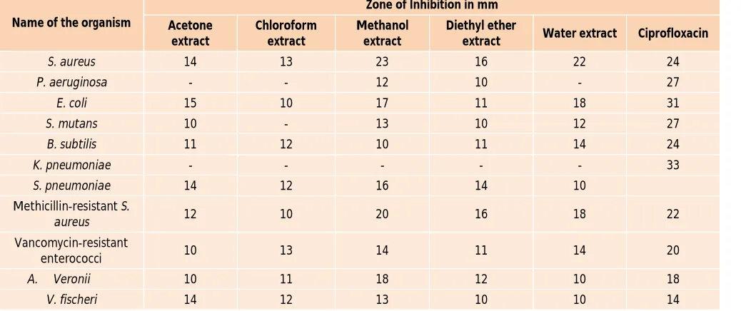
Table 2: The extract of Ocimum sanctum leaves in different solvents showing the zone of inhibition (in mm) against pathogenic bacterial strains
Name of the organism
Zone of Inhibition in mm Acetone
extract
Chloroform extract
Methanol extract
Diethyl ether
extract Water extract Ciprofloxacin
S. aureus 14 13 23 16 22 24
P. aeruginosa - - 12 10 - 27
E. coli 15 10 17 11 18 31
S. mutans 10 - 13 10 12 27
B. subtilis 11 12 10 11 14 24
K. pneumoniae - - - 33
S. pneumoniae 14 12 16 14 10
Methicillin-resistant S.
aureus 12 10 20 16 18 22
Vancomycin-resistant
enterococci 10 13 14 11 14 20
A. Veronii 10 11 18 12 10 18
V. fischeri 14 12 13 10 10 14
Table 3: MIC of OA, UA and BA against different pathogenic bacterial strains
Bacterial strains MIC (µg/ml)
Oleanolic acid Ursolic acid Betulinic acid Ciprofloxacin
S. aureus 16 8 >128 3.4
P. aeruginosa >128 >128 >128 7.4
E. coli >128 >128 >128 2.6
S. mutans 4 2 >128 8.5
B. subtilis 8 4 >128 2.5
K. pneumoniae >256 >256 >256 15
S. pneumoniae 16 8 >128 6.8
MRSA 32 16 >128 6
VRE 8 4 >128 2.5
Aeromonas veronii >128 >128 >128 7.9
Vibrio fischeri 4 8 >128 1.65
Antibacterial activity of different plant extracts
The methanolic extract showed maximum antibacterial activity followed by aqueous and diethyl extracts against
S. aureus, P. aeruginosa, E. coli, S. mutans and K. pneumonia. etc. (Table 2). However, the extracts were
MIC of OA, UA and BA
mutans, S. pneumoniae, MRSA and VRE, suggesting its
good antimicrobial efficacy. However, these were not found effective against gram negative bacteria.
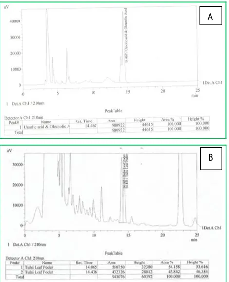
Estimation of Ursolic/Oleanolic acid concentration in Ocimum Sanctum leaf by HPLC
Examining the antibacterial potential of OA and UA, the estimation of the concentration of these bioactive compounds in Ocimum sanctum leaves was calculated as:
Total UA/OA % = AUC of Sample/AUC of Standard × Concn. of Std/Concn. Of Sample × potency of Std
Where,
AUC : Area under curve
Concentration in mg/ml Potency- Purity of Std (56.66%) =943076/1549045 × 0.102/10 × 56.66 = 0.3518%
= 0.352%
Thus, the total concentration of UA/OA present in the leaf of Ocimum sanctum was 0.352% which is quite a considerable amount (fig 1).
Figure 1: Chromatogram of the Ursolic acid/ Oleanolic acid obtained by HPLC (A) standard and (B) the one extracted from Ocimum sanctum leaves.
DISCUSSION
The current scenario of antibiotics is very threatening with significant emergence of resistance among bacterial pathogens against available antibiotics. The present investigation reveals that the Ocimum sanctum could be a
major source for metabolites with greater efficacy against resistant pathogenic bacterial strains. Antimicrobial characteristics of the herbs are due to various chemical compounds including volatile oils, alkaloids, tannins and lipids that are presented in their tissue. Keeping these points in view, the present study was focused to undergo the phytochemical screening and to investigate the antibacterial efficacy of the plant O. sanctum by using different extracts. Phytochemical analysis revealed that
O. sanctum is a rich source of bioactive compounds, as
the tests were found to be positive for tannins, saponins, steroids, triterpenoids, flavonoids, glycosides, carbohydrates and amino acids, however alkaloids were found to be absent in all the extracts. The results are in line with other researchers confirming the presence of these phytochemicals.15, 16
We observed that O. sanctum have a potent antibacterial activity and proved medicinal herb for both its application and efficacy. The methanolic extract came out to be most effective against the selected pathogenic strains followed by aqueous, diethyl ether and chloroform extracts. However, none of them were found to be effective against K. pneumonia. Also gram positive organisms were found to be more susceptible to the extracts than gram negative organisms. In accordance to other studies those also observed the antimicrobial efficacy of O. sanctum leaves in various extracts.17, 18
The HPLC analysis of leaf extracts confirmed the presence of Urocelic acid and Oleanolic acid in high amount (0.352%) which is a considerable amount. The minimum inhibitory concentration of UA, OA and BA was determined. The UA was found most effective against the pathogenic bacteria followed by OA and BA. Thus, observing the potent antibacterial efficacy of UA and OA. These compounds possess potent antibacterial activity, thereby proving their efficacy as a medicinal herb.
CONCLUSION
The present study clearly indicates that Ocimum sanctum is a rich source of phyto-chemical constituents. The antimicrobial efficacy of Ocimum sanctum leaves indicates that the plant possess potent antimicrobial properties as well. As Ocimum is widespread in India, it can be recommended as an easily available and renewal source of antimicrobial agent instead of synthetic chemicals. Furthermore, the concentration of UA and OA bioactive compounds in Ocimum sanctum leaves was also determined by HPLC analysis. The presence of these bioactive Compounds in high concentration justifies that the leaves can be used for various ailments by traditional practitioners. However, isolation of individual phytochemical constituents and subjecting it to pharmacological activity will definitely give fruitful results. A
REFERENCES
1. Lowry FD, Staphylococcus aureus infections, N Engl J Med, 20, 1998, 520–532.
2. Austin DJ, Bonten MJ, Weinstein RA, Slaughter S, Anderson RM, Vancomycin-resistant Enterococci in intensive-care hospital settings: Transmission dynamics, persistence, and the impact of infection control programs, Proc Natl Acad Sci., 96, 1999, 6908-6913
3. Engemann JJ, Carmeli Y, Cosgrove SE, Fowler VG, Bronstein MZ, Trivette SL, Briggs JP, et al., Adverse clinical and economic outcomes attributable to methicillin resistance among patients with Staphylococcus aureus surgical site infection, Clin Infect Dis., 36, 2003, 592-598.
4. Karaman I, Sahin F, Fulluce M, Ogutch S, Sengul M, Adiguzel A, Antimicrobial activity of aqueous and methanol extract of Juniperus oxycedrus L., J. Ethnopharmacol, 2838, 2003, 1-5.
5. Srivastava J, Lambert J, Vietmeyer N, Medicinal plants: An expanding role in development, World Bank Technical, 1996, 320.
6. Joshi B, Sah GP, Basnet BB, Bhatt MR, Sharma D, Subedi K, et al., Phytochemical extraction and antimicrobial properties of different medicinal plants: Ocimum sanctum (Tulsi), Eugenia caryophyllata (Clove), Achyranthes
bidentata (Datiwan) and Azadirachta indica (Neem),
Journal of Microbiology and Antimicrobials, 3, 2011, 1-7.
7. Liu J, Pharmacology of oleanolic acid and ursolic acid, J. Ethnopharmacol, 49, 1995, 57–68.
8. Fontanay S, Grare M, Mayer J, Finance C, Duval RE, Ursolic, oleanolic and betulinic acids: antibacterial spectra and selectivity indexes. J. Ethnopharmacol., 120, 2008, 272– 276.
9. Kim MJ, Kim CS, Ha WH, Kim BH, Lim YK, Park SN, Cho YJ, Kim M, Ko JH, Kwon SS et al., Antimicrobial effects of
oleanolic acid against Streptococcus mutans and
Streptococcus sobrinus isolated from a Korean population,
Int. J. Oral Biol, 35, 2010, 191–195.
10. Kim MJ, Kim CS, Park JY, Lim YK, Park SN, Ahn SJ, Jin DC, Kim TH, Kook JK, Antimicrobial effects of ursolic acid against mutans streptococci isolated from Koreans, Int. J. Oral Biol, 36, 2011, 7–11.
11. Sofowara A, Medicinal plants and Traditional medicine in Africa, Spectrum Books Ltd., Ibadan, Nigeria, 1993, 289.
12. Trease GE, Evans WC, Pharmacognsy, 11th edn., Brailliar Tiridel Can, Macmillan Publishers, 1989.
13. Perez C, Pauli M, Bazerque P, An antibiotic assay by the agar-well diffusion method, Acta Biologiae et Medecine Experimentalis, 15, 1990, 113-115.
14. NCCLS, Methods for dilution antimicrobial susceptibility tests for bacteria that grow aerobically, Approved standard, 5th ed. NCCLS document M7-A5, NCCLS, Wayne, Pa, 2000.
15. Devendran G, Balasubramanian U, Qualitative phytochemical screening and GC-MS analysis of Ocimum
sanctum L. leaves, Asian J Plant Sci and Res, 1(4), 2011,
44-48.
16. Shafqatullah, Khurram M, Asadullah, Khaliqurrehman and Khan F A, Comparative Analyses of Ocimum santum Stem and Leaves for Phytochemicals and Inorganic Constituents, Middle-East J Sci Res, 13 (2), 2013, 236-240.
17. Baskaran X, Preliminary Phytochemical Studies and Antibacterial Activity of Ocimum sanctum L., Ethnobotanical Leaflets, 12, 2008, 1236-1239.
18. Prasad MP, Jayalakshmi k and Rindhe GG, Antibacterial activity of Ocimum species and their phytochemical and antioxidant potential, Int J Micrbiol Res, 4(8), 2012, 302-307.