Scholarship@Western
Scholarship@Western
Electronic Thesis and Dissertation Repository
4-24-2014 12:00 AM
Effects of an Angiotensin II Type 1 Receptor Blocker on
Effects of an Angiotensin II Type 1 Receptor Blocker on
Cardiovascular Calcification
Cardiovascular Calcification
Zachary B. Armstrong
The University of Western Ontario
Supervisor
Dr. Kem A. Rogers
The University of Western Ontario Joint Supervisor Dr. Derek R. Boughner
The University of Western Ontario
Graduate Program in Anatomy and Cell Biology
A thesis submitted in partial fulfillment of the requirements for the degree in Doctor of Philosophy
© Zachary B. Armstrong 2014
Follow this and additional works at: https://ir.lib.uwo.ca/etd
Part of the Cardiovascular Diseases Commons
Recommended Citation Recommended Citation
Armstrong, Zachary B., "Effects of an Angiotensin II Type 1 Receptor Blocker on Cardiovascular Calcification" (2014). Electronic Thesis and Dissertation Repository. 1988.
https://ir.lib.uwo.ca/etd/1988
This Dissertation/Thesis is brought to you for free and open access by Scholarship@Western. It has been accepted for inclusion in Electronic Thesis and Dissertation Repository by an authorized administrator of
(Thesis format: Integrated Article)
by
Zachary Brian Armstrong
Graduate Program in Anatomy & Cell Biology
A thesis submitted in partial fulfillment of the requirements for the degree of
Doctor of Philosophy
The School of Graduate and Postdoctoral Studies The University of Western Ontario
London, Ontario, Canada
Aims: Three types of cardiovascular calcification are commonly found in humans: arterial
calcification, intimal calcification, and calcific aortic valve disease. Very little is known about
the mechanisms driving cardiovascular calcification despite serious clinical implications and
a clear association with morbidity and mortality. Indeed, it is even unclear whether the same
factors are involved in arterial, intimal, and valvular calcification. The objective of this study
was to elucidate the effects of an angiotensin II type 1 receptor blocker (ARB) on the
progression of cardiovascular calcification in male New Zealand White rabbits. Where
appropriate, statins were examined in conjunction and in combination with ARBs.
Methods and Results: In vivo and ex vivo techniques were used to assess overall disease
burden and the extent of calcification including magnetic resonance imaging,
micro-computed tomography, histology, and immunohistochemistry. ARB administration
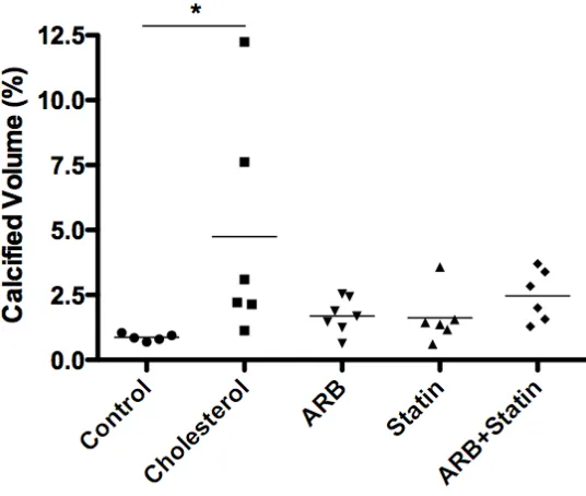
significantly inhibited progression of arterial calcification (2.80 ± 1.17 versus 0.01 ± 0.01 %
calcified tissue in Cholesterol and ARB-treated, respectively; P < 0.05), but not intimal or
valvular calcification. ARB treatment significantly reduced atherosclerotic lesion area when
delivered alone (95.50 ± 1.94 versus 61.61 ± 10.17 % lesion area in Cholesterol and
ARB-treated, respectively; P < 0.05), but not when combined with statin therapy (92.39 ± 3.25 %
in ARB+Statin; P < 0.05 when compared to ARB monotherapy). Finally, ARB-treated
animals had significantly increased valvular calcium.
Conclusions: This study provides evidence that ARBs robustly inhibit arterial calcification
and is the first to suggest ARBs as a novel treatment option for those at risk for
cardiovascular calcification. It also suggests that ARBs may not be beneficial for those at risk
for intimal or valvular calcification. These disparate results suggest that the three types of
cardiovascular calcification are distinct from one another and provides impetus to further
examine the underlying molecular mechanisms at play in these debilitating disease processes.
aortic valve sclerosis, angiotensin receptor blocker, arterial calcification, atherosclerosis,
calcific aortic valve disease, pre-clinical model, renin-angiotensin system, statin, valvular
interstitial cell, vascular smooth muscle cell
Chapter 2:
Entitled “Angiotensin II type 1 receptor blocker inhibits arterial calcification in a pre-clinical
model” was written by Zachary B. Armstrong with inputs from Dr. Derek R. Boughner, Dr.
Maria Drangova, and Dr. Kem A. Rogers, experimental procedures and data analyses were
performed by Zachary B. Armstrong. Dr. Derek R. Boughner, Dr. Maria Drangova, and Dr.
Kem A. Rogers provided intellectual input.
Chapter 3:
Entitled “Potential negative interaction between statin therapy and angiotensin receptor
blockade in atherosclerotic lesion regression” was written by Zachary B. Armstrong with
inputs from Dr. Derek R. Boughner, Dr. Maria Drangova, and Dr. Kem A. Rogers,
experimental procedures and data analyses were performed by Zachary B. Armstrong and
Colin P. Carruthers. Dr. Derek R. Boughner, Dr. Maria Drangova, and Dr. Kem A. Rogers
provided intellectual input.
Chapter 4:
Entitled “Effects of an angiotensin II type 1 receptor blocker on aortic valve sclerosis in a
pre-clinical model” was written by Zachary B. Armstrong with inputs from Dr. Derek R.
Boughner, Dr. Maria Drangova, and Dr. Kem A. Rogers, experimental procedures and data
analyses were performed by Zachary B. Armstrong and Colin P. Carruthers. Dr. Derek R.
Boughner, Dr. Maria Drangova, and Dr. Kem A. Rogers provided intellectual input.
“If I have seen further it is by standing on the shoulders of giants.”
— Isaac Newton, 1676
My time at The University of Western Ontario, both undergraduate and graduate, has been a
wholly enriching experience. For nearly a decade I have lived and breathed Western,
interacting with thousands of students, staff, and faculty. Not once have I been disappointed
by the energy, enthusiasm, and drive of the Western community; it truly is an amazing place.
My time in the Rogers Lab has been equally rewarding. This thesis would not have been
possible without the assistance and encouragement of hardworking technicians, fellow
students, and good friends. In particular, thank you to Kim Thomaes and Colin Carruthers for
making the lab especially interesting.
To my supervisors, Kem and Derek:
Your advice and support has been invaluable to my development as a scientist and as a
person. I have always appreciated your counsel and will continue to do so in the months and
years to come.
Finally, to my parents, Jim and Kim:
You have supported me every step of the way, even when you could no longer see where I
was going. There is no way for me to express my gratitude except to say that, without
question, I would not be where I am today if not for you. Thank you.
...
List of Tables x
...
List of Figures xi
...
List of Appendices xii
...
List of Abbreviations xiii
...
General Introduction 1
...
Renin-Angiotensin System 1
...
Arterial Calcification 5
...
Initiation and Progression of Arterial Calcification 5
...
Osteoblast Transdifferentiation of VSMCs 5
...
RAS and Osteoblast Transdifferentiation 7
...
Clinical Implications 7
...
Pharmaceutical Management 8
...
Intimal Calcification 9
...
Initiation and Progression of Atherosclerosis 9
...
The Vulnerable Plaque 9
...
Initiation and Progression of Calcification in Atherosclerosis 10
...
Calcification and Vulnerability 10
...
Clinical Implications 11
...
Pharmaceutical Management 11
...
Calcific Aortic Valve Disease 13
...
The Aortic Valve 13
...
Calcific Aortic Valve Disease 13
...
Angiotensin II in Aortic Valve Disease 14
...
Clinical Implications 15
...
Study Rationale and Hypothesis 17
...
Objectives and Future Directions 17
. Investigate Effects of an ARB on Arterial Calcification in a Pre-Clinical Model 17
.. Investigate Effects of an ARB on Intimal Calcification in a Pre-Clinical Model 17
Investigate Effects of an ARB on Calcific Aortic Valve Disease in a Pre-Clinical ...
Model 18
...
Conclusions and Future Directions 18
...
References 20
Angiotensin II Type 1 Receptor Blocker Inhibits Arterial Calcification in a Pre-...
Clinical Model 52
...
Methods 53
...
Pre-Clinical Model 53
...
Plasma Chemistry 53
...
Micro-Computed Tomography 54
...
Histological and Immunohistochemical Analysis 54
...
Statistical Analysis 55
...
Results 55
...
Animals and Plasma Chemistry 55
...
Arterial Calcification is Abolished After Treatment with the ARB 55
Calcified Regions Express the Bone-Related Proteins BMP2 and Osteocalcin and ...
Dramatically Increase Expression of the AT1R 57
... Calcified Areas of the Media are not Associated with SMCs or Macrophages 57
...
Discussion 61
...
References 63
...
Methods 70
...
Pre-Clinical Model 70
...
Micro-Computed Tomography 70
...
Quantification of Lesion Area 71
...
Histological and Immunohistochemical Analysis 71
...
Statistical Analysis 71
...
Results 72
Twelve Months Cholesterol Feeding Induces Advanced Atherosclerotic Lesions 72
ARBs Cause Significant Regression of Advanced Atherosclerotic Lesions when ...
Delivered Alone, but not when Combined with Statin Therapy 72
ARBs and Statin Therapy Prevent Progression of Atherosclerotic Plaque
...
Calcification 76
... ARBs Do No Affect the Cellular Composition of Atherosclerotic Plaques 76
...
Discussion 81
...
References 83
Effects of an Angiotensin II Type 1 Receptor Blocker on Aortic Valve Sclerosis in a ...
Pre-Clinical Model 89
...
Methods 90
...
Pre-Clinical Model 90
...
Physiological Data 91
...
Magnetic Resonance Imaging 91
...
Histological and Immunohistochemical Analysis 91
...
Statistical Analysis 92
...
Results 92
...
Dietary Hypercholesterolemia Induced Significant AVS at 12 Months 92
In Vivo Monitoring of AVS Progression did not Reveal Significant Treatment ...
Effects 95
Histological Analysis of Non-Coronary Cusps Revealed Significant
... Morphological Changes in Response to Pharmaceutical Intervention 97
...
Discussion 97
...
References 103
...
General Discussion 108
...
Summary of Results and Conclusions 108
...
Arterial Calcification 108
...
Intimal Calcification 108
...
Calcific Aortic Valve Disease 110
...
Three Distinct Mechanisms 110
...
Clinical Implications 112
...
Limitations 113
...
Conclusions 115
...
Future Directions 116
...
References 118
...
Appendices 128
...
Curriculum Vitae 138
Table 3.1: Atherosclerotic lesion composition analysis of thoracic aorta in rabbits ...79
Figure 1.1: The sinus of valsalva and three types of cardiovascular calcification ...2
Figure 1.2: The canonical pathway of the renin-angiotensin system (RAS)...4
Figure 1.3: The cellular interactions underlying cardiovascular calcification...6
Figure 2.1: Treatment with the angiotensin II type 1 receptor blocker (ARB) inhibited arterial calcification...56
Figure 2.2: Arterial calcification versus micro-calcification of atherosclerotic plaques ...58
Figure 2.3: Characterization of calcified regions indicates an osteoblast-like phenotype...59
Figure 2.4: Calcified regions of the media are not associated with smooth muscle cells or macrophages ...60
Figure 3.1: Significant atherosclerosis progression is achieved after 12 months...73
Figure 3.2: ARB treatment causes regression of established atherosclerosis...75
Figure 3.3: Pharmaceutical intervention slows progression of atherosclerotic calcium...77
Figure 3.4: Atherosclerotic lesion composition is unaffected by pharmaceutical intervention ...78
Figure 3.5: Negative correlation between calcium and macrophage ratio in advanced atherosclerotic lesions...80
Figure 4.1: Significant AVS progression is achieved after 12 months...93
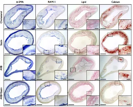
Figure 4.2: In vivo monitoring of AVS does not reveal treatment effect of ARBs, alone or in combination with statin therapy...96
Figure 4.3: Lipid insudation is significantly increased in Statin treated animals, but not Cholesterol, ARB, or ARB+Statin treated animals...98
Figure 4.4: Macrophage infiltration is significantly increased in Cholesterol, ARB, and ARB +Statin treated animals, but not Statin treated animals...99
Figure 4.5: Calcification is significantly increased in ARB and ARB+Statin treated animals, but not Cholesterol or Statin treated animals...100
Figure 5.1: An angiotensin II type 1 receptor blocker effectively inhibits arterial calcification, but not intimal calcification or calcific aortic valve disease...109
Supplementary Results ...128
Arterial Calcification is Detected After Eight Weeks on the Atherogenic Diet ...128
Supplementary Figure 2.1: Arterial calcification, localized to the IEL and medial layer, is present after 8 weeks on the atherogenic diet...129
Supplementary Figure 2.2: Treatment with the angiotensin II type 1 receptor blocker has no effect on systemic disease parameters ...130
Supplementary Figure 2.3: Characterization of calcified regions indicates an osteoblast-like phenotype...131
Supplementary Figure 2.4: Calcified regions of the media are not associated with smooth muscle cells or macrophages ...132
Supplementary Figure 4.1: Experimental design...133
Supplementary Table 4.1: Physiological data...134
License Agreement (Oxford University Press)...135
License Agreement (Elsevier)...136
Animal Protocol Approval...137
ACE, angiotensin converting enzyme
ACEI, angiotensin converting enzyme inhibitor AngI, angiotensin I
AngII, angiotensin II
ARB, angiotensin II type 1 receptor blocker AT1R, angiotensin II type 1 receptor
AVS, aortic valve sclerosis
BMP, bone morphogenetic protein
Cbfα1/Runx2, core-binding factor alpha1/runt-related transcription factor 2 CKD, chronic kidney disease
DM, diabetes mellitus DRI, direct renin inhibitor
HMG-CoA, 3-hydroxy-3-methylglutaryl Co-enzyme A HU, hounsfield units
IEL, internal elastic lamina LDL, low-density lipoprotein MI, myocardial infarction
MRI, magnetic resonance imaging Msx2, muscle segment homeobox 2
NADPH, nicotinamide adenine dinucleotide phosphate NF-κB, nuclear factor kappa B
OCN, osteocalcin
PPARɣ, peroxisomal proliferator-activated receptor-gamma RANKL, receptor activator of NF-κB
RAS, renin-angiotensin system ROS, reactive oxygen species SMA, smooth muscle actin SMC, smooth muscle cell VIC, valvular interstitial cell
VSMC, vascular smooth muscle cell
Chapter 1
1
General Introduction
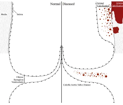
Cardiovascular calcification can be broadly grouped into three categories: arterial
calcification, intimal calcification, and calcific aortic valve disease (Figure 1.1). Each
class of cardiovascular calcification is associated with unique pathologies. While arterial
calcification is mainly associated with diabetes mellitus (DM) and chronic kidney disease
(CKD),1 intimal calcification and calcific aortic valve disease is found more commonly in
the elderly and associated with typical risk factors of atherosclerosis.2-5
Clinical consequences of cardiovascular calcification are numerous and diverse. In
dialysis patients, arterial calcification is responsible for calcific uremic arteriolopathy, a
condition causing necrosis of the skin which has a very high mortality rate.6 Moreover,
arterial calcification is correlated with future cardiovascular events in patients with DM
and is a strong predictor of mortality in patients with CKD.7,8 Intimal calcification is
associated with an increased risk of myocardial infarction (MI)9,10 and may promote
plaque instability.11,12 Patients who have aortic valve disease without concomitant
coronary artery disease have a 50% increased risk of MI and cardiovascular death
compared to patients who have a normal aortic valve,13-16 and calcification of valvular
tissue is recognized as the primary mode of valve failure in both native and bio-prosthetic
valves.17
Despite the myriad of clinical implications and the clear association with morbidity and
mortality, very little is known about the underlying molecular mechanisms leading to
cardiovascular calcification. Indeed, it is even unclear whether the same mechanisms are
at play in arterial, intimal, and valvular calcification. Most importantly, there is no
preventive therapy available to physicians or patients.
1.1 Renin-Angiotensin System
Dysregulation of the Renin-Angiotensin System (RAS) is well known to promote
Figure 1.1: The sinus of valsalva and three types of cardiovascular calcification.
Arterial calcification presents as large masses localized along the internal elastic lamina
and within the tunica media. Intimal calcification typically begins as micro-calcifications
within the cholesterol-rich atherosclerotic plaque, which develops in the cellular tunica
intima. Calcific aortic valve disease also involves micro-calcifications, typically near the
maintaining fluid volume and preventing ischemia during fluid loss. The main vasoactive
agent, angiotensin II (AngII), induces vasoconstriction and sympathetic activation, raises
aldosterone levels, and promotes salt and water retention via the AngII type 1 receptor
(AT1R).21 The canonical RAS cascade is rather simple (Figure 1.2). Angiotensinogen, the
precursor peptide that is produced in the liver, is cleaved by renin, an enzyme produced
by juxtaglomerular cells in the kidney in response to low blood pressure or low sodium
levels.22 Cleavage of angiotensinogen by renin produces angiotensin I (AngI), which
appears to have no biological activity.23 AngI is further cleaved by angiotensin converting
enzyme (ACE), usually in the endothelium,24 to produce AngII.21 However, ACE is not
the only AngII-producing enzyme in the cardiovascular system. Several groups have
shown that mast cell-derived chymase and cathepsin G can also produce AngII in blood
vessels,25 the heart,26,27 and heart valves.28-30 Angiotensin(1–12), which contains two
extra amino acids on the C-terminus of AngI, is the substrate for chymase production of
AngII.31-33 Regardless of its source, AngII exerts its cellular effects via the ATIR. Certain
cell types, including much of the cardiovascular system, also express the AngII type 2
receptor which, when bound by AngII, generally opposes the effects of the AT1R.34
There are a number of clinically available pharmaceuticals that modulate the RAS
(Figure 1.2). The direct renin inhibitor aliskiren first became available in 2007 and
inhibits the rate limiting step of the RAS cascade, the conversion of angiotensinogen to
AngI, thereby reducing the synthesis of all downstream components.21 ACE inhibitors
(ACEIs), as their name implies, directly inhibit ACE and prevent the conversion of AngI
into AngII.24 They also prevent ACE-mediated degradation of bradykinin which elicits
positive cardiovascular effects.24 Unfortunately, chronic administration of ACEIs
sometimes leads to reactivation of AngII, which is linked to poorer outcomes.35 Finally,
AngII type 1 receptor blockers (ARBs) inhibit the binding of AngII to the AT1R and thus
they are able to inhibit the function of AngII regardless its source, which is particularly
important given the capability of mast cell-derived chymase to produce AngII.
Furthermore, the affinity of ARBs for the AT1R provide an opportunity for AngII to bind
Figure 1.2: The canonical pathway of the renin-angiotensin system (RAS).
Angiotensinogen, produced in the liver, is cleaved by renin, an enzyme produced by
juxtaglomerular cells in the kidney, to form angiotensin I (AngI). Angiotensin converting
enzyme (ACE), typically found in the endothelium, cleaves AngI to form angiotensin II
(AngII). AngII acts through the AngII type 1 receptor (AT1R) to elicit vasoconstriction,
sympathetic activation, salt retention, and water retention. Pharmaceutical inhibitors of
the RAS (shown in red) include direct renin inhibitors (DRIs), ACE inhibitors (ACEIs),
generally more tolerable than other antihypertensives, with significantly less cough and
angioedema.18,36,37
1.2 Arterial Calcification
Arterial calcification, also known as medial artery calcification or Mönckeberg sclerosis,
38 involves calcification of the internal elastic lamina and elastic fibers within the medial
layer of the artery resulting in hardening and increased pulse pressure (Figure 1.1).
Commonly associated with advanced age, hypertension, CKD, DM, and osteoporosis,
arterial calcification is closely related to cardiovascular morbidity and mortality.8,39,40
1.2.1 Initiation and Progression of Arterial Calcification
Originally considered a passive, degenerative and, most importantly, irreversible process,
arterial calcification is now considered to be a highly regulated process resembling
natural bone formation (Figure 1.3).41,42 The initiating event appears to be the deposition
of hydroxyapatite-like material on degraded or damaged elastin fibers. Vascular smooth
muscle cells (VSMCs) cultured in a pre-calcified elastin matrix down-regulated their
typical biological markers (α-smooth muscle actin and myosin heavy chain) and
up-regulated markers of osteogenic differentiation including core-binding factor
alpha1/runt-related transcription factor 2 (Cbfα1/Runx2), alkaline phosphatase, and osteocalcin
(OCN).43 When the calcified conditions were removed, VSMCs reverted to their original
phenotype, which suggests some potential for regression. In response to elevated levels of
extracellular phosphate, VSMCs release matrix vesicles that contain calcium and
phosphate ions, especially if local (matrix gla-protein) or circulating (feutin-A) inhibitors
are lost.44 It is likely, therefore, that VSMCs transdifferentiate to an osteoblast-like
phenotype after the local microenvironment is altered.
1.2.2 Osteoblast Transdifferentiation of VSMCs
An elegant fate-mapping study by Speer et al.45 has shown VSMCs are capable of
osteoblast transdifferentiation in calcifying arteries. This transdifferentiation was
associated with downregulation of smooth muscle cell (SMC) markers and upregulation
Figure 1.3: The cellular interactions underlying cardiovascular calcification. Arterial
calcification (top right) involves calcification of the internal elastic lamina and tunica
media, a cellular environment consisting of vascular smooth muscle cells (VSMCs).
Intimal calcification (top centre) occurs within the cholesterol-rich atherosclerotic plaque,
and environment rich with VSMCs, macrophages, lymphocytes, and mast cells. Calcific
aortic valve disease (bottom left) exists primarily in the collagenous fibrosa of the aortic
valve, involving native valvular interstitial cells (VICs) as well as macrophages,
lymphocytes, and mast cells. Solid arrows represent known interactions; dashed arrows
factors, including muscle segment homeobox 2 (Msx2) and osterix, have also been
implicated in VSMC transdifferentiation and in the progression of arterial calcification.
47,48 A number of factors have been shown to induce VSMC differentiation and promote
an osteoblast-like phenotype including fibroblast growth factor-2,49 tumor necrosis
factor-alpha,50 oxidized-low density lipoprotein (LDL),47 and bone morphogenetic
protein (BMP) 2.51 Although tremendous progress has been made in this area, the
molecular mechanisms underlying this process remain to be fully defined.
1.2.3 RAS and Osteoblast Transdifferentiation
There is growing evidence that vasoactive agents are important modulators of vascular
calcification. Naturally existing peptides such as endothelin-1 and urotensin II can
promote arterial calcification, while others – adrenomedullin and C-type natriuretic
peptide – act to inhibit its progression.52-54 Until recently, the role of the RAS and its
vasoactive agent, AngII, had not been thoroughly investigated. AngII plays a number of
roles in vascular pathology, and was thought to exert its effects by inducing nicotinamide
adenine dinucleotide phosphate (NADPH) oxidase and increasing cellular reactive
oxygen species (ROS).19 In turn, ROS stimulate the expression of BMP2 and the
osteoblast transcription factor Cbfα1/Runx2, thereby inducing osteoblast
transdifferentiation.46 The first evidence that AngII affected calcification in VSMCs was
from Jaffe and Mendelsohn,55 who suggested it (along with aldosterone) acted through
the mineralocorticoid receptor to promote fibrosis and calcification.56 More recently, an
in vitro study by Jia et al.57 showed that AngII promoted vascular calcification via Cbfα1/
Runx2 and nuclear factor κB (NF-κB). The receptor activator of NF-κB (RANKL) and
BMP2 axis has been long implicated in arterial calcification, and a subsequent study
suggested that AngII induced vascular calcification in vitro and in vivo via RANKL
activation. In turn, RANKL promoted ACE and AT1R, members of the RAS pathway,
creating a feedback loop.58
1.2.4 Clinical Implications
There is evidence to suggest that arterial calcification, at least in the peripheral arteries,
CKD, arterial calcification tends to be more advanced and is associated with increased
morbidity and mortality.60-63 Indeed, the leading cause of death in CKD patients is
cardiovascular disease.64 In patients with non-insulin-dependent DM, arterial calcification
is a strong independent predictor of total, cardiovascular, and coronary heart
disease-related mortality. It is also associated with increased risk for MI, stroke, and amputation.
7,65 Tibial artery calcium score also predicts the short-term risk of amputation in patients
with peripheral artery disease.66 There is also some evidence to suggest that calcification
of small blood vessels can lead to necrosis and ulceration of the skin.67,68 Clearly, arterial
calcification is prevalent in Western society and in dire need of preventive therapy.
1.2.5 Pharmaceutical Management
Blockade of the RAS has been shown to reduce morbidity and mortality in patients with
hypertension, atherosclerosis, heart failure, stroke, DM, and CKD,69-73 often independent
of changes in blood pressure.69,71,73,74 To date, there have been no clinical studies
examining the role of RAS blockade on arterial calcification. However, pre-clinical
studies do provide some encouraging evidence. Recent studies have shown that ACEIs –
specifically, perindopril and captopril – can prevent the progression of arterial
calcification in rat models of CKD56,75 and hypertension.76 An older study in a 5/6
nephrectomized rat model found that enalapril could not suppress arterial calcification
but did decrease mortality.77 The ARB irbesartan has also been shown to prevent arterial
calcification in a rat model of hypertension as long as therapy is initiated alongside insult,
which in this case was warfarin and vitamin K1.78 Irbesartan is also capable of blocking
AngII-induced expression of BMP2 in human endothelial cells.79
The effects of other pharmaceuticals have also been examined in relation to arterial
calcification, with some success. The calcium channel blocker amlodipine, another
antihypertensive, was able to induce regression of arterial calcification in a pre-clinical
model.80 The endothelin receptor antagonist darusentan may also be able to induce
regression.78,81 Interestingly, there is some evidence to suggest that osteoporosis
calcification.82 Ultimately, however, no pharmaceuticals are clinically indicated for the
specific prevention of arterial calcification.
1.3 Intimal Calcification
Intimal calcification occurs within the cholesterol-rich lesions characteristic of
atherosclerosis which can result in MI, stroke, or limb ischemia (Figure 1.1). Commonly
associated with old age, male sex, hypertension, smoking, and hypercholesterolemia,83
intimal calcification is a reliable marker of plaque burden84,85 and may contribute to
plaque instability.86
1.3.1 Initiation and Progression of Atherosclerosis
Atherosclerosis is a chronic, progressive disease of the vascular system. In areas
predisposed to atherosclerosis, variations in hemodynamic forces can result in adaptive
intimal thickening defined as an increase in SMCs and extracellular matrix lacking any
inflammatory infiltrate.87 The initial atherosclerotic plaque (Type I or II), appearing in
those as young as age 2,88 is described as a fatty streak or a visible accumulation of
lipid-laden macrophages (foam cells) and is capable of regressing naturally.89 The advanced
atherosclerotic plaque (Type IV, V, or VI), or fibroatheroma, is characterized by a
lipid-rich necrotic core covered by a SMC-lipid-rich fibrous cap.90 The rupture of these advanced
plaques can lead to either downstream arterial occlusion or localized thrombus formation
and subsequent ischemic death of the tissue supplied.
1.3.2 The Vulnerable Plaque
The difference between a stable and a vulnerable plaque is primarily related to the
thickness and composition of the fibrous cap. The concept of a vulnerable plaque, or a
plaque that is prone to rupture, was first introduced by Muller and Tofler.91 The
vulnerable plaque was described as having a lipid-rich necrotic core and a generally thin
fibrous cap,92 until Burke et al.93 refined the classification to those plaques with a fibrous
cap less than 65 µm thick. They also noted that the fibrous cap of vulnerable plaques
often had macrophage infiltration and a loss of SMCs, characteristics which would
1.3.3 Initiation and Progression of Calcification in Atherosclerosis
Calcification of the atherosclerotic plaque begins as micro-calcifications, typically less
than 15 µm in diameter.95,96 Micro-calcifications are present in all types of lesions,
including fatty streaks, and their abundance increases as atherosclerosis advances.97,98
They also precede the appearance of the bone-promoting proteins BMP2 and OCN which
suggests that, at least initially, the calcification process in atherosclerotic plaques is
distinct from typical bone formation.98 Micro-calcifications may be initiated by matrix
vesicles of apoptotic SMCs99-101 or macrophages (Figure 1.3).95,102 Once initiated, there is
evidence to suggest a highly regulated process which involves several bone-related
proteins that promote (BMP2, BMP4, osteopontin, and osteonectin) and inhibit (matrix
gla-protein and bone sialoprotein) atherosclerotic calcification.103-106 Furthermore, the
mineral composition of calcific atherosclerotic plaques is chemically similar to that
observed in bone.107-111
1.3.4 Calcification and Vulnerability
There is controversy as to whether plaque calcification stabilizes advanced lesions or
promotes rupture. Generally, the presence of calcification is correlated with the incidence
of cardiovascular disease,112 and is associated with increased atherosclerosis progression.
85,113 While some have reported an association between calcification and stability,114,115
others suggest calcification is a marker of vulnerability86,116 and may even promote
rupture.94 These disparate reports suggest that the localization of calcification, rather than
the volume, may be an important indicator of plaque vulnerability. Indeed, an
intravascular ultrasound study suggested that calcification near the base of the lesion
increased stability,117 and a mathematical modeling study suggested that calcification in
the fibrous cap may as well.118 However, several studies have suggested that
micro-calcifications within the fibrous cap directly promote plaque rupture.95,96,119 Taken
together, these reports suggest that the relationship between calcification and plaque
rupture is biphasic. Abedin et al.120 argue that the principal site of stress is the interface
between hard, calcified areas and soft, cellular areas within the plaque; therefore, stress
areas enlarge and coalesce, thereby reducing interface area and overall stress. This
argument was validated by Motoyama et al.,121 who observed spotty calcification more
frequently in ruptured plaques from acute coronary syndrome patients than stable ones
from angina pectoris patients. Conversely, they observed large calcification more
frequently in stable plaques.122 Clearly, a deeper understanding of the processes
underlying atherosclerotic calcification and its relationship to vulnerability is required.
1.3.5 Clinical Implications
For almost a century, cardiovascular disease has accounted for more deaths than any
other major cause in the United States, and a majority of these deaths are a result of
atherosclerosis.83 Coronary arterial calcification is present in 52.9% of men and 32.0% of
women over the age of 45, and its severity is predictive of overall cardiovascular risk.123
Obviously, the acute clinical implications of atherosclerosis depend on the location of
individual vulnerable plaques. Rupture of a plaque in the coronary arteries or the carotid
can lead to MI or stroke, respectively. Furthermore, the implications of intimal
calcification rely on its relationship to plaque vulnerability. This is an area that requires
considerably more study, including a reliable mechanism for blockade of calcification.
1.3.6 Pharmaceutical Management
Current clinical guidelines recommend an LDL-cholesterol goal of less than 100 mg/dL
in patients at risk for cardiovascular disease,124,125 and statins play a role in achieving that
goal. Statins, or 3-hydroxy-3-methylglutaryl Co-enzyme A (HMG-CoA) reductase
inhibitors, inhibit the rate limiting enzyme in endogenous cholesterol production and thus
are frontline drugs for the management of hypercholesterolemia. It is well known that
statins reduce the incidence of cardiovascular events,126 including MI and stroke,127 and
studies have shown that statins alter the progression of subclinical atherosclerosis. Statins
are effective at reducing atherosclerotic lesion volume,128-132 reducing the size of the lipid
core,133 and reducing progression of carotid intima-media thickness.134,135 Pre-clinical
models have provided more information about the specific effects of statins, including
pleiotropic effects (those not related to cholesterol-lowering). Statins have been shown to
and vascular cell adhesion molecule-1,136,137 down-regulate cyclooxygenase 2,138 improve
nitric oxide bioavailability,139 suppress oxidation of LDL140, and reduce the overall
inflammatory burden within atherosclerotic plaques.141 The effects of statins on intimal
calcification have not been thoroughly examined, although Kizu et al.142 showed that
cerivastatin could inhibit calcification of VSMCs in vitro. In contrast, a recent clinical
study found progression of coronary calcium was significantly higher in frequent statin
users versus those who used statins infrequently.143
Inhibition of the RAS may also have benefits in atherosclerosis. Three of the four
prospective clinical trials examining the effect of ARBs on cardiovascular outcomes
showed little144 or no145,146 benefit; however, the OLIVUS (Impact of OLmesartan on
progression of coronary atherosclerosis: evaluation by IntraVascular UltraSound) trial
found a lower rate of coronary atherosclerosis progression and decreased incidence of
major adverse cardio- and cerebrovascular events in patients treated with olmesartan.
147,148 More recently, Zhao et al.149 showed that telmisartan reduced plaque size,
macrophage infiltration, lipid deposition, and apoptosis in atherosclerotic plaques in a
pre-clinical model. Three clinical trials examining ACEIs showed improved
cardiovascular outcomes;70,150,151 however, two more recent trials showed no benefit.
152,153 Recently, meta-analysis suggested that ARBs and ACEIs reduce the incidence of
cardiovascular death, non-fatal MI, and non-fatal stroke, even in normotensive
atherosclerosis patients.154 Other clinical studies have shown that ARBs reduce plaque
volume155 and decrease inflammation.156 A pre-clinical study in monkeys also observed a
regressive effect of ARBs, albeit on fatty streaks.157 Interestingly, aliskiren, a renin
inhibitor, was associated with increased progression of atherosclerosis.158
There have also been a number of studies examining various combinations of statin
therapy and RAS blockade. Several pre-clinical studies examining statins in combination
with ARBs have found an additive reduction of atherosclerosis burden in mice159-161 and
rabbits.162,163 Another pre-clinical study examined the effects of simvastatin and a
peroxisomal proliferator-activated receptor-gamma (PPARɣ) agonist and showed that the
regression;164 which is of particular interest since some ARBs have been shown to have
PPARɣ agonist activity.165-167
It remains unclear whether aggressive medical management of intimal calcification will
provide a reduction in cardiovascular events. However, improving our understanding of
the mechanisms underlying calcification could provide an opportunity to prevent it
which, in turn, could help reveal its relationship to plaque vulnerability.
1.4 Calcific Aortic Valve Disease
Calcific aortic valve disease, encompassing early aortic valve sclerosis (AVS) and clinical
aortic stenosis, results in calcification of the aortic valve cusps and hardened, non-pliable
valve tissue, decreased orifice area, and increased aortic jet velocity (Figure 1.1).168
Commonly associated with old age, male sex, hypertension, smoking, increased plasma
LDL, increased plasma lipoprotein (a), and DM,13,20,169-178 calcific aortic valve disease is
associated with an increased risk of MI and cardiovascular death.13-16
1.4.1 The Aortic Valve
The normal human aortic valve has a three-layered structure measuring 1 mm in
thickness169,179 with the fibrosal layer on the aortic side, the ventricularis on the
ventricular side, and the spongiosa centrally (Figure 1.1). The fibrosa primarily contains
highly organized collagen bundles between which lie valvular interstitial cells (VICs).
The ventricularis is less organized and generally less cellular; it contains elastin sheets
and some collagen. In contrast, the spongiosa is rich in cells and proteoglycans, but
contains little collagen.
1.4.2 Calcific Aortic Valve Disease
A chronic progressive disease, AVS is characterized by five factors: endothelial
dysfunction, lipid deposition, chronic inflammation, activation of a local RAS and
eventually, tissue calcification. The development of AVS results in distinctive anatomical
changes first described by Otto et al.169 as sub-endothelial thickening and fibrosis,
lipids. In the same year, Olsson et al.171 independently reported large numbers of
activated T-lymphocytes located in subendothelial areas in close proximity to calcium
deposits.
The initiating event in AVS appears to be endothelial cell activation in areas of
mechanical stress, thereby predisposing the tissue to infiltration by plasma lipoproteins,
180,181 a course similar to the development of atherosclerosis (Figure 1.3). In the aortic
valve, lipid deposition tends to be localized to the fibrosa where it co-localizes with
components of atherogenic apolipoproteins.170,182 Over time, lipid particles become
oxidized and are taken up by infiltrating macrophages to form foam cells.169,171 Oxidized
LDL is highly cytotoxic for many cells including endothelial cells and VICs; in addition,
native LDL has been shown to co-localize with ACE both in human plasma and aortic
valve lesions,20 creating an environment for the local production of AngII. More recently,
Helske et al.29,30 described the participation of two additional AngII-forming enzymes:
mast cell-derived chymase and cathepsin G.
1.4.3 Angiotensin II in Aortic Valve Disease
The pro-inflammatory and pro-fibrotic effects of AngII are well understood183 and are
often cited as its main contribution to AVS.184-186 It is likely, however, that AngII is
affecting all aspects of aortic valve disease, including lipid retention, inflammation,
endothelial integrity, and calcification. In cardiac fibroblasts and VSMCs, AngII has been
shown to induce the production of biglycan, a proteoglycan with enhanced LDL binding
properties.187,188 Production of biglycan in aortic valves would promote retention of LDL
and associated ACE, creating a positive feedback loop for AngII production. Blockade of
AngII improves endothelial integrity in a rabbit model of AVS, and also up-regulated
endothelial nitric oxide synthase, an enzyme that is generally considered to be
cardioprotective.189 Most importantly, AngII has been shown to transactivate NADPH
oxidase in VSMCs, leading to production of ROS. In turn, ROS have been shown to
promote vascular calcification,190 and specifically localize around calcifying foci in
1.4.4 Clinical Implications
The prevalence of AVS is estimated to be 25–30% in those over age 65 and up to 40% in
those over age 75,4,192 typically progressing to severe aortic stenosis in 6% of patients
over a period of 7 years.193 Even early AVS without concomitant coronary artery disease
is predictive of a 50% increase in cardiovascular death.13 Considering that the primary
cause of valve failure is extensive calcium deposits, it is no surprise that calcified volume
correlates with the severity of AVS194 and is associated with the incidence of coronary
events and all-cause mortality.195-197
1.4.5 Pharmaceutical Management
Once AVS is initiated, increased physical activity or a change in diet is not sufficient to
alter the disease process.198 However, an excellent environment exists for preventive,
pharmaceutical management. The average time interval from subjective diagnosis of AVS
to the development of severe stenosis is eight years,193,199 and it is increasingly clear that
intervention should start as early as possible.200
The overlap in clinical factors associated with AVS and atherosclerosis had suggested a
shared disease process,201 thus the demonstrable benefits of statins on atherosclerosis
provided support for their use in AVS. Several pre-clinical studies also promoted the
effectiveness of statins for inhibiting hypercholesterolemia-induced cellular
proliferation202 and calcification.203,204 In all, twelve clinical studies have evaluated the
role of statins on AVS progression but the results have been conflicting.205-216 Six small
retrospective studies, with a mean follow up of 6–44 months, showed a lower rate of AVS
progression in patients treated with statins.205-209,211 Conversely, a larger retrospective
study with a mean follow up of 66 months did not observe slower progression.210 Five
prospective trials observed little213 or no212,214-216 effect of statins on the progression of
AVS. The SEAS (intensive lipid lowering with Simvastatin and Ezetimibe in Aortic
Stenosis) trial was the largest to date and the only one to measure clinical outcomes.
Patients treated with simvastatin and ezetimibe showed a decrease in ischemic cardiac
217 confirmed the ineffectiveness of statins in a pre-clinical model, observing a reduction
of inflammation but retention of lipids and the continuation of calcium deposition.
Compared to statins, blockade of the RAS has received relatively little clinical
examination as a preventive therapy for AVS. A retrospective study found that ACEIs
could not slow the increase in aortic jet velocity associated with AVS;209 however, a
separate retrospective study, specifically examining aortic valve calcium, showed a
significant reduction in progression.218 The ACEI ramipril was effective in reducing
progression of AVS,219 and the ARB olmesartan has been shown to reduce atherosclerotic
changes and endothelial disruption in short-term animal models;189 however, neither
study examined effects on calcification. More recently, a clinical study of excised aortic
valves observed lower remodeling scores and decreased weight in those valves from
patients who had been treated with ARBs.220,221 A retrospective study compared the
effects of ARBs and ACEIs on AVS and found that only ARBs were effective at reducing
progression.222 A small clinical study of the aldosterone receptor blocker epleronone
showed no effect on AVS,223 but a pre-clinical study examining the early stages of the
disease suggested some effects, including a small, qualitative reduction in
micro-calcification.224
There has been limited examination of other potential therapies for AVS. In an
observational clinical trial, a cohort of patients receiving osteoporosis therapy –
bisphosphonates, calcitonin, or estrogen receptor modulators – had a lower rate of AVS
progression.225 A pre-clinical trial also had success with bisphosphonates, suggesting they
are able to robustly inhibit valve calcification.82 An experimental recombinant
apolipoprotein, Apo-A1Milano, was successful in reversing AVS in a pre-clinical model,
including a qualitative reduction of micro-calcification.226
To date, clinical guidelines provide no recommendation for the managements of AVS,
suggesting additional trials with patients earlier in the disease continuum and with longer
follow-up periods are required.168 In addition, the multitude of mechanisms involved in
1.5 Study Rationale and Hypothesis
In summary, little is known about the mechanisms driving cardiovascular calcification
despite serious clinical implications and a clear association with morbidity and mortality.
Indeed, it is even unclear whether the same mechanisms are at play in arterial, intimal,
and valvular calcification. There is some evidence to suggest that the RAS and its
vasoactive agent, AngII, are involved in promoting cardiovascular calcification; however,
ARBs have not been robustly examined as potential therapies. As such, we set out to test
the hypothesis that the ARB olmesartan medoxomil inhibits progression of established
cardiovascular calcification in vivo.
1.6 Objectives and Future Directions
1.6.1 Investigate Effects of an ARB on Arterial Calcification in a
Pre-Clinical Model
In Chapter 2, a version of which has been published in Cardiovascular Research,228 we
use New Zealand White rabbits to investigate the effects of the ARB olmesartan
medoxomil on established arterial calcification. This pre-clinical model has been used
successfully by others to investigate potential pharmaceutical therapies.229 Using
micro-computed tomography (micro-CT), histology, and immunohistochemistry, we are the first
to show robust inhibition of arterial calcification by an ARB. Calcified areas in our
animals displayed a down-regulation of α-smooth muscle actin, a smooth muscle cell
marker; up-regulation of BMP2 and the ATIR; and expression of the osteoblast-specific
protein OCN.
1.6.2 Investigate Effects of an ARB on Intimal Calcification in a
Pre-Clinical Model
In Chapter 3, a version of which has been submitted for publication in the Canadian
Journal of Cardiology, we use New Zealand White rabbits to investigate the effects of the
ARB olmesartan medoxomil, alone or in combination with atorvastatin, on established
atherosclerosis and intimal calcification. The long-term, low-level cholesterol rabbit
shown to produce significant atherosclerosis over a period of six months with 58% of the
aortic surface covered by atheroma and up to 75% of those lesions considered to be
advanced.230,231 Previous examination of these lesions revealed their similarity to human
atherosclerosis; VSMCs and collagen extracellular matrix formed a fibrous cap over a
core of lipids, cholesterol crystals, and necrotic debris.231 Using classical lesion area
analysis, micro-CT, histology, and immunohistochemistry, we show a significant
reduction of atherosclerotic burden in animals treated with ARB monotherapy, but not in
combination therapy (ARB+Statin). In addition, both ARBs and statins may have slowed
progression of intimal calcification.
1.6.3 Investigate Effects of an ARB on Calcific Aortic Valve Disease in
a Pre-Clinical Model
In Chapter 4, a version of which has been accepted for publication in the Canadian
Journal of Cardiology,232 we use the same animals as in Chapter 3 but examine the
effects of the ARB olmesartan medoxomil, alone or in combination with atorvastatin, on
established calcific aortic valve disease. Previously, Cimini et al.233 examined the
development of calcific aortic valve disease in our long-term, low-level cholesterol rabbit
model.230,231 Rabbits have been shown to develop aortic valve thickening, inflammation,
and lipid deposition over a period of ten months. While calcification was not observed in
the initial report, it was present in aortic valves after 30 months of cholesterol feeding.217
We did not observe any demonstrable treatment effects using in vivo magnetic resonance
imaging. However, ex vivo histological and immunohisochemical analyses revealed
structural changes to the aortic valve. In particular, ARB-treated animals had significantly
increased levels of valvular calcification.
1.6.4 Conclusions and Future Directions
Finally, in Chapter 5, I summarize the results and conclusions of my work. Namely, the
disparate effects of the ARB in treating arterial, intimal, and valvular calcification suggest
distinct cellular and molecular mechanisms are at play in each disease process. More
work needs to be done to fully understand these differences but the robust inhibitory
prospective, randomized trial in patients with chronic kidney disease, a group prone to
rapid development of arterial calcification. Such a trial would be valuable, given there is
currently no preventive therapy for arterial calcification. In all, the studies herein have
advanced our understanding of cardiovascular calcification and, together with future
1.7
References
1. Chen NX, Moe SM. Arterial calcification in diabetes. Curr Diab Rep 2003;3:28–
32.
2. Hisar I, Ileri M, Yetkin E, Tandoğan I, Cehreli S, Atak R, Senen K, Demirkan D.
Aortic valve calcification: its significance and limitation as a marker for coronary
artery disease. Angiology 2002;53:165–9.
3. Ortlepp JR, Schmitz F, Bozoglu T, Hanrath P, Hoffmann R. Cardiovascular risk
factors in patients with aortic stenosis predict prevalence of coronary artery
disease but not of aortic stenosis: an angiographic pair matched case-control
study. Heart 2003;89:1019–22.
4. Stewart BF, Siscovick D, Lind BK, Gardin JM, Gottdiener JS, Smith VE,
Kitzman DW, Otto CM. Clinical factors associated with calcific aortic valve
disease. Cardiovascular Health Study. J Am Coll Cardiol 1997;29:630–4.
5. Yamamoto H, Shavelle D, Takasu J, Lu B, Mao SS, Fischer H, Budoff MJ.
Valvular and thoracic aortic calcium as a marker of the extent and severity of
angiographic coronary artery disease. Am Heart J 2003;146:153–9.
6. Coates T, Kirkland GS, Dymock RB, Murphy BF, Brealey JK, Mathew TH,
Disney AP. Cutaneous necrosis from calcific uremic arteriolopathy. Am J Kidney
Dis 1998;32:384–91.
7. Lehto S, Niskanen L, Suhonen M, Rönnemaa T, Laakso M. Medial artery
calcification. A neglected harbinger of cardiovascular complications in
non-insulin-dependent diabetes mellitus. Arterioscler Thromb Vasc Biol 1996;16:978–
83.
8. Blacher J, Guerin AP, Pannier B, Marchais SJ, London GM. Arterial
calcifications, arterial stiffness, and cardiovascular risk in end-stage renal disease.
9. Beadenkopf WG, Daoud AS, Love BM. Calcification in the coronary arteries and
its relationship to arteriosclerosis and myocardial infarction. Am J Roentgenol
Radium Ther Nucl Med 1964;92:865–71.
10. Loecker TH, Schwartz RS, Cotta CW, Hickman JR. Fluoroscopic coronary artery
calcification and associated coronary disease in asymptomatic young men. J Am
Coll Cardiol 1992;19:1167–72.
11. Fitzgerald PJ, Ports TA, Yock PG. Contribution of localized calcium deposits to
dissection after angioplasty. An observational study using intravascular
ultrasound. Circulation 1992;86:64–70.
12. Burke AP, Taylor A, Farb A, Malcom GT, Virmani R. Coronary calcification:
insights from sudden coronary death victims. Z Kardiol 2000;89 Suppl 2:49–53.
13. Otto CM, Lind BK, Kitzman DW, Gersh BJ, Siscovick DS. Association of
aortic-valve sclerosis with cardiovascular mortality and morbidity in the elderly. N Engl
J Med 1999;341:142–7.
14. Olsen MH, Wachtell K, Bella JN, Gerdts E, Palmieri V, Nieminen MS, Smith G,
Ibsen H, Devereux RB, LIFE substudy. Aortic valve sclerosis relates to
cardiovascular events in patients with hypertension (a LIFE substudy). Am J
Cardiol 2005;95:132–6.
15. Taylor HA, Clark BL, Garrison RJ, Andrew ME, Han H, Fox ER, Arnett DK,
Samdarshi T, Jones DW. Relation of aortic valve sclerosis to risk of coronary
heart disease in African-Americans. Am J Cardiol 2005;95:401–4.
16. Walsh CR, Larson MG, Kupka MJ, Levy D, Vasan RS, Benjamin EJ, Manning
WJ, Clouse ME, O'Donnell CJ. Association of aortic valve calcium detected by
electron beam computed tomography with echocardiographic aortic valve disease
and with calcium deposits in the coronary arteries and thoracic aorta. Am J
17. O'Keefe JH, Lavie CJ, Nishimura RA, Edwards WD. Degenerative aortic
stenosis. One effect of the graying of America. Postgrad Med 1991;89:143–6–
151–4.
18. Burnier M, Vuignier Y, Wuerzner G. State-of-the-art treatment of hypertension:
established and new drugs. Eur Heart J 2014;35:557–62.
19. Bataller R, Schwabe RF, Choi YH, Yang L, Paik YH, Lindquist J, Qian T,
Schoonhoven R, Hagedorn CH, Lemasters JJ, Brenner DA. NADPH oxidase
signal transduces angiotensin II in hepatic stellate cells and is critical in hepatic
fibrosis. J Clin Invest 2003;112:1383–94.
20. O'Brien KD, Shavelle DM, Caulfield MT, McDonald TO, Olin-Lewis K, Otto
CM, Probstfield JL. Association of angiotensin-converting enzyme with
low-density lipoprotein in aortic valvular lesions and in human plasma. Circulation
2002;106:2224–30.
21. Robles NR, Cerezo I, Hernandez-Gallego R. Renin-angiotensin system blocking
drugs. J Cardiovasc Pharmacol Ther 2014;19:14–33.
22. Schmid J, Oelbe M, Saftig P, Schwake M, Schweda F. Parallel regulation of renin
and lysosomal integral membrane protein 2 in renin-producing cells: further
evidence for a lysosomal nature of renin secretory vesicles. Pflugers Arch
2013;465:895–905.
23. Keidar S, Kaplan M, Gamliel-Lazarovich A. ACE2 of the heart: From angiotensin
I to angiotensin (1-7). Cardiovasc Res 2007;73:463–9.
24. Fleming I, Kohlstedt K, Busse R. New fACEs to the renin-angiotensin system.
Physiology (Bethesda) 2005;20:91–5.
25. Company C, Piqueras L, Naim Abu Nabah Y, Escudero P, Blanes JI, Jose PJ,
endogenous angiotensin II generation and leucocyte recruitment in vivo.
Cardiovasc Res 2011;92:48–56.
26. Ferrario CM, Varagic J, Habibi J, Nagata S, Kato J, Chappell MC, Trask AJ,
Kitamura K, Whaley-Connell A, Sowers JR. Differential regulation of
angiotensin-(1-12) in plasma and cardiac tissue in response to bilateral
nephrectomy. Am J Physiol Heart Circ Physiol 2009;296:H1184–92.
27. Prosser HCG, Forster ME, Richards AM, Pemberton CJ. Cardiac chymase
converts rat proAngiotensin-12 (PA12) to angiotensin II: effects of PA12 upon
cardiac haemodynamics. Cardiovasc Res 2009;82:40–50.
28. Katwa LC, Tyagi SC, Campbell SE, Lee SJ, Cicila GT, Weber KT. Valvular
interstitial cells express angiotensinogen and cathepsin D, and generate
angiotensin peptides. Int J Biochem Cell Biol 1996;28:807–21.
29. Helske S, Lindstedt KA, Laine M, Mäyränpää M, Werkkala K, Lommi J, Turto H,
Kupari M, Kovanen PT. Induction of local angiotensin II-producing systems in
stenotic aortic valves. J Am Coll Cardiol 2004;44:1859–66.
30. Helske S, Syväranta S, Kupari M, Lappalainen J, Laine M, Lommi J, Turto H,
Mäyränpää M, Werkkala K, Kovanen PT, Lindstedt KA. Possible role for mast
cell-derived cathepsin G in the adverse remodelling of stenotic aortic valves. Eur
Heart J 2006;27:1495–504.
31. Ahmad S, Simmons T, Varagic J, Moniwa N, Chappell MC, Ferrario CM.
Chymase-dependent generation of angiotensin II from angiotensin-(1-12) in
human atrial tissue. PLoS ONE 2011;6:e28501.
32. Ahmad S, Wei C-C, Tallaj J, Dell'italia LJ, Moniwa N, Varagic J, Ferrario CM.
Chymase mediates angiotensin-(1-12) metabolism in normal human hearts. J Am
33. Ahmad S, Varagic J, Westwood BM, Chappell MC, Ferrario CM. Uptake and
metabolism of the novel peptide angiotensin-(1-12) by neonatal cardiac myocytes.
PLoS ONE 2011;6:e15759.
34. Steckelings UM, Kaschina E, Unger T. The AT2 receptor--a matter of love and
hate. Peptides 2005;26:1401–9.
35. Swedberg K, Eneroth P, Kjekshus J, Wilhelmsen L. Hormones regulating
cardiovascular function in patients with severe congestive heart failure and their
relation to mortality. CONSENSUS Trial Study Group. Circulation
1990;82:1730–6.
36. Greathouse MK, Weir MR. The role of ARBs alone or with HCTZ in the
treatment of hypertension and prevention of cardiovascular and renal
complications. Postgrad Med 2012;124:40–52.
37. Makani H, Messerli FH, Romero J, Wever-Pinzon O, Korniyenko A, Berrios RS,
Bangalore S. Meta-analysis of randomized trials of angioedema as an adverse
event of renin-angiotensin system inhibitors. Am J Cardiol 2012;110:383–91.
38. Mönckeberg JG. Über die reine Mediaverkalkung der Extremitätenarterien und
ihr Verhalten zur Arteriosklerose. Virchows Arch Path Anat 1903;171:141–67.
39. Jeffcoate WJ, Rasmussen LM, Hofbauer LC, Game FL. Medial arterial
calcification in diabetes and its relationship to neuropathy. Diabetologia
2009;52:2478–88.
40. Takasu J, Budoff MJ, Katz R, Rivera JJ, O'Brien KD, Shavelle DM, Probstfield
JL, O'Leary D, Nasir K. Relationship between common carotid intima-media
thickness and thoracic aortic calcification: the Multi-Ethnic Study of
41. Virchow R. Cellular pathology. As based upon physiological and pathological
histology. Lecture XVI--Atheromatous affection of arteries. 1858. Nutr Rev
1989;47:23–5.
42. Shroff RC, Shanahan CM. The vascular biology of calcification. Semin Dial
2007;20:103–9.
43. Lei Y, Sinha A, Nosoudi N, Grover A, Vyavahare N. Hydroxyapatite and calcified
elastin induce osteoblast-like differentiation in rat aortic smooth muscle cells. Exp
Cell Res 2014.
44. Reynolds JL, Joannides AJ, Skepper JN, McNair R, Schurgers LJ, Proudfoot D,
Jahnen-Dechent W, Weissberg PL, Shanahan CM. Human vascular smooth
muscle cells undergo vesicle-mediated calcification in response to changes in
extracellular calcium and phosphate concentrations: a potential mechanism for
accelerated vascular calcification in ESRD. J Am Soc Nephrol 2004;15:2857–67.
45. Speer MY, Yang H-Y, Brabb T, Leaf E, Look A, Lin W-L, Frutkin A, Dichek D,
Giachelli CM. Smooth muscle cells give rise to osteochondrogenic precursors and
chondrocytes in calcifying arteries. Circ Res 2009;104:733–41.
46. Byon CH, Javed A, Dai Q, Kappes JC, Clemens TL, Darley-Usmar VM,
McDonald JM, Chen Y. Oxidative stress induces vascular calcification through
modulation of the osteogenic transcription factor Runx2 by AKT signaling. J Biol
Chem 2008;283:15319–27.
47. Taylor J, Butcher M, Zeadin M, Politano A, Shaughnessy SG. Oxidized
low-density lipoprotein promotes osteoblast differentiation in primary cultures of
vascular smooth muscle cells by up-regulating Osterix expression in an
48. Shao J-S, Cheng S-L, Pingsterhaus JM, Charlton-Kachigian N, Loewy AP, Towler
DA. Msx2 promotes cardiovascular calcification by activating paracrine Wnt
signals. J Clin Invest 2005;115:1210–20.
49. Nakahara T, Sato H, Shimizu T, Tanaka T, Matsui H, Kawai-Kowase K, Sato M,
Iso T, Arai M, Kurabayashi M. Fibroblast growth factor-2 induces osteogenic
differentiation through a Runx2 activation in vascular smooth muscle cells.
Biochem Biophys Res Commun 2010;394:243–8.
50. Lee H-L, Woo KM, Ryoo H-M, Baek J-H. Tumor necrosis factor-alpha increases
alkaline phosphatase expression in vascular smooth muscle cells via MSX2
induction. Biochem Biophys Res Commun 2010;391:1087–92.
51. Balderman JAF, Lee H-Y, Mahoney CE, Handy DE, White K, Annis S, Lebeche
D, Hajjar RJ, Loscalzo J, Leopold JA. Bone morphogenetic protein-2 decreases
microRNA-30b and microRNA-30c to promote vascular smooth muscle cell
calcification. J Am Heart Assoc 2012;1:e003905.
52. Wu SY, Zhang BH, Pan CS, Jiang HF, Pang YZ, Tang CS, Qi YF. Endothelin-1 is
a potent regulator in vivo in vascular calcification and in vitro in calcification of
vascular smooth muscle cells. Peptides 2003;24:1149–56.
53. Huang Z, Li J, Jiang Z, Qi Y, Tang C, Du J. Effects of adrenomedullin, C-type
natriuretic peptide, and parathyroid hormone-related peptide on calcification in
cultured rat vascular smooth muscle cells. J Cardiovasc Pharmacol 2003;42:89–
97.
54. Tintut Y, Demer LL. Recent advances in multifactorial regulation of vascular
calcification. Curr Opin Lipidol 2001;12:555–60.
55. Jaffe IZ, Mendelsohn ME. Angiotensin II and aldosterone regulate gene
transcription via functional mineralocortocoid receptors in human coronary artery
56. Wu SY, Yu Y-R, Cai Y, Jia L-X, Wang X, Xiao C-S, Tang CS, Qi YF. Endogenous
aldosterone is involved in vascular calcification in rat. Exp Biol Med (Maywood)
2012;237:31–7.
57. Jia G, Stormont RM, Gangahar DM, Agrawal DK. Role of matrix Gla protein in
angiotensin II-induced exacerbation of vascular calcification. Am J Physiol Heart
Circ Physiol 2012;303:H523–32.
58. Osako MK, Nakagami H, Shimamura M, Koriyama H, Nakagami F, Shimizu H,
Miyake T, Yoshizumi M, Rakugi H, Morishita R. Cross-talk of receptor activator
of nuclear factor-κB ligand signaling with renin-angiotensin system in vascular
calcification. Arterioscler Thromb Vasc Biol 2013;33:1287–96.
59. Rocha-Singh KJ, Zeller T, Jaff MR. Peripheral arterial calcification: Prevalence,
mechanism, detection, and clinical implications. Catheter Cardiovasc Interv 2014.
60. Schwarz U, Buzello M, Ritz E, Stein G, Raabe G, Wiest G, Mall G, Amann K.
Morphology of coronary atherosclerotic lesions in patients with end-stage renal
failure. Nephrol Dial Transplant 2000;15:218–23.
61. Câmpean V, Neureiter D, Nonnast-Daniel B, Garlichs C, Gross M-L, Amann K.
CD40-CD154 expression in calcified and non-calcified coronary lesions of
patients with chronic renal failure. Atherosclerosis 2007;190:156–66.
62. Nitta K. Vascular calcification in patients with chronic kidney disease. Ther Apher
Dial 2011;15:513–21.
63. Amann K. Media calcification and intima calcification are distinct entities in
chronic kidney disease. Clin J Am Soc Nephrol 2008;3:1599–605.
64. Foley RN, Parfrey PS, Sarnak MJ. Epidemiology of cardiovascular disease in
65. Everhart JE, Pettitt DJ, Knowler WC, Rose FA, Bennett PH. Medial arterial
calcification and its association with mortality and complications of diabetes.
Diabetologia 1988;31:16–23.
66. Guzman RJ, Brinkley DM, Schumacher PM, Donahue RMJ, Beavers H, Qin X.
Tibial artery calcification as a marker of amputation risk in patients with
peripheral arterial disease. J Am Coll Cardiol 2008;51:1967–74.
67. Pulitzer DR, Martin PC, Collins PC, Reitmeyer WJ. Cutaneous vascular
calcification with ulceration in hyperparathyroidism. Arch Pathol Lab Med
1990;114:482–4.
68. Mehregan DA, Winkelmann RK. Cutaneous gangrene, vascular calcification, and
hyperparathyroidism. Mayo Clin Proc 1989;64:211–5.
69. Schiffrin EL, Park JB, Intengan HD, Touyz RM. Correction of arterial structure
and endothelial dysfunction in human essential hypertension by the angiotensin
receptor antagonist losartan. Circulation 2000;101:1653–9.
70. Yusuf S, Sleight P, Pogue J, Bosch J, Davies R, Dagenais G. Effects of an
angiotensin-converting-enzyme inhibitor, ramipril, on cardiovascular events in
high-risk patients. The Heart Outcomes Prevention Evaluation Study
Investigators. N Engl J Med 2000;342:145–53.
71. Henriksen EJ, Jacob S, Kinnick TR, Teachey MK, Krekler M. Selective
angiotensin II receptor receptor antagonism reduces insulin resistance in obese
Zucker rats. Hypertension 2001;38:884–90.
72. Jacobsen P, Andersen S, Rossing K, Jensen BR, Parving H-H. Dual blockade of
the renin-angiotensin system versus maximal recommended dose of ACE
inhibition in diabetic nephropathy. Kidney Int 2003;63:1874–80.
73. Hoogwerf BJ. Renin-angiotensin system blockade and cardiovascular and renal
74. Igarashi M, Hirata A, Yamaguchi H, Tsuchiya H, Ohnuma H, Tominaga M,
Daimon M, Kato T. Candesartan inhibits carotid intimal thickening and
ameliorates insulin resistance in balloon-injured diabetic rats. Hypertension
2001;38:1255–9.
75. Ng K, Hildreth CM, Avolio AP, Phillips JK. Angiotensin-converting enzyme
inhibitor limits pulse-wave velocity and aortic calcification in a rat model of
cystic renal disease. Am J Physiol Renal Physiol 2011;301:F959–66.
76. Ng K, Butlin M, Avolio AP. Persistent effect of early, brief angiotensin-converting
enzyme inhibition on segmental pressure dependency of aortic stiffness in
spontaneously hypertensive rats. J Hypertens 2012;30:1782–90.
77. Tokumoto M, Mizobuchi M, Finch JL, Nakamura H, Martin DR, Slatopolsky E.
Blockage of the renin-angiotensin system attenuates mortality but not vascular
calcification in uremic rats: sevelamer carbonate prevents vascular calcification.
Am J Nephrol 2009;29:582–91.
78. Dao HH, Essalihi R, Graillon J-F, Larivière R, De Champlain J, Moreau P.
Pharmacological prevention and regression of arterial remodeling in a rat model
of isolated systolic hypertension. J Hypertens 2002;20:1597–606.
79. Zhang M, Zhou S-H, Zhao S-P, Liu Q-M, Li X-P, Shen X-Q. Irbesartan attenuates
Ang II-induced BMP-2 expression in human umbilical vein endothelial cells.
Vasc Med 2008;13:239–45.
80. Essalihi R, Zandvliet ML, Moreau S, Gilbert L-A, Bouvet C, Lenoël C, Nekka F,
McKee MD, Moreau P. Distinct effects of amlodipine treatment on vascular
elastocalcinosis and stiffness in a rat model of isolated systolic hypertension. J