YEASTBOOK
CELL SIGNALING & DEVELOPMENT
Response to Hyperosmotic Stress
Haruo Saito*,1and Francesc Posas†,1
*Division of Molecular Cell Signaling, Institute of Medical Science, The University of Tokyo, Minato-ku, Tokyo 108-8638, Japan, andyCell Signaling Unit, Departament de Ciències Experimentals i de la Salut, Universitat Pompeu Fabra, E-08003 Barcelona, Spain
ABSTRACTAn appropriate response and adaptation to hyperosmolarity,i.e., an external osmolarity that is higher than the physio-logical range, can be a matter of life or death for all cells. It is especially important for free-living organisms such as the yeast
Saccharomyces cerevisiae. When exposed to hyperosmotic stress, the yeast initiates a complex adaptive program that includes tem-porary arrest of cell-cycle progression, adjustment of transcription and translation patterns, and the synthesis and retention of the compatible osmolyte glycerol. These adaptive responses are mostly governed by the high osmolarity glycerol (HOG) pathway, which is composed of membrane-associated osmosensors, an intracellular signaling pathway whose core is the Hog1 MAP kinase (MAPK) cascade, and cytoplasmic and nuclear effector functions. The entire pathway is conserved in diverse fungal species, while theHog1 MAPK cascade is conserved even in higher eukaryotes including humans. This conservation is illustrated by the fact that the mammalian stress-responsive p38 MAPK can rescue the osmosensitivity ofhog1Δmutations in response to hyperosmotic challenge. As the HOG pathway is one of the best-understood eukaryotic signal transduction pathways, it is useful not only as a model for analysis of osmostress responses, but also as a model for mathematical analysis of signal transduction pathways. In this review, we have summarized the current understanding of both the upstream signaling mechanism and the downstream adaptive responses to hyper-osmotic stress in yeast.
TABLE OF CONTENTS
Abstract 289
Introduction 290
Upstream Signaling Mechanisms 291
Overview of the HOG pathway 291
Sln1 branch of the HOG pathway 292
Two-component signal transduction system: 292
Sln1-Ypd1-Ssk1 multistep phosphorelay: 292
Regulation of Sln1 HK activity: 293
HPt protein Ypd1: 293
Activation of the Ssk2/Ssk22 MAPKKKs by Ssk1: 293
Ssk2/Ssk22-Pbs2-Hog1 kinase cascade: 294
Sln1-Ypd1-Skn7 multistep phosphorelay: 294
Sho1 branch of the HOG pathway 295
Continued
Copyright © 2012 by the Genetics Society of America doi: 10.1534/genetics.112.140863
Manuscript received March 30, 2012; accepted for publication June 11, 2012 Available freely online through the author-supported open access option.
CONTENTS,continued
Overview: 295
Putative osmosensors Msb2 and Hkr1: 295
Co-osmosensor Sho1: 296
Adaptor protein Ste50: 296
Membrane anchor Opy2: 297
Activation of Ste20/Cla4: 297
Activation of Ste11 by Ste20/Cla4: 298
Activation of Pbs2 by Ste11: 298
Activation of the HOG pathway by non-osmotic stresses 298
Nuclear transport of activated Hog1 298
Dynamics of HOG pathway signaling 299
Negative feedback by glycerol accumulation: 299
Negative feedback by protein phosphatases: 300
Negative feedback by phosphorylation of upstream elements: 300
Inhibition of crosstalk among MAPK signaling pathways: 300
Single-cell dynamics: 301
In silico simulation: 301
Downstream Adaptive Responses 301
Reestablishment of osmotic balance 301
Compatible osmolytes: 301
Glycerol accumulation: 301
Metabolic adjustments: 302
Glycerol transport: 302
General stress responses 302
Regulation of gene expression by osmostress 303
Global analysis of gene expression upon osmostress: 303
Hog1 controls gene expression by regulating transcription factors: 304
Hog1 controls gene expression by associating with chromatin: 304
Transcription initiation at osmostress-responsive promoters: 305
Transcription elongation of osmostress-responsive genes: 305
Remodeling of chromatin in response to osmostress: 306
Control of mRNA processing and stability by Hog1: 306
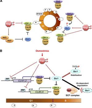
Regulation of cell-cycle progression by osmostress 306
G1/S transition: 307
S phase: 308
G2phase: 308
Exit from mitosis: 309
Other downstream effectors of the Hog1 MAPK 309
Ion channels: 309
Protein kinases regulated by Hog1: 309
Perspectives 310
S
ACCHAROMYCES(literally, sugar yeast) thrive, in their natural habitat, on decomposing fruits, including grape, where sugar (such as glucose, fructose, and sucrose) is abundant. As the fruits dry, the sugar concentration may approach its saturation point. This high sugar concentration poses a dilemma to the yeast, as the abundant food also brings unfavorable osmotic conditions that are a potential threat to their survival. Increased external osmolarity in-duces water efflux, an increased concentration of cytosolic ions (especially Na+), and cell shrinkage, which are all detrimentalto cell growth [for general biological effects of osmostress,
see Wood (1999, 2011)]. Amazingly, yeast can grow and vigorously ferment in media containing as much as 40% (2.2 M) glucose (Watanabeet al.2010), which is obviously a highly dangerous osmotic condition.
whose core is theHog1MAP kinase (MAPK) cascade. In this review, we have summarized the current, often fragmentary, understanding of both the upstream signaling mechanism of osmostress and the downstream adaptive responses. Be-cause the HOG pathway is highly conserved across fungal species, elucidation of the signaling and effector mecha-nisms inSaccharomyces cerevisiaewill be highly relevant to the studies of other yeasts and fungi (Krantzet al.2006a,b). We endeavored to be as comprehensive as possible, but due to space limitations, many interesting subjects had to be left out. Readers who are interested in various aspects of yeast osmostress responses are encouraged to consult a number of excellent review articles (Gustinet al.1998; Sprague 1998; Chellappan 2001; Hohmann 2002a,b, 2009; O’Rourkeet al.
2002; Saito and Tatebayashi 2004; Schwartz and Madhani 2004; Sheikh-Hamad and Gustin 2004; Chen and Thorner 2007; Hohmannet al.2007; de Nadal and Posas 2010).
Upstream Signaling Mechanisms
Overview of the HOG pathway
The central core of the HOG pathway is the Hog1 MAPK cascade. MAPK cascades are evolutionarily conserved signal-ing units that are utilized in many intracellular signal trans-duction pathways in diverse eukaryotic organisms, including fungi and yeast (Chen et al.2001). Each MAPK cascade is composed of three sequentially activating kinases (Figure 2). A MAPK is activated by a MAPK kinase (MAPKK) by dual
phosphorylation of the conserved Thr and Tyr residues in the TXY motif within the activation loop. A MAPKK is similarly activated by a MAPKK kinase (MAPKKK) by phos-phorylation of the Ser/Thr residues in its activation loop. Thefirst kinase of the cascade, MAPKKK, is activated either by phosphorylation by an upstream kinase, sometimes called MAPKKKK, or by binding of an activator protein, depending on the pathway. Each MAPK module is activated by specific types of stimuli and induces specific adaptive responses.
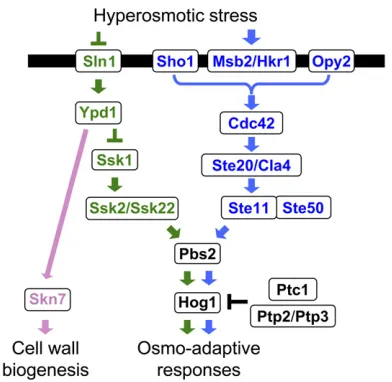
The upstream part of the HOG pathway comprises the functionally redundant, but mechanistically distinct, Sln1
and Sho1 branches (Figure 3). A signal emanating from either branch converges on a common MAPKK,Pbs2, which is the specific activator of the Hog1MAPK (Brewsteret al.
1993; Maeda et al. 1994). The Sln1 branch activates the redundant Ssk2and Ssk22 MAPKKKs, which then activate
Pbs2 (Maeda et al. 1995). TheSho1 branch activates the
Ste11MAPKKK, which also activatesPbs2(Posas and Saito 1997). Thus, a mutant that lacks both theSSK2andSSK22
genes (anssk2Dssk22Dmutant) is totally dependent on the
Sho1 branch for activation of the Hog1 MAPK, whereas a mutant that lacksSTE11is dependent on theSln1branch. Once activated, a substantial fraction of theHog1MAPK is transported into the nucleus where it regulates transcription and the cell cycle, although there are also Hog1targets in the cytoplasm. As adaptation proceeds, and osmotic balance is re-established, Hog1 activity goes down to near basal
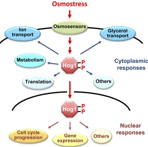
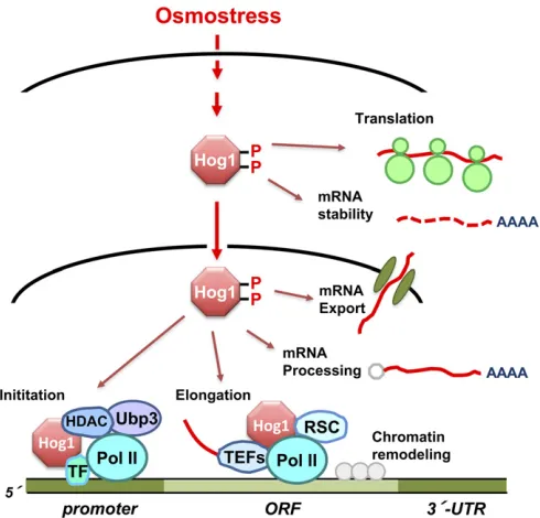
Figure 1 Osmo-adaptive responses in yeast. In response to an increase in extracellular osmolarity, the Hog1 MAPK is activated, which leads to the induction of cytoplasmic and nuclear adaptive responses. Cytoplasmic responses include the control of ionicfluxes and glycerol transport, met-abolic enzymes, and protein translation. Nuclear responses include the
levels, and Hog1 is exported back to the cytoplasm. Thus, there are mechanisms that control Hog1 nuclear import/ export, as well as downregulation ofHog1activity.
There are several other signal pathways that utilize a MAPK cascade in yeast, which are involved in the mating response, filamentous and invasive growth (FIG), and regu-lation of cell-wall biogenesis. Surprisingly, three of these pathways (HOG, mating, and FIG) share many of the same signaling elements, including theSte11MAPKKK. Thus, it is important to prevent signal leakage from one pathway into another pathway. This aim seems to be attained by insulation and exquisite network of reciprocal cross-regulation among the signaling pathways.
Sln1 branch of the HOG pathway
Two-component signal transduction system: The Sln1
branch of the HOG pathway is a variation of the so-called two-component system. Two-component systems are ubiq-uitous in prokaryotes, plants, and fungi (for comprehensive reviews, see Stocket al.2000; Gao and Stock 2009; Casino
et al.2010; Schalleret al.2011). As the name implies, the prototypical two-component system is composed of two pro-teins (Figure 4A): thefirst is a sensor histidine kinase (SHK) that contains an input (or sensor) domain, a HK catalytic domain, and a histidine auto-phosphorylation site, and the second is a response regulator (RR) that contains an output (or effector) domain and a receiver (REC) domain. When the input domain senses a relevant stimulus, the HK is acti-vated (or inactiacti-vated), and a histidine residue located near the HK domain is phosphorylated (or dephosphorylated). This phosphoryl group is then transferred to the acceptor aspartate residue in the REC domain of a cognate RR. This phosphotransfer reaction is termed the His-Asp
phosphore-lay. Because both histidine phosphate and aspartate phos-phate are energetically activated, they are often symbolized as HisP and AspP. In bacteria, numerous simple two-component systems exist that are composed of an SHK and a cognate RR. However, there are also more complex variations of this theme, where the basic His-Asp phosphor-elay reaction is repeated twice so that a phosphoryl group is transferred sequentially through a His-Asp-His-Asp multi-step phosphorelay (Figure 4B). In a complex two-component system, a phosphoryl group is initially transferred from a HK domain to a cognate REC domain as in the simple systems. This phosphoryl group, however, is then transferred to an intermediate phospho-carrier termed histidine-containing phospho-transfer (HPt) protein, which catalyzes specific phospho-transfer reactions between two REC domains. The phosphoryl group is then transferred from HPt to a second REC domain. TheSln1branch of the yeast HOG pathway is an example of complex two-component systems (Posaset al.
1996; Saito 2001). In the budding yeast, there are three REC proteins (Sln1, Ssk1, and Skn7), but only one SHK (Sln1) and one HPt (Ypd1). In fact, Sln1governs two dis-tinct signaling pathways: the Sln1-Ypd1-Ssk1 multistep phosphorelay, which regulates hyper-osmolarity responses, and the Sln1-Ypd1-Skn7 multistep phosphorelay, which makes a contribution to hypo-osmolarity responses.
Sln1-Ypd1-Ssk1 multistep phosphorelay: The N-terminal half of Sln1 is the sensor domain that is composed of an extracellular domain (ECD) flanked by two transmembrane segments, TM1 and TM2 (Ota and Varshavsky 1993; Maeda
et al.1994). The C-terminal half is composed of a HK domain and a REC domain; henceSln1is termed a“hybrid histidine kinase.” When activated, the Sln1 HK auto-phosphorylates His-576 near the HK domain, using ATP as a phospho-donor (Posaset al.1996). This phosphoryl group is then transferred to Asp-1144 in theSln1REC domain. It is likely that the HK
Figure 4 Schematic diagram of two-component signaling systems. (A) The prototypical two-component system that is characterized by the conserved phosphotransfer reaction between a histidine residue and an aspartate residue. (B) The Sln1-Ypd1-Ssk1 multistep phosphorelay. SHK, sensor histidine kinase; RR, response regulator; HK histidine kinase do-main; REC, receiver dodo-main; HPt, histidine-containing phospho-transfer protein; TM, transmembrane segment; P, phosphoryl group.
catalytic site of one molecule phosphorylates the His phos-phorylation site in another molecule in an Sln1 dimer. The phosphate is then transferred to His-64 ofYpd1, an HPt pro-tein. The phosphoryl group onYpd1is finally transferred to Asp-554 in the REC domain ofSsk1.
Regulation of Sln1 HK activity:Genetic analyses of various mutants in the Sln1pathway suggest that the Sln1HK do-main is catalytically active under normal osmotic conditions, whereas it is inactivated when the environmental osmolarity is increased (Maeda et al. 1994; Fassler and West 2010).
In vitroreconstitution of theSln1-Ypd1-Ssk1multistep phos-phorelay reactions supports the same conclusion (Posas
et al. 1996). As expected, the ECD and its flanking trans-membrane (TM) domains are important for regulation of the HK activity. For example, deletion of TM1 constitutively activates, whereas removal of both TM1 and ECD inacti-vates, Sln1 HK (Ostrander and Gorman 1999). In vivo,
Sln1 seems to respond to changes in turgor pressure (the pressure exerted by water inside the cell against the cell wall). When yeast is exposed to high external osmolarity, turgor pressure decreases as the cytoplasm shrinks. An ear-lier study suggested that turgor change rather than water loss activates the HOG pathway (Tamáset al.2000), which was later supported by biophysical analyses (Schaberet al.
2010). Consistent with these findings, Sln1 HK activity is inhibited when turgor is reduced by the antifungal antibiotic nystatin or by enzymatic removal of the cell wall (Reiser
et al. 2003). Conversely, Sln1 HK activity is enhanced by increased turgor pressure caused by raised intracellular glyc-erol concentration (Taoet al.1999). In a more recent study, it was found that the presence of the abundant GPI-anchored cell-wall mannoproteinCcw12has a role inSln1
HK activation (Shankarnarayan et al. 2008). These results suggest thatSln1responds to osmolarity-induced changes in the cell wall. On the other hand, it was also found that the
Sln1 branch of the HOG pathway is activated when mem-brane fluidity is reduced by a rapid downshift in tempera-ture to ,10° or by dimethyl sulfoxide treatment (Hayashi and Maeda 2006; Panaderoet al.2006). Hypoxia also acti-vates the Sln1 branch, perhaps by an altered membrane
fluidity caused by depletion of heme and ergosterol (Hickman
et al.2011). These results suggest thatSln1might respond to changes in the plasma membrane. Cold activation of the HOG pathway might be physiologically important because
Hog1-dependent accumulation of glycerol would protect yeast from freezing. In any case, it is clear that further stud-ies are needed to establish the biophysical nature of the stimuli that controlSln1activity.
HPt protein Ypd1:Ypd1is a small protein of 167 aa and is composed of a four-helix bundle with the phospho-accepting histidine (His-64) in the middle of the third helix (Song
et al.1999; Xu and West 1999). Ypd1interacts with three different REC domains, one each inSln1,Ssk1, andSkn7. A systematic Ala-scanning mutagenesis of Ypd1 coupled to
two-hybrid interaction analyses indicated that the REC domains ofSln1,Ssk1, andSkn7interact withYpd1at over-lapping binding sites (Porter et al. 2003; Porter and West 2005). Thea1 helix of theSsk1REC domain was identified as the interaction site with Ypd1 by isolation of Ssk1
mutants that cannot interact with Ypd1(Horieet al.2008). The structure of a complex between Ypd1and the REC do-main ofSln1is consistent with these mutational studies (Xu
et al.2003; Zhaoet al.2008).
Phosphotransfer reactions involving wild-type Ypd1 are very rapid, reaching steady-state levels in ,5 sec in vitro
(Janiak-Spens and West 2000). Thus, detailed kinetic anal-yses are possible only by using a rapid quenchflow appara-tus (Kaserer et al. 2010). Perhaps the most important
finding is that phosphotransfer fromYpd1P toSsk1is both very rapid (160 sec21) and irreversible, whereas that from Ypd1P toSkn7is slower (1.4 sec21) and readily reversible
(Janiak-Spenset al.2005). These and other kinetic proper-ties ofYpd1are consistent with the notion thatSsk1is con-stitutively phosphorylated under normal osmotic conditions.
Activation of the Ssk2/Ssk22 MAPKKKs by Ssk1:Ssk1 acti-vates a pair of homologous, and functionally redundant, MAPKKKs termedSsk2andSsk22(Maedaet al.1995). Like many other members of the MAPKKK family, the kinase cat-alytic domain ofSsk2/Ssk22is near the C-terminal end, and there is an auto-inhibitory domain (AID) in the N-terminal region.Ssk1binds to the N-terminal region ofSsk2/Ssk22, and, perhaps by conformational change, relieves the cata-lytic domain from inhibition by the AID (Posas and Saito 1998). Since the Sln1 HK is active under normal osmotic conditions,Ssk1is constitutively phosphorylated byYpd1P. However, under hyperosmotic conditions, unphosphorylated
Ssk1-OH will accumulate, and it binds and activatesSsk2/
Ssk22. Consistent with this notion, expression of unphos-phorylatable Ssk1 mutants such as Ssk1-D544S or Ssk1
mutants that cannot interact with Ypd1 (and thus cannot accept phosphate fromYpd1P), such asSsk1-I514T, hyper-activate theHog1MAPK cascade (Horieet al.2008).
AspP is chemically unstable and is spontaneously hy-drolyzed. Indeed, the half-life of purified Ssk1P is only
13 minin vitro(Janiak-Spenset al.2000). If it is similarly unstable in cells, then it is unlikely that all of the Ssk1 is stably converted toSsk1P, and therefore there is a possi-bility that persistent Ssk1-OH would activate the Hog1
MAPK cascade in the absence of any osmotic stimulation. However, several mechanisms exist that prevent erroneous activation of theHog1MAPK cascade. First, the half-life of
Ssk1P dramatically increases to 40 hr when Ypd1 is in-cluded in the incubation reaction in vitro (Janiak-Spens
et al.1999). It was proposed thatYpd1forms a stable com-plex withSsk1P and sterically shields the phosphorylated Asp residue from hydrolysis (Janiak-Spenset al.2000). Such enhanced stability of Ssk1P would maintain the levels of
prevented. Second, any residualSsk1-OH that may still exist would not contribute significantly toSsk2/Ssk22activation because only a doubly dephosphorylatedSsk1dimer, (Ssk1 -OH)2, can activateSsk2andSsk22(Horieet al.2008). For
example, when 1% ofSsk1is dephosphorylated, only 0.01% ofSsk1dimer is doubly dephosphorylated. Third,Ssk1-OH is degraded by a ubiquitin-proteasome-dependent mecha-nism, which may serve as an additional safeguard against spontaneous activation of Ssk2/Ssk22 in the absence of osmostress (Satoet al.2003). Finally, it should be noted that there is in fact a low basal signaling in theSln1pathway in the absence of any external stimulation, which may allow more rapid response upon osmostress (Maciaet al.2009).
Although stable Ssk1P is required to prevent spontane-ousHog1activation under nonstimulated conditions, it causes another difficulty under high-osmolarity conditions. When yeast is exposed to hyper-osmolarity, activation of theHog1
MAP kinase cascade occurs within minutes, which requires a much faster dephosphorylation of Ssk1P than the ob-served half-life of 40 hr in vitro in the presence ofYpd1. Higher osmolyte concentrations decrease the Ssk1P half-life by two-fold in in vitroreactions, but this modest effect alone would not be sufficient to account for the rapid
in vivo activation of the MAPK cascade (Kaserer et al.
2009). Therefore, the dephosphorylation ofSsk1P might be accelerated under stress conditionsin vivo, perhaps by an as-yet-unidentified phosphatase.
The actin cytoskeleton is important for the survival of yeast under osmostress, as many mutations in actincause osmosensitivity (Wertmanet al.1992). Hyperosmotic stress causes a rapid disassembly ofactincables, followed by de-polarization of actin patches leading to a cell-cycle delay (Chowdhuryet al.1992). The reassembly of theactin cyto-skeleton occurs only after osmotic balance is re-established (Brewster and Gustin 1994). During osmostress,Ssk2 con-centrates in the neck of budding cells and forms a complex with actin, and following reestablishment of osmotic bal-ance, Ssk2 promotes actin cytoskeleton recovery (Yuzyuk
et al.2002). This recovery mechanism requires a polarized distribution of Ssk2, its actin-interacting activity and its kinase catalytic activity, but, interestingly, does not require
Ssk1 (Yuzyuk and Amberg 2003; Bettinger et al. 2007). Although Ssk1is the only known activator ofSsk2/Ssk22, osmostress does cause slight activation of theHog1MAPK in
ssk1Δsho1Δmutants, whereas no activation is observed in
ssk2Δssk22Δsho1Δmutants (Maedaet al.1994; Reiseret al.
2000). Thesefindings suggest that there may be an as-yet-unknown mechanism that can activateSsk2/Ssk22without
Ssk1.
Ssk2/Ssk22-Pbs2-Hog1 kinase cascade:Once activated, the
Ssk2/Ssk22MAPKKK initiates a kinase cascade reaction that involves thePbs2MAPKK and theHog1MAPK (Boguslawski 1992; Brewster et al. 1993). Although there are several other MAPKKs and MAPKs in yeast with similar sequences, activatedSsk2/Ssk22exclusively phosphorylates, and thereby
activates, Pbs2, and activated Pbs2 phosphorylates only
Hog1. These specific interactions are due to the presence of specific docking sites in Pbs2. An Ssk2/Ssk22-specific docking site is located in the Pbs2 N-terminal regulatory region (Tatebayashiet al.2003). Fusion of thisPbs2docking site to the Ste7 MAPKK, which is not a substrate of Ssk2/
Ssk22, allows phosphorylation ofSte7bySsk2/Ssk22.Pbs2
has two specific binding sites for Hog1: one is in the N-terminal regulatory region, and another is near the C ter-minus (Murakami et al.2008).
The activity of wild-typeHog1is absolutely dependent on double phosphorylation of its TGY motif byPbs2. However, several Hog1mutants that are partially active without any phosphorylation by Pbs2 have been isolated (Bell et al.
2001; Bell and Engelberg 2003). By using these mutants,
Hog1-dependent effects can be studied without exposing cells to osmostress, which would induce bothHog1-dependent and -nondependent effects (Yaakovet al.2003).
Stress-responsive MAPK cascades that are homologous to theHog1MAPK cascade are found in both lower and higher eukaryotes (Sheikh-Hamad and Gustin 2004). For example, the mammalian stress-responsive p38 MAPK is structurally highly similar to Hog1, and p38 can complement mutant strains of yeast that lack theHog1MAPK (Hanet al.1994). Also, the kinase domain of the mammalian stress-responsive MAPKKK termed MTK1 (also known as MEKK4) is highly similar to the kinase domains ofSsk2andSsk22, and expres-sion of constitutively active MTK1-ΔN can complement the
ssk2Δssk22Δdouble mutation (Takekawaet al.1997). MTK1 is activated by binding of its specific activator, Gadd45, in a manner similar to activation of Ssk2 and Ssk22by Ssk1, although these activators are unrelated and not functionally exchangeable (Takekawa and Saito 1998; Mitaet al. 2002; Miyakeet al.2007).
Sln1-Ypd1-Skn7 multistep phosphorelay:Ypd1donates its phosphoryl group not only toSsk1but also toSkn7(Figure 4B). Skn7 is composed of an N-terminal DNA-binding do-main and a C-terminal REC dodo-main and is highly conserved among fungi (Brown et al.1994). A phosphotransfer reac-tion fromSln1toSkn7via the intermediaryYpd1was dem-onstratedin vitro(Liet al.1998; Aultet al.2002). Although
Skn7 is exclusively localized in the nucleus and Ssk1 is mostly in the cytoplasm,Ypd1is found in both the nucleus and the cytoplasm, which is consistent with its ability to transfer phosphate to both Skn7 and Ssk1(Lu et al.2003). TheSln1-Ypd1-Skn7phosphorelay regulates a response that is complementary to that of the Sln1-Ypd1-Ssk1 phosphorelay: whereas Ssk1is activated under hyperosmotic conditions,
Skn7is activated under hypo-osmotic conditions.Skn7 reg-ulates oxidative stress-responsive genes, andskn7Δmutants are hypersensitive to oxidative stresses such as exposure to hydrogen peroxide (Krems et al.1996; Raittet al.2000a). However, the role ofSkn7in oxidative responses is not de-pendent on Sln1, and the phospho-accepting Asp-427 of
In contrast, induction of hypo-osmostress responsive genes, such as OCH1, is dependent on Sln1 and requires the Asp-427 ofSkn7(Ketelaet al.1998; Liet al.2002; Shankarnarayan
et al. 2008). OCH1 encodes the mannosyltransferase in thecis-Golgi apparatus that initiates N-linked glycosylation of secreted/membrane proteins and thus is a key enzyme in cell-wall maintenance. Although theskn7Δmutants are not osmosensitive, the suppression of the hypo-osmotic stress sensitivity of apkc1Δmutant bySKN7overexpression sug-gests thatSkn7and the PKC pathway coordinately regulate cell-wall integrity that is critical for growth under hypo-osmotic conditions (Brown et al. 1994). For more details on Skn7, see a recent comprehensive review by Fassler and West (2011).
Sho1 branch of the HOG pathway
Unlike the Sln1 branch, which is a variation of the well-understood two-component paradigm, the activation mech-anism of the Sho1 branch is still only vaguely defined. Although many important observations have been made, there is still a lack of a unifying mechanism that incorporates all of the separate facts. Thus, we willfirst present an over-view of the current hypothesis of how the Sho1 branch might be activated and will then discuss the details of in-dividual steps in the following sections.
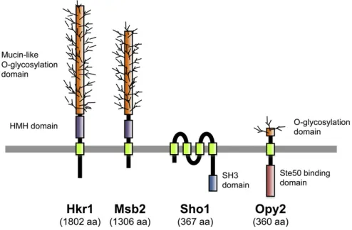
Overview:A signaling response in theSho1branch is initi-ated by the putative osmosensorsMsb2andHkr1, which are highly glycosylated single-pass TM proteins (Tatebayashi
et al. 2007). Through an as-yet-undefined mechanism that seems to involve an interaction between the Msb2/Hkr1
osmosensors and the Sho1 co-osmosensor, this response leads to activation of the PAK-like kinases Ste20 andCla4
by inducing their association with the membrane-bound small G-protein Cdc42 (Lamson et al. 2002). Activated
Ste20/Cla4 then phosphorylates and activates the Ste11
MAPKKK (Raittet al.2000b; van Drogenet al.2000), which in turn phosphorylates and activates thePbs2MAPKK that is associated with the Sho1 membrane anchor (Maeda et al.
1995; Tatebayashi et al. 2006). Because both the Cdc42
-Ste20 and the Sho1-Pbs2 complexes are localized on the membrane, Ste11 must also be localized to the membrane so that efficient activator/substrate interactions between
Ste20 and Ste11, as well as betweenSte11 and Pbs2, can take place. Membrane localization of Ste11 is mediated by theSte50adaptor protein, which forms a stable complex with
Ste11(Posaset al.1998; Wuet al.1999), primarily via asso-ciation of Ste50 with the membrane anchor protein Opy2
(Ekiel et al. 2009; Yamamotoet al. 2010), and secondarily by Ste50–Cdc42 and Ste50–Sho1 interactions (Tatebayashi
et al. 2006; Truckses et al. 2006). Activation of the Hog1
MAPK byPbs2seems to proceed as in theSln1branch.
Putative osmosensors Msb2 and Hkr1: Both Msb2 and
Hkr1 are highly glycosylated single-path transmembrane proteins (Figure 5). The extracellular domains of these
proteins are highly Ser/Thr rich and contain numerous
O-glycosylation sites that are glycosylated by the protein
O-mannnosyl transferase Pmt4 (Yang et al. 2009). The
MSB2gene was originally identified as a multicopy suppres-sor of a cdc24 mutant (Bender and Pringle 1989). Since
Cdc24is a guanine exchange factor forCdc42, it is believed that Msb2 somehow regulates the activity of Cdc24 or
Cdc42. Indeed, a weak binding between Msb2 andCdc42
has been observed (Cullenet al.2004). However, howMsb2
controlsCdc42activity is unclear.
The possible involvement ofMsb2 in the HOG pathway was initially suggested by the observation that the weak osmo-tolerance of the ssk1Δ sho1Δ mutant was abolished in the ssk1Δ sho1Δ msb2Δ triple mutant (O’Rourke and Herskowitz 2002). This observation was interpreted at that time as indicating that Msb2 is a third osmosensor in the HOG pathway (Sln1andSho1being the other two). A later study, however, revealed thatMsb2and another transmem-brane glycoprotein, Hkr1, are the more likely osmosensors in the Sho1branch, but thatSho1itself has a downstream function as a co-osmosensor (Tatebayashiet al.2007). This conclusion is partly based on genetic epistasis tests that in-dicated that MSB2/HKR1 functions upstream of SHO1: a constitutively active SHO1 mutant can activate Hog1
MAPK even in the msb2Δ hkr1Δ double-mutant cells, but a constitutively active MSB2 or HKR1 mutant cannot acti-vateHog1in asho1Δmutant.
The Ser/Thr-rich glycosylation domains of Msb2 and
Hkr1have a negative regulatory function, as their deletion converts Msb2 and Hkr1 into constitutively active forms (Cullenet al.2004; Tatebayashiet al. 2007). Furthermore, inhibition of O-glycosylation by pmt4Δ mutation, together with inhibition of N-glycosylation by tunicamycin, activates the Hog1 MAPK cascade in an Msb2-dependent manner (Yanget al.2009). Based on these observations, two possi-ble mechanisms of activating these osmosensors have been proposed. One is by proteolytic cleavage in the extracellular domain by the aspartyl proteaseYps1, which eliminates the
Ser/Thr-rich glycosylation domain (Vadaieet al.2008). An-other is by an osmostress-induced conformational change in the oligosaccharide structure (Tatebayashi et al. 2007). However, the actual mechanism remains unclear.
Co-osmosensor Sho1: The SHO1 gene was initially
identi-fied by isolation of mutants that are synthetically high
osmolarity sensitive in the presence of mutations that inac-tivate the Sln1branch of the HOG pathway (Maeda et al.
1995). Sho1 is a relatively small protein (367 aa) that is composed of an N-terminal bundle of four transmembrane segments (TM1–TM4) and a C-terminal, cytoplasmic SH3 domain (Figure 5). TheSho1 SH3 domain binds to a Pro-rich motif (KPLPPLPV) in the N-terminal regulatory region ofPbs2and serves to localizePbs2to the membrane (Maeda
et al. 1995). Of the 27 SH3 domains found in the yeast proteome, only theSho1-SH3 bindsPbs2, indicating a very high level of selectivity (Zarrinpar et al.2003). TheSho1– Pbs2 interaction is required for activation of Pbs2 by the
Ste11 MAPKKK. The Sho1 SH3 domain can also bind to
Pro-rich motifs in Fus1 (KPLPLTPN) (Nelson et al. 2004) and in Ste20(QPLPPIPP) (K. Tanaka, K. Tatebayashi, H.-Y. Yang, and H. Saito, unpublished results). Thus, during a mat-ing response, induced Fus1 might downregulate the Sho1
branch by competitively inhibiting the Sho1–Pbs2 interac-tion. The role of the Sho1–Ste20 interaction seems to be redundant with that of other signaling elements in the
Sho1branch because this Pro-rich motif inSte20is required for activation of theSho1branch only in some mutants, but not in wild-type cells.
A few lines of evidence suggest that Sho1 might serve additional roles in signaling other than membrane targeting of Pbs2 and Ste20. First, Pbs2 appears to dissociate from
Sho1 upon activation of the Pbs2 MAPKK, as suggested by decreased membrane localization of Pbs2 following osmostress stimulation, and this dissociation is hindered in aste20Δor aste11Δmutant or in apbs2Δmutant in which a catalytically inactivePbs2-K389M is expressed, but inter-estingly not in ahog1Δmutant (Reiser et al.2000). These observations suggest that theSho1–Pbs2interaction might be dynamically regulated by a feedback phosphorylation by activatedPbs2. Second, an experimental replacement of the
Sho1 SH3 domain with another SH3 domain derived (and modified) from the Fyn kinase resulted in a hybridSho1that bound toPbs2just as well as the wild-typeSho1. Nonethe-less, such a hybrid Sho1is functionally defective, implying that theSho1SH3 domain has other functions in addition to
Pbs2 binding (Marleset al. 2004). Third, and possibly re-lated to the previous point, several proteins, notably Ste11
andSte50, have been shown to interact withSho1, but this binding is independent of the Pro-rich-motif-binding ability of theSho1SH3 domain (Zarrinparet al.2004; Tatebayashi
et al.2006). These interactions might enable Ste11to effi -ciently interact with Pbs2 that is associated with Sho1. Finally, there are a number ofSho1mutants that are consti-tutively activated in the sense that their expression will
activate the Hog1MAPK in the absence of any osmostress (Tatebayashi et al.2006, 2007; Vadaie et al.2008). These mutations are found both in the TM region and in the cyto-plasmic region, suggesting that Sho1 might engage in dy-namic interaction with other molecules through both its TM and cytoplasmic regions. Thus, the potentially dynamic functions ofSho1are still far from being understood.
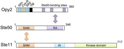
Adaptor protein Ste50: STE50was originally identified as a gene that is required for an efficient mating response, as its deletion mutants are moderately sterile (Ramezani Rad
et al. 1992; Xuet al. 1996).Ste50is essential for theSho1
branch of the HOG pathway (Posas et al. 1998; Wu et al.
1999) and is also necessary for thefilamentous and invasive growth pathway that activates the Kss1 MAPK (Ramezani Rad et al.1998; Jansenet al.2001). Thus, all three signal pathways that involveSte11are dependent onSte50. Struc-turally, Ste50 is composed of an N-terminal sterile-amotif (SAM) domain and a C-terminal Ras association (RA) do-main (Ramezani-Rad 2003) (Figure 6). A SAM dodo-main is a protein interaction module of 70 amino acids that can homo-dimerize and hetero-oligomerize with other SAM domains (Qiao and Bowie 2005). In vivo binding studies have shown that the Ste50SAM domain binds to the SAM domain inSte11(Posaset al.1998; Wuet al.1999; Jansen
et al. 2001), while in vitro studies demonstrated that the
Ste50 SAM domain can homo-dimerize as well as
hetero-dimerize with Ste11 SAM (Bhattacharjya et al. 2004; Grimshaw et al.2004; Kwanet al.2004, 2006). The SAM-mediated Ste50–Ste11 interaction is essential for all the known activities ofSte50(Ramezani-Rad 2003).
In spite of its name, theSte50RA domain does not seem to interact with Ras proteins. Genetic evidence suggests that the RA domain might interact with the Cdc42 GTPase, which is supported by a coprecipitation assay that showed that the Ste50 RA domain interacted equivalently with ei-ther GTP- or GDP-bound Cdc42 (Tatebayashi et al. 2006; Truckses et al. 2006). A Ste50 mutant that lacks the RA domain (Ste50-ΔRA) is functionally defective and cannot activate the Hog1 MAPK in response to osmostress. How-ever, forced localization of Ste50-ΔRA to the plasma mem-brane, by attachment of a membrane-targeting signal, results in efficient activation of theHog1MAPK, indicating that an essential function of the RA domain is to aidSte50
membrane localization (Tatebayashi et al. 2006; Truckses
et al.2006; Wuet al.2006). In wild-type cells,Ste50 mem-brane localization could be attained, in principle, by an interaction of the Ste50 RA domain with the membrane-associated Cdc42 GTPase. However, the major factor that recruitsSte50to the membrane appears to be the membrane anchor protein Opy2 (Wu et al. 2006; Yamamoto et al.
2010). Importantly, membrane-targeting of Ste50-ΔRA, us-ing the Ras C-terminal prenylation signal, can rescue the osmostress-induced Hog1 activation in the absence of
Ste50RA domain (Tatebayashiet al.2007).Ste50has also been shown to interact with the membrane protein Sho1
(Tatebayashi et al. 2006), but the roles of Ste50–Sho1 in-teraction in signaling remain to be determined. In summary, the main function ofSte50seems to be to serve as an adap-tor between theSte11MAPKKK and the membrane anchor
Opy2, so thatSte11is efficiently recruited to the membrane.
Membrane anchor Opy2:TheOPY2gene was initially iden-tified as a multicopy suppressor that downregulates the mat-ing MAPK signal pathway (Edwardset al. 1997). However, disruption ofOPY2does not have any significant impact on the mating pathway. It was later found that the opy2Δ mu-tation, together with a defect in the Sln1 branch, causes synthetic osmosensitivity, indicating thatOpy2has an essen-tial function in the Sho1branch of the HOG pathway (Wu
et al.2006).
Opy2is a single-path transmembrane protein of 360 aa. Its short extracellular domain is composed of, from the N terminus, a highly Ser-rich (SR1) domain, a Cys-rich (Cys-R) domain, and another Ser-rich (SR2) domain followed by the TM segment (Figure 6). The SR1 domain, but not SR2, is highlyO-glycosylated by the proteinO-mannnosyl transfer-asePmt4, but deletion of SR1 does not have any observable effect on Opy2functions (Hutzler et al. 2007; Yang et al.
2009). The Cys-R domain is characterized by an arrange-ment of eight cysteine residues, and genes that encode a sim-ilar Cys-rich motif are found in a wide range of fungal species. The cytoplasmic region of Opy2is intrinsically dis-ordered as revealed by NMR spectroscopy (Ekielet al.2009) and comprises four short well-conserved regions (CR-A to CR-D) interspersed among nonconserved sequences (Yamamotoet al.2010).
The essential function of Opy2in theSho1branch is to recruit theSte50/Ste11complex to the plasma membrane. Earlier studies suggested that there is more than oneSte50 -binding site inOpy2(Wuet al.2006; Ekielet al.2009). A more recent study extended this hypothesis and showed that there are actually three independentSte50-binding sites in
Opy2, which correspond to the conserved regions CR-A, CR-B, and CR-D. CR-A and CR-D seem to constitutively bind
Ste50, whereas CR-B (DIRSHITLGSSIL) binds Ste50 only
when the Ser and Thr residues are phosphorylated by the casein kinase I isoforms, Yck1and Yck2(Yamamoto et al.
2010).Yck1/Yck2are activated when glucose availability is high (Zamanet al.2008). In fact,Opy2CR-B is phosphory-lated only when there is abundant glucose in the media.
Opy2 is required not only for the Sho1 branch, but also for the FIG pathway, which is activated under limited nutri-tion and activates theKss1MAPK. Interestingly, CR-B seems to function only in the Hog1 pathway, but not in the FIG pathway. Thus, it is possible that under glucose-rich environ-ments the phosphorylation of CR-B shiftsOpy2activity away fromKss1and towardHog1.
In summary, the main function of Opy2 is to serve as a membrane anchor for theSte11MAPKKK through its bind-ing to the adaptor proteinSte50.Opy2also integrates sig-nals from the osmosensors and the glucose sensors.
Activation of Ste20/Cla4: Ste20 is a member of the p21-activated kinase (PAK) family of protein kinases that are activated by the small GTPase Cdc42 (Bokoch 2003). In the absence of stimuli, PAK family kinases are inhibited by their N-terminal auto-inhibitory domain that binds to their C-terminal kinase domain (Lei et al. 2000). This auto-inhibition is relieved when GTP-bound (activated) Cdc42
binds to the p21-binding domain termed“CRIB”that is close to the auto-inhibitory domain (Peter et al. 1996; Leberer
et al. 1997; Lamson et al. 2002; Ash et al. 2003). Ste20
was initially identified as a kinase that is required to activate
theSte11 MAPKKK in the mating signal pathway (Leberer
et al. 1992). Later, Ste20 was shown to participate in two other signal pathways, the FIG and the Sho1branch of the HOG pathway (Möschet al.1996; O’Rourke and Herskowitz 1998; Raittet al.2000b).Cla4is another PAK family kinase and is involved mainly in cell-cycle regulation, such as septin formation and polarized growth (Tjandra et al. 1998). Al-though bothste20Δandcla4Δmutants are viable, theste20Δ cla4Δdouble mutation is lethal (Cvrckováet al.1995). Thus, it is believed thatSte20andCla4share at least one essential function, although the nature of that essential function is not known.
The growth ofste20Δmutants of a parental strain that is defective in theSln1branch, such asssk2Δssk22Δ, is sensi-tive to high osmolarity, but these mutants can tolerate mod-erate osmostress (Raitt et al. 2000b). In contrast, ste20Δ cla4ts double mutants of the same strain are highly
osmo-sensitive and are completely unable to activateHog1, indi-cating that Cla4 partially compensates for the function of
Ste20 (Tatebayashi et al. 2006). The finding that ste20
(ΔCRIB) mutants are more osmosensitive than the STE20
wild-type parental cells seems to indicate that Cdc42 bind-ing to Ste20 is required for activation and/or membrane localization of Ste20 (Raitt et al. 2000b; Winters et al.
2005). However, overexpression of constitutively active
cdc42(G12V)only very moderately activatesHog1, suggest-ing that an additional factor might be necessary for full activation of Ste20(Raitt et al. 2000b). Although it is
frequently assumed that GTP association of Cdc42 is in-creased and that Ste20 kinase is activated in response to osmostress, there is no direct evidence for these assump-tions. An alternative mechanism, in which osmostress in-duces the association of active Ste20 (which has been activated by an osmostress-independent manner) andSte11, might betterfit the available data. Indeed, the mating MAPK pathway is activated by an analogous mechanism, i.e., by pheromone-induced association ofSte20andSte11(Pryciak and Huntress 1998; Lamsonet al.2002).
Activation of Ste11 by Ste20/Cla4:Activation of theSte11
MAPKKK by osmostress requires at least two events. Thefirst event is the binding ofSte50to theSte11N-terminal SAM domain. This interaction helps to dissociate the N-terminal inhibitory domain from the C-terminal kinase catalytic do-main, thus relieving inhibition of the kinase (Wu et al.
1999). However, as theSte11–Ste50interaction is constitu-tive, this effect is not likely to play an active role in regulat-ingSte11activity during osmostress. The second event that is required is phosphorylation ofSte11bySte20/Cla4. It has been demonstrated that, in response toa-mating factor, ac-tivatedSte20phosphorylates Ser-302, Ser-306, and Thr-307 in the N-terminal regulatory region of Ste11 (van Drogen
et al.2000). Based on the effects of phospho-mimetic muta-tions, it is believed that theseSte11sites are also phosphor-ylated bySte20/Cla4upon osmostress stimulation (Lamson
et al.2006).
Ste50 binding and phosphorylation by Ste20/Cla4 are important, but not sufficient for Ste11 to transmit signals to downstream elements. Phospho-mimetic substitutions at the phosphorylation sites, or mutations in the auto-inhibitory domain, or even a deletion of the entire N-terminal regula-tory region, all constitutively activateSte11. Overexpression of one of these constitutively activeSte11mutants activates both the Ste11-Pbs2-Hog1 and the Ste11-Ste7-Fus3/Kss1
MAPK cascades, without any stimulation (Posas and Saito 1997; Lamson et al.2006; Tatebayashiet al. 2006). How-ever, expression of the same constitutively active Ste11
mutants using the native STE11promoter does not signifi -cantly activate the Hog1 MAPK or the Fus3/Kss1 MAPK (Lamsonet al.2006; Tatebayashiet al.2006). Constitutively active Ste11 mutants do activate the Hog1MAPK cascade and the mating MAPK cascade in aSte20/Cla4-independent manner upon respective stimulation (Lamson et al. 2006; Tatebayashi et al. 2006). Thus, it is clear that, in addition to activation of Ste11 by Ste20/Cla4, another stimulus-dependent signal amplification step is required to transmit sufficient signal to the downstream component (Pbs2in the case of the HOG pathway andSte7in the cases of the mating and FIG pathways). The nature of this amplification step is unclear, but one possibility is a stimulus-induced membrane localization of activatedSte11(Lamsonet al.2006).
Activation of Pbs2 by Ste11:Ste11can be activated by any of the three MAPK cascades: the osmoregulatory HOG
path-way, the mating pathpath-way, and the FIG pathway. When acti-vated by osmostress, however,Ste11activates only thePbs2
MAPKK, while in the other pathways Ste11 activates the
Ste7MAPKK. Thus, there must be a mechanism that allows only Pbs2to be activated bySte11during osmotic stimula-tion. As discussed earlier, Pbs2 is recruited to the plasma membrane by the membrane-associated scaffold protein
Sho1 (Maeda et al. 1995; Reiser et al. 2000), and the
Ste11/Ste50 complex is recruited to the membrane by the membrane anchor proteinOpy2(Wuet al.2006; Ekielet al.
2009; Yamamoto et al.2010). However, efficient activation ofPbs2bySte11seems to require, in addition to their mem-brane localization, direct and indirect docking interactions betweenSte11andPbs2. It is known thatSte11andPbs2,
Ste11 and Sho1, Ste50 and Sho1, and possiblyOpy2 and
Sho1 bind to each other (Posas and Saito 1997; Zarrinpar
et al. 2004; Tatebayashi et al.2006). Thus, multiple inter-actions between the Opy2/Ste50/Ste11 complex and the
Sho1/Pbs2complex bringSte11in close contact withPbs2
for efficient activation. The relative contributions of these interactions to Pbs2activation, as well as their regulation by osmostress, remain to be determined.
Activation of the HOG pathway by non-osmotic stresses
A number of non-osmotic stresses are known to activate the HOG pathway, including cold stress (Hayashi and Maeda 2006; Panadero et al. 2006), heat stress (Winkler et al.
2002), hypoxia (Hickman et al. 2011), arsenite (Sotelo and Rodríguez-Gabriel 2006; Thorsen et al. 2006), acetic acid (Mollapour and Piper 2006, 2007), low pH (Kapteyn
et al.2001), inhibition of glycosylphosphatidylinositol (GPI) anchor synthesis (Toh-E and Oguchi 2001), and inhibition of sphingolipid synthesis (Tanigawa et al. 2012). In most cases, Hog1 is only moderately activated, and the kinetics of Hog1 phosphorylation is different from those observed upon osmostress. Although it is unclear how Hog1 is acti-vated by these stresses, such stresses often activate either theSln1branch or the Sho1branch, but not both. Adapta-tion to these diverse stresses, in addiAdapta-tion to osmostress, might explain why yeast has apparently redundant osmostress-signaling branches. In this context, it is worth noting that theAspergillus nidulansHogA MAPK (a homolog ofHog1) is activated only by the two-component signaling pathway ho-mologous to the Sln1 branch, even though the mold has aSho1homolog (Furukawaet al.2005).
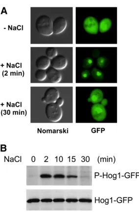
Nuclear transport of activated Hog1
Hog1rapidly accumulates in the nucleus following osmotic stress (Figure 7A).Hog1is then exported back to the cyto-plasm after return to an iso-osmotic environment or after adaptation to high osmolarity (Ferrigno et al.1998; Reiser
1998; Reiseret al.1999).Hog1phosphorylation itself, how-ever, is not sufficient for its nuclear localization because the constitutively phosphorylatedHog1molecules in the ptp2Δ ptc1Δdouble-mutant cells do not accumulate in the nucleus (Mattison and Ota 2000). Catalytically inactive Hog1 mu-tants, such as D144A, cannot translocate into the nucleus after hyper-osmotic stimulation (Westfall and Thorner 2006). In contrast, other catalytic site mutants that retain partial activity, such as K52R or K52M, not only translocate into the nucleus, but also even fail to be exported out of the nucleus (Ferrignoet al.1998; Mattison and Ota 2000). Thus,Hog1
catalytic activity seems to be required for its nuclear import and/or export, but its precise role remains unclear. Strains that lack the general stress activators Msn2andMsn4, the related transcription factors Msn1andHot1, or the nuclear protein tyrosine phosphatase Ptp2accumulate less Hog1in the nucleus than wild-type cells, suggesting that these mole-cules bind and retainHog1in the nucleus (Reiseret al.1999; Repet al.1999b; Mattison and Ota 2000).
Nuclear import of Hog1 is partially dependent on the activity of Gsp2 (homolog of mammalian Ran GTPase) and Nmd5 (homolog of importin b), but not on that of
Srp1andRsl1, which encode the nuclear localization signal (NLS)-binding importin a/b heterodimer (Ferrigno et al.
1998). This result is consistent with the fact thatHog1does not contain a classical NLS. Nuclear export ofHog1requires the activity of the nuclear export signal (NES) receptor
Xpo1/Crm1(Ferrignoet al.1998).
Nuclear localization is necessary forHog1to phosphory-late its nuclear substrates, including transcription factors and
cell-cycle regulators. Indeed, cells that express plasma membrane-tethered Hog1 (Hog1-CCAAX), which cannot translocate to the nucleus, seem to have deficient expression of theHog1-dependent genes (Westfallet al.2008). Strikingly, however, membrane-tetheredHog1permits robust growth un-der conditions of hyper-osmotic stress, suggesting thatHog1 -mediated cytoplasmic modulation of metabolic activities, perhaps those that are necessary for glycerol synthesis and accumulation, are more important for long-term cell sur-vival than alteration of the gene expression pattern (Bouwman
et al.2011).
Unlike Hog1, the Hog1-activating kinase Pbs2 is found mostly in the cytoplasm of both unstressed and osmostress-stimulated cells (Ferrigno et al. 1998). Nevertheless, Pbs2
has an NES at its N terminus (residues 4–18) and an NLS at its C terminus (residues 636–639).Pbs2ΔNES mutants ac-cumulate in the nucleus, whereasPbs2ΔNESΔNLS double mutants are found in the cytoplasm (Tatebayashi et al.
2003). Thus, it is likely that Pbs2 shuttles between the two compartments, but the function of such shuttling is unknown.
Dynamics of HOG pathway signaling
The Hog1 MAPK is only transiently activated following osmostress stimulation. Phosphorylation of theHog1 activa-tion sites (TGY) increases rapidly, reaches a maximal level at
5 min, and then gradually decreases to near basal levels within 30 min (Maedaet al.1995; Haoet al.2007) (Figure 7B). This negative regulation is dependent on the kinase activity ofHog1itself because phosphorylation of catalytically inactive Hog1 persists much longer than that of wild-type
Hog1 (Wurgler-Murphy et al. 1997). Several negative-feedback mechanisms are known in the HOG pathway. Fur-thermore, the Hog1 MAPK pathway is part of a complex signaling network that involves at least two other MAPK pathways. The dynamic characteristics of this signal net-work are intensely investigated both by conventional genetic/ biochemical approaches and by more recent systems bio-logical and computational approaches.
Negative feedback by glycerol accumulation: The most important negative feedback mechanism of Hog1 pathway signaling is removal of the osmostress by induced accumu-lation of the compatible solute glycerol (Brewster et al.
1993; Albertynet al.1994; Klippet al.2005; Muzzeyet al.
2009). Although transcriptional induction of GPD1 and other genes necessary for glycerol accumulation is important for long-term downregulation of the Hog1 pathway, such induction takes too long (at least 15 min) to account for the rapid decline of Hog1 activity (Hirayama et al. 1995). It has been proposed thatHog1might more rapidly regulate glycerol accumulation by directly modulating the activities of the glycerol channel Fps1 and metabolic enzymes in-volved in glycerol biosynthesis (Dihazi et al. 2004; Klipp
et al. 2005; Mollapour and Piper 2007; Westfall et al.
2008; Beeseet al.2009; Bouwmanet al.2011).
Negative feedback by protein phosphatases: Although signaling from the upstream osmosensors stops when osmotic imbalance is eliminated by glycerol accumulation, it is still necessary to inactivate the kinases by dephosphorylation to bring the system to the prestimulation state. The two acti-vating phosphorylation sites in Hog1, namely Thr-174 and Tyr-176, are dephosphorylated by different enzymes (for reviews, see Saito and Tatebayashi 2004; Martínet al.2005). Members of the type 2C Ser/Thr phosphatase family,Ptc1,
Ptc2, andPtc3, dephosphorylate Thr-174. Of these phospha-tases,Ptc1is the most important for de-activation ofHog1, as the ptc1Δmutant retains highHog1activity even after 1 hr (Warmkaet al.2001). The specificity ofPtc1towardHog1is indirectly conferred by the adaptor proteinNbp2(Mapes and Ota 2004). Nbp2binds to bothPtc1andPbs2, and as Pbs2
also has a high affinity forHog1,Ptc1is indirectly recruited to
Hog1by theNbp2–Pbs2complex. In contrast,Ptc2andPtc3
seem to have more of a subsidiary role of limiting the maxi-mal activity ofHog1during activation (Younget al.2002).
Members of the protein tyrosine phosphatase family,Ptp2
and Ptp3, dephosphorylate Tyr-176 (Jacoby et al. 1997; Wurgler-Murphyet al.1997). Although these tyrosine phos-phatases are partially redundant,Ptp2is primarily responsible forHog1dephosphorylation, whereasPtp3is more important for Fus3 dephosphorylation (Zhan and Guan 1999). Ptp2 is found in the nucleus, whereas Ptp3 is localized in the cyto-plasm (Mattison and Ota 2000). This localization of Ptp2
seems to ensure that tyrosine dephosphorylation of Hog1
occurs only after Hog1 has entered into the nucleus. Be-cause Hog1is inactivated when either Thr-174 or Tyr-176 is dephosphorylated, theptc1Δptp2Δdouble-mutant strain is lethal because of Hog1 hyperactivation (Maeda et al.
1993). Phosphatases that inactivate other kinases in the
Hog1pathway have not been identified confidently.
Negative feedback by phosphorylation of upstream ele-ments: Activated Hog1 also negatively feedback regulates the Hog1 pathway by phosphorylating upstream signaling elements. Osmostress-activated Hog1phosphorylates Sho1
at Ser-166, which is located within the cytoplasmic linker region between the four TM domains and the C-terminal SH3 domain (Hao et al. 2007). Hog1 activation is slightly diminished in cells expressing the phosphomimetic Sho1 -S166E. It has been shown that some mutations at Ser-166 disrupt Sho1oligomerization. However, neither the role of Ser-166 phosphorylation in Sho1 oligomerization, nor the role of Sho1oligomerization in Hog1activation, is clear.
Activated Hog1 phosphorylates several amino acids in
Ste50 (Ser-155, Ser-196, Ser-202, Thr-244, Ser-248, and
Thr-341) (Haoet al.2008). Phosphorylation ofSte50reduces its affinity for the membrane anchorOpy2(Yamamotoet al.
2010). Because theOpy2–Ste50 interaction is essential for
Hog1 activation via the SHO1 branch, phosphorylation of
Ste50 by Hog1 serves as a negative feedback mechanism.
Indeed, the duration ofHog1activation by osmotic stress is longer in cells that express a phosphorylation-deficientSte50
mutant than in the control cells. Pheromone-activatedFus3
and Kss1also phosphorylate the sameSte50 residues, sug-gesting that Ste50 phosphorylation may also serve as a cross-regulatory mechanism between the mating and HOG pathways (Yamamotoet al.2010).
Inhibition of crosstalk among MAPK signaling pathways:
In general, each MAPK module is activated by specific types of stimuli and induces specific adaptive responses. To achieve this specificity would be easy if each MAPK module was composed of only unique and dedicated components. In yeast, however, three MAPK modules (the Sho1 branch of HOG pathway, the mating pathway, and the FIG pathway) share many components, including theSte11MAPKKK, and still maintain their individuality. Leakage of signal, or cross-talk, from one MAPK pathway to another is prevented by a number of mechanisms, in addition to the negative regu-lation that involves protein phosphatases (Saito 2010).
One mechanism is insulation of each MAPK pathway from the others by docking interactions and scaffold proteins (Reményiet al.2005; Bardwell 2006; Dard and Peter 2006). Activation of the mating MAPK module (Ste11/Ste7/Fus3) is dependent on the presence of the Ste5scaffold (Elion 2001; Flatauer et al.2005; Winters et al. 2005; Garrenton
et al. 2006; Good et al. 2009). In contrast, activation of the Sho1 branch of the Hog1 MAPK module (Ste11/ Pbs2/Hog1) is dependent on the presence of the Sho1
scaffold (Maedaet al.1995; Zarrinparet al.2004). Indeed, when a wild-type cell is costimulated with osmostress and a mating factor, dual activation of the HOG and the mating MAPK pathways occurred, indicating that these two MAPK modules are practically insulated and activated indepen-dently of each other (Patterson et al. 2010). The impor-tance of docking and scaffold interactions in determining pathway specificity has also been demonstrated by
arti-ficially forcing interaction between non-native pairs of signaling elements, thus diverting the signaling flow into preselected directions (Harriset al.2001; Parket al.2003; Tatebayashiet al.2003; Modyet al.2009).
Another mechanism is cross-inhibition by one MAPK pathway of other MAPK pathways. Although theHog1MAPK module (Ste11/Pbs2/Hog1) shares many upstream com-ponents with the FIG Kss1 MAPK module (Ste11/Ste7/ Kss1), osmostress activates theKss1MAPK of the FIG path-way only very weakly and transiently (Shock et al. 2009; Wang et al. 2009), and glycosylation defects that activate
Kss1 do not activateHog1 (Cullen et al. 2000; Yanget al.
2009). In the absence ofPbs2orHog1, however, osmostress activates Kss1robustly andFus3to a lesser degree, induces
Kss1/Fus3-dependent genes, and induces FIG/mating-like po-larized cell growth (O’Rourke and Herskowitz 1998, 2004; Pitoniak et al. 2009). Using an ATP analog-sensitive Hog1
mutant, it was shown that inhibition of this crosstalk requires
nucleus (Shocket al.2009), even a membrane-tethered ver-sion ofHog1, which, in principle, cannot enter the nucleus, can prevent this crosstalk, implying that a cytoplasmic sub-strate might be involved in this process (Westfallet al.2008). However, cells expressing mutants of the known or suspected
Hog1 substrate proteins (Sho1, Ste50, Opy2, Ste7, Tec1,
Dig1/Dig2, andRck1/Rck2) that lackHog1-dependent phos-phorylation sites do not display constitutive crosstalk (Hao
et al. 2007, 2008; Shock et al. 2009; Yamamoto et al.
2010). Thus, the mechanism of cross-inhibition between the HOG and FIG/mating pathways remains obscure.
Single-cell dynamics: Conventional methods used to detect MAPK activity such as immunostaining of fixed cells or im-munoblotting of cell extracts using phospho-MAPK-specific antibodies can show only static snapshots and/or population averages of MAPK activation. To study the systems dynamics of a signaling pathway, it is necessary to monitor the behavior of single cells under controlled environmental conditions. The
Hog1 MAPK pathway is particularly suited for this type of analysis. By using a microfluidic device to change the osmo-larity of media (input), and by monitoring the nuclear trans-location of fluorescent protein-tagged Hog1 (output), two groups have reported the frequency responses of HOG path-way activation (Hersen et al.2008; Mettetalet al.2008). At low frequency (,1/200 sec21), the HOG pathway faithfully
follows the input changes, whereas at higher frequency, it responds only to the average input osmolarity. Other aspects of HOG-signaling properties have also been studied using var-ious single-cell monitoring methods (McClean et al. 2007; Muzzeyet al.2009; Pattersonet al.2010; Peletet al.2011).
In silico simulation:The HOG-signaling pathway is also an intense subject ofin silicosimulation, or mathematical mod-eling, that aims to elucidate system architecture, dynamics, and regulation based on data sets in the literature. Modeling is rapidly evolving from a simple tool that describes and summarizes the known facts into a more advanced predic-tive facility that can test the validity of various hypotheses (Klipp et al. 2005; Gat-Viks and Shamir 2007; Zou et al.
2007; Krantzet al.2009; Rensing and Ruoff 2009; Ziet al.
2010; Parmar et al.2011; Schaberet al.2011). The popu-larity of the HOG pathway for such studies is undoubtedly because of its relative simplicity together with the availability of detailed mechanistic knowledge regarding this pathway and abundant quantitative and qualitative data. Thus, the HOG pathway will continue to be an excellent testing ground for algorithms that attempt to simulate and analyze more complex signal transduction networks in higher eukaryotes.
Downstream Adaptive Responses
Reestablishment of osmotic balance
Compatible osmolytes: Activation of Hog1 in response to osmostress elicits a program for cell adaptation that includes
short- and long-term responses. Long-term adaptation involves transcriptional and translational regulation of the genome, whereas short-term adaptation is accomplished by changes in glycerol accumulation (Albertynet al.1994) and the reestablishment of ionic balance (Proft and Struhl 2004). Exposure to increased osmolarity is known to result in loss of water, shrinkage in cell size, and a temporary arrest of growth until adaptation occurs. The major strategy for survival under high osmolarity is to produce and accumulate compatible osmolytes such as glycerol to maintain the water balance and reestablish the volume and the turgor of the cells (Blomberg and Adler 1989; Hohmann et al. 2007; Westfallet al.2008; de Nadalet al.2011). The accumulation of compatible osmolytes is a ubiquitous mechanism in cel-lular osmoregulation. Although there are a number of com-patible osmolytes such as trehalose, amino acids, and ions that contribute differently to adaptation to osmostress, glyc-erol seems to be the most important compatible osmolyte for the growth ofS. cerevisiaein the presence of high osmolarity (Hohmannet al.2007).
Intracellular accumulation of glycerol is an essential response for survival under high-osmolarity conditions, and the Hog1 MAPK is responsible mainly for the accumula-tion of glycerol in the presence of high osmolarity (Albertyn
et al. 1994). There are several mechanisms to control glyc-erol accumulation: regulation of gene expression, meta-bolic adjustment, and control of glycerol export and import (Hohmann 2002b).
Glycerol accumulation: The expression of key metabolic enzymes that are involved in glycerol, trehalose, and glycogen metabolism is upregulated in response to Hog1activation. The enzymes directly responsible for the synthesis of glyc-erol,i.e., glycerol-3-phosphate dehydrogenase (Gpd1) and glycerol-3-phosphatases (Gpp1 and Gpp2), are upregu-lated upon osmostress (see below), and the lack of these genes severely impairs growth at high osmolarity (Figure 8) (Hohmann 2002a). Expression of sugar transporters and genes involved in sugar metabolism are also upregulated in response to osmostress (Rep et al.1999a, 2000; Gasch
et al.2000; Tomás-Coboset al.2004; Capaldiet al.2008). However, some studies indicated that regulation of gene expression by Hog1is not absolutely required for cell sur-vival under certain high-osmolarity conditions, especially at the initial phases of the stress and at medium osmolarity (Mettetalet al.2008; Westfallet al.2008). In contrast, other studies indicated thatHog1-dependent regulation of the ex-pression of specific genes involved in glycerol metabolism is important for cell survival at high osmolarity over an ex-tended period of time (Hohmann 2002b; de Nadal and Posas 2010; Martínez-Montañéset al.2010).
the transcription of particular genes, and therefore addi-tional mechanisms other than transcripaddi-tional regulation must exist that permit such a rapid response. There are two main mechanisms to achieve such a rapid initial increase in glycerol concentration: changes in carbon metabolism and changes in glycerol transport.
Metabolic adjustments: Adaptation to osmotic stress requires direct metabolic adjustments. Cells must redirect carbon resources toward enhanced production of glycerol, and thus there is significant modulation of central carbon metabolism during osmo-adaptation. There are indications that regulation of glycolysis is crucial for osmotic adapta-tion; for example, cells deficient in glycerol synthesis are highly osmosensitive. The control of glycolysis and glycerol production appears to be distributed among several en-zymes through allosteric control by different metabolites (Hohmann et al. 2007). However, there is direct evidence indicating that the activity of the 6-phosphofructo-2-kinase,
Pfk2, which is responsible for controlling the levels of fructose-2,6-bisphosphate (F2,6BP), a key activator of glycol-ysis, is regulated by the Hog1 MAPK (Dihazi et al. 2004). Therefore, Hog1may directly control the metabolic flux in response to stress. Along the same lines, recent studies using aerobic, glucose-limited cultures suggest that metabolic reg-ulation rather thande novoenzyme synthesis dominates the initial phase of the adaptive process, at least in the presence of moderately high osmolarity (1 M sorbitol) (Bouwmanet al.
2011). Therefore, the regulation of metabolic flux is an im-portant component inHog1-regulated glycerol accumulation.
Glycerol transport: Because the lipid bilayer has low permeability for glycerol, specific channel proteins mediate the rapid import and export of glycerol. As a consequence, the control of import and export rates is one mechanism by which the glycerol content inside of the cell can be altered. Thus, the control of the flux of glycerol through the membrane is another key factor for the initial accumulation
of glycerol upon osmostress. Stl1, a sugar transporter-like protein whose expression is strongly induced byHog1upon stress, might contribute to glycerol accumulation by import-ing glycerol from the environment in response to stress. However, the fastest mechanism to alter glycerol concentra-tion is via Fps1-mediated glycerol export (Tamás et al.
1999). Fps1is a member of the aquaporin family of trans-membrane channels, and cells that express Fps1 mutant proteins that are constitutively open do not accumulate glyc-erol and grow poorly in the presence of high osmolarity (Hohmann et al. 2007). In response to osmostress, the
Fps1 channel closes to maintain internal glycerol, but this effect seems to be independent ofHog1(Tamáset al.1999). On the other hand, direct regulation of Fps1transport ca-pacity and protein stability byHog1has been described for arsenite transport and in response to weak acid treatment (Thorsenet al.2006; Mollapour and Piper 2007; Beeseet al.
2009). In addition, the stress-induced phosphorylation of
Rgc2, a novel regulator ofFps1channel activity, is also par-tially controlled by the Hog1 MAPK (Mollapour and Piper 2007; Beeseet al.2009). The precise mechanism by which
Fps1is controlled upon osmostress remains unclear. The combined data indicate that the accumulation of glycerol is a key adaptive response to high osmolarity that is modulated by several mechanisms with different kinetics and different quantitative contributions to achieve proper adaptation to osmostress.
General stress responses
In addition to glycerol, a number of other organic osmolytes, including trehalose, protect yeast from osmostress, not only by counteracting water efflux and reestablishing osmotic balance, but also by playing unique roles in antioxidation, detoxification, and the stabilization of cellular proteins and structures (Mager and Varela 1993; Yancey 2005). Notably, a number of genes that are upregulated by osmostress have similar protective functions as these osmolytes (de Nadal and Posas 2010; Martínez-Montañés et al. 2010). For
example, in response to osmostress, a number of genes that protect cells from oxidative damage are upregulated, includ-ing genes involved in redox metabolism, mitochondrial func-tion, and the biosynthesis of antioxidative compounds (e.g.,
TRX2,CTT1,GRE3, andSOD2). Genes that encode the chap-erones (e.g.,HSP12,HSP104, andHSP42) that protect cells from damage by protein denaturation are also upregulated. It is worth noting thatHog1has also been implicated in ER stress protection, which is induced in response to the accu-mulation of unfolded proteins (Bicknellet al.2010; Torres-Quirozet al.2010; Erasoet al.2011), and in the control of mitophagy, the specific autophagic elimination of mitochon-dria (Aoki et al.2011; Maoet al.2011).
One role of the transcriptional response to a specific stress is to generate a cross-protection to other types of stresses. Osmostress induces many genes that are consid-ered to be part of general stress responses. Conversely, when cells are subjected to a mild stress (e.g., oxidative stress or heat stress), stress response element (STRE)-mediated responses are induced even in the absence of Hog1(Berry and Gasch 2008). Thus, at 37°,hog1Δcells can survive on moderate osmostress, such as 0.8 M sorbitol, better than at 30°(Sideriuset al.2000). This protection is not sufficient for
hog1Δcells to survive higher levels of osmolarity.
Regulation of gene expression by osmostress
Global analysis of gene expression upon osmostress:
Exposure of yeast to high osmolarity results in profound changes in the physiology of the cell and has a major impact on the capacity of the cell for gene expression. Analysis of the transcriptional changes mediated byHog1in response to osmostress may lead to a general understanding of how cells rapidly, precisely, and extremely efficiently adjust the full complement of a transcriptional program in response to ex-tracellular stimuli. Indeed, theHog1MAPK plays a key role in the regulation of mRNA biogenesis by controlling several steps in the transcription process (Figure 9) (Hohmann 2002b; de Nadal and Posas 2010; Martínez-Montañéset al.
2010; de Nadal et al. 2011). Although the role of Hog1 -dependent gene expression in osmo-adaptation is still in-completely understood, it is clear that long-term adaptation to high osmolarity requires regulated transcription, as a number of mutants in the transcriptional machinery render cells osmosensitive (de Nadal et al. 2004; Zapater et al.
2007; Mas et al. 2009). On the other hand, it has been shown that a membrane-tetheredHog1construct abolishes short-term transcription responses at certain osmolarities (so that it cannot enter the nucleus). Nevertheless, this
Hog1 construct is still able to suppress the osmosensitivity of ahog1Δstrain (Westfallet al.2008). Therefore, cytoplas-mic events caused by the rapid and transient activation of the Hog1 MAPK in response to osmostress—such as the control of glycerol production by direct modulation of met-abolic enzymes (Dihaziet al.2004; Bouwman et al.2011) and the altered mRNA stability (Molinet al.2009; Romero-Santacreu et al. 2009; Miller et al.2011)—might be suffi
-cient for the maintenance of osmotic balance under these experimental conditions without invoking induced gene ex-pression in the nucleus.
Global transcriptional responses to diverse stresses in
S. cerevisiaehave been studied in detail using gene expres-sion profiling. There are a large number of genes whose transcription is induced in response to osmostress; of these genes, there is one subset of genes that specifically responds to osmostress, whereas another subset of genes responds indiscriminately to diverse types of stresses. Induction of the latter group of genes is known as the environmental stress response (ESR). The ESR consists of 300 to 600 genes whose expression is upregulated or downregulated by stresses such as DNA damage, heat shock, osmostress, or oxidative stress (Gasch et al.2000; Caustonet al.2001; Capaldiet al.
2008). The extent and kinetics of the ESR appear to be de-pendent on the severity of the stress, since cells exposed to increasing stress often display broader changes in gene expres-sion. This general stress response has been implicated in the phenomenon of cross-protection, whereby exposure to a non-lethal dose of one stress can protect cells against unrelated stresses (Berry and Gasch 2008). The genes upregulated by the ESR include genes involved in carbohydrate metabolism, protein metabolism, intracellular signaling, and defense against reactive oxygen species and DNA damage. On the other hand, most of the genes downregulated by the ESR are involved in protein synthesis and in growth-related processes (Gasch 2007; Martínez-Montañéset al. 2010).