INVESTIGATION
Architectural and Functional Diversity of Polycomb
Group Response Elements in
Drosophila
J. Lesley Brown and Judith A. Kassis1 Program on Genomics of Differentiation, Eunice Kennedy Shriver National Institute of Child Health and Human Development, National Institutes of Health, Bethesda, Maryland 20892-2785
ABSTRACTPolycomb group response elements (PREs) play an essential role in gene regulation by the Polycomb group (PcG) repressor proteins inDrosophila. PREs are required for the recruitment and maintenance of repression by the PcG proteins. PREs are made up of binding sites for multiple DNA-binding proteins, but it is still unclear what combination(s) of binding sites is required for PRE activity. Here we compare the binding sites and activities of two closely linked yet separable PREs of theDrosophilaengrailed(en) gene, PRE1 and PRE2. Both PRE1 and PRE2 contain binding sites for multiple PRE–DNA-binding proteins, but the number, arrangement, and spacing of the sites differs between the two PREs. These differences have functional consequences. Both PRE1 and PRE2 mediate pairing-sensitive silencing of mini-white, a functional assay for PcG repression; however, PRE1 requires two binding sites for Pleioho-meotic (Pho), whereas PRE2 requires only one Pho-binding site for this activity. Furthermore, for full pairing-sensitive silencing activity, PRE1 requires an AT-rich region not found in PRE2. These two PREs behave differently in a PRE embryonic and larval reporter construct inserted at an identical location in the genome. Our data illustrate the diversity of architecture and function of PREs.
T
HE Polycomb group (PcG) and Trithorax group (TrxG) proteins are key regulators of genomic programming and differentiation in multicellular organisms. InDrosophila, PcG proteins work as multiprotein complexes recruited to spe-cific regions in the DNA [Polycomb group response elements (PREs)] to heritably repress target gene expression through post-translational covalent modifications of histones and modulation of chromatin structure. Three major complexes have been extensively discussed [Polycomb repressive com-plex 1 (PRC1), PRC2, and Pho-RC but as more types of tissue, developmental stages, and purification techniques have been used, a number of other complexes have been identified: PR-DUB (Scheuermannet al.2010), dRAF, and Pcl-PRC2 (reviewed in Kerppola 2009; Müller and Verrijzer 2009; Simon and Kingston 2009).Of the characterizedDrosophilaPcG complexes, only Pho-RC contains a DNA-binding PcG protein [Pho or Pho-like (Phol)] that binds directly to PREs (Brown et al. 1998, 2003). Pho-RC consists of Pho or Phol and dSfmbt (Klymenko
et al.2006; Grimmet al.2009). Scm (Sex combs on midleg)
has genetically and physically been shown to interact with dSfmbt (Grimm et al.2009), and it has been proposed that Scm may interact with an as-yet-unidentified DNA-binding pro-tein and cooperate with Pho-RC to recruit PRC1 and PRC2 (Wanget al.2010). PRC2 is composed of E(z) (Enhancer of zeste), Esc (Extra sex combs), Su(z)12 (Suppressor of zeste 12), and Nurf55. PRC2 trimethylates H3K27 through the histone methyltransferase activity of the E(z) SET do-main to set a repressive mark (Caoet al.2002; Czerminet al.
2002; Müller et al.2002). A variant of PRC2, Pcl-PRC2, is required for high levels of H3K27me3 (Nekrasov et al.
2007). The PRC1 core complex is composed of Ph (Polyho-meotic), Psc (Posterior sex combs), Pc (Polycomb), and Sce (Sex combs extra), also known as dRing (Shaoet al.1999; Fritsch et al.2003). dRing/Sce has a ubiquitin ligase func-tion (Wanget al.2004). dRaf is a variant of PRC1 that has demethylase activity and removes an active mark from histone H3K36me2 and replaces it with repressive ubiquitylation at H2A-K118 (Lagarou et al.2008). PR-DUB has deubiquitinase activityin vitro(Scheuermannet al.2010). Multiple orthologs of the PcG genes are present and play major roles in differenti-ation, stem cell maintenance, and cancer in mammals (reviewed in Gieni and Hendzel 2009).
Central to PcG repression is the recruitment of PcG proteins to the PRE (reviewed in Müller and Kassis 2006;
Copyright © 2013 by the Genetics Society of America doi: 10.1534/genetics.113.153247
Manuscript received May 15, 2013; accepted for publication July 30, 2013 1Corresponding author: 6 Center Dr. MSC 2785, Bldg. 6B, Rm. 3B-331, Bethesda, MD
Schuettengruber,et al.2007; Kassis and Brown 2013). PREs from a number of genes have been closely examined, includ-ing four PREs from the engrailed (en) and invected (inv) genes (Americoet al. 2002; Cunninghamet al.2010);bxd,
iab2, andiab7(also known asFab-7) PREs from the bithorax complex (BX-C; Hagstrom et al. 1997; Shimellet al. 2000; Busturiaet al.2001; Mishraet al.2001; Dejardinet al.2005); and anevenskipped(eve) PRE (Fujiokaet al.2008). PREs are made up of binding sites for many different proteins includ-ing Pho/Phol (Brownet al.1998, 2003; Fritschet al.1999), Spps (Sp1 factor forpairing-sensitive silencing) (Brown and Kassis 2010); GAGA factor (GAF); and Dsp1. Grainy head (Grh), and Zeste have also been implicated in PRE function (reviewed in Müller and Kassis 2006; Schuettengruberet al.
2007; Kassis and Brown 2013). Genome-wide studies show that not all of these factors are bound to every PRE although Pho/Phol seems to be a key component of most PREs. Genome-wide studies of Spps have not yet been reported. Despite knowing a number of factors that bind directly to PREs, it is not possible to identify PREs based on DNA sequence alone. PRE predictions based on clustering of DNA-binding sites within a given region identify only10–15% of PREs identified by genome-wide chromatin immunoprecipi-tation (ChIP) experiments with components of the PcG com-plexes (Ringroseet al.2003; Fiedler and Rehmsmeier 2006; Schuettengruber et al. 2009). Some PRE-binding proteins are still not identified (Americoet al.2002). It may be that there is no one size thatfits all formula for PREs. Differences in DNA sequence and transcription factor-binding site com-position might be important for different types of PREs and allow PcG activity to respond to different cell-type-specific cues. Genome-wide ChIP has shown differences in the bind-ing patterns of PcG proteins/complexes in different cell types (Negreet al.2006; Schwartzet al.2006; Tolhuiset al.2006; Kwonget al.2008; Oktabaet al.2008; Schuettengruberet al.
2009). Initially thought to act as a simple off/on switch, regulation by the PcG proteins has the ability to dynamically respond to changing needs during development.
enis a segment polarity gene that is a target of PcG re-pression (Moazed and O’Farrell 1992). The regulatory sequences ofenspan70 kb. Embryonically,enis expressed in a complex manner, in stripes, in the peripheral and central nervous system, the fat body, and portions of the head. In larvae, en is expressed in the posterior compartments of imaginal discs and in a subset of cells of the nervous system. A 2-kb region extending from22.4 kb to2395 bp upstream of the entranscription start site contains two PREs (PRE1 and PRE2) (DeVido et al. 2008; see Figure 1). This 2-kb piece of DNA has PRE, pairing-sensitive silencing (PSS), and homing ability (Kassis 1994, 2002; DeVido et al.
2008; Cheng et al. 2012). In addition to silencing, these PREs can act with distant enhancers to facilitate transcrip-tional activation (DeVido et al. 2008; Kwon et al. 2009). Here we focus our efforts on analyzing different aspects of the en PREs and determining what DNA sequences are needed to specifically constitute an enPRE. Our data show
that there are differences in the DNA sequence requirements of the two closely linked PREs in the PSS assay. Further-more, these two PREs behave differently in embryonic and larval functional assays. Our data refute the idea that all PREs are the same, an important point in understanding recruitment and functioning of the PcG system of repression at such complex developmental target genes.
Materials and Methods
P-element transgene constructs
These constructs were made as described in Americoet al.
(2002). Mutations of individual sites within the PREs were generated by PCR with mutated oligonucleotides. The oli-gonucleotides incorporatedEcoRI andBamHI restriction en-donuclease site ends to facilitate cloning. The PCR products were cloned into theEcoR1-BamHI sites ofpCaSpeR(Pirrotta 1988). The clones were confirmed by sequencing. Transgenic
fly lines were generated by Genetic Services Inc.
Cloning and injection offC31 constructs
PRE1 or PRE2 DNA was amplified using oligonucleotides with XbaI restriction site ends and cloned into theXbaI site of the PRE300-bxd-Ubx-Z plasmid (Fujioka et al. 2008) in place of the eve PRE. The PREs were cloned in the same orientation with respect to the Ubx promoter as they are with respect to theenpromoter at the endogenousenlocus. Recombinase-mediated cassette exchange (RMCE) was per-formed as described by Bateman et al.(2006) into the attP site located at 52D (injections by Genetic Services). The in-tegrity of the RMCE products were confirmed by PCR, and the constructs used here were all inserted in the same ori-entation to minimize variability.
Gel mobility shift assays
Gel mobility shift assays were performed as described in Americoet al.(2002) except that they were carried out using 3ml of commercially prepared SL2 extracts from Active Motif in place of embryonic nuclear extracts or with full-length recombinant Pho protein synthesized in vitro using the TNT-coupled transcription/translation system from Promega. The sequences of the Pho1–4 probes are Pho1 AAAGGCAGC CATTTTCC, Pho2 CACATGGCCATCTCTTTC, Pho3 GGCAGC CATTGTTGTCA, and Pho4 GTCAGCCATTAAAAGTC.
Searching known PREs for predicted protein-binding sites
The en, eve, and iab7/Fab-7 PREs were searched for the presence of consensus binding sites using GenePalette (http://www.genepalette.org) (Rebeiz and Posakony 2004).
Cross-linked chromatin immunoprecipitation
were fixed in 2% formaldehyde (Ted Pella, Redding, CA)
fixing solution (50 mM HEPES, pH 7.6, 100 mM NaCl, 0.1 mM EDTA, 0.5 mM EGTA) for 15 min and then rinsed in stop solution (PBS, 0.01% Triton X-100, 0.125 M glycine) for 10 min, followed by 2 3 10 min washes with wash solution (50 mM Tris–HCl, pH 8.0, 10 mM EDTA, 0.5 mM EGTA, 0.25% Triton X-100). Fixed and washed discs were stored at280°in storage solution (10 mM Tris HCl, pH 8.0, 1 mM EDTA, 0.5 mM EGTA). The storage buffer was replaced with 300 ml of action buffer (50 mM Tris, pH 7.4, 1 mM EDTA), supplemented with Complete Protease Inhibitor Cocktail (Roche) in a 1.5-ml microcentrifuge tube, and sonicated in BioRuptor (Diagenode, Denville, NJ) for 30 sec on/30 sec off (20 cycles) at high power. The sonication resulted in chromatin fragments tightly concentrating at 200 bp, with a diminishing smear up to 1500 bp. Remaining insoluble material was removed by centrifugation, and the chromatin supernatant was transferred to a new tube. One percent of total volume was saved from each sample for input reactions. ChIP was performed with 1:200 dilution of the required antibody (Grh antibody, Kim and McGinnis 2011; H3K27me3 antibody, Millipore) and the Protein A agarose Chromatin Immunoprecipitation Assay Kit from Millipore according to the manufacturer’s instructions.
Cross-links were reversed by incubating at 65°for 4 hr with 100 mM NaCl. All samples were then purified with standard phenol/chloroform extraction. DNA samples were ethanol-precipitated overnight, washed with 75% ethanol, and resuspended in 100ml of water.
Quantitative PCR analysis of cross-linked chromatin immunoprecipitation
ChIP samples were analyzed by quantitative PCR (qPCR) using a Lightcycler 480 Real-Time PCR System (Roche Applied Science) and Lightcycler 480 DNA SYBR Green I Master Mix (Roche Applied Science) according to manufac-turer instructions. All samples are given as percentage input. Each sample was analyzed in triplicate.
Primer sets were the following:
PRE1 F: TGAACAGCTCAGATGCATAAATTG PRE1 R: CAGACTGGAAGGTGCGTTC PRE2 F: GCTTATGAAAAGTGTCTGTG PRE2 R: GGGGCTTGTTAGGCAGCAAT En control F: CGCCTTAAGGTGAGATTCAGTT En control R: GGCGGTGTCAATATTTTGGT PREDF: CGAAATGCTACTGCTCTCTA
PREDR: GCGTAGTCTTATCTGTATCT
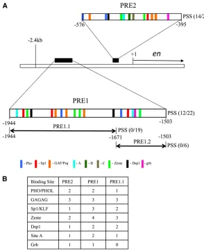
Figure 1 Predicted binding sites in PRE1 and PRE 2. (A) A schematic drawing of the genomic region of theenlocus showing theentranscription unit and the two upstream PREs, PRE1 and PRE2 (DeVidoet al.2008). Each PRE is shown as an enlargement with predicted binding sites/ consensus sequences represented by color-coded bars us-ing the followus-ing consensus sequences: Pho-GCCAT; GAF/ Psq-GAGAG; Zeste-BGAGTGV or YGAGYG (Mohrmann
et al.2002; Ringroseet al.2003) (B = C,T, or G; V = A,
C, or G; Y = C or T); Sp1/Klf-RRGGYG (R = A or G) (Shields and Yang 1998); Dsp1-GAAAA (Dejardin et al. 2005), Grh-TGTTTTTT or WCHGGTT (W = A or T; H = A, T, or C) (Blastyaket al.2006; Venkatesanet al.2003; Almeida and Bray 2005); site A-GAACNG. (N = A, C, T, or G). PRE1 (21944 to21503) when subdivided into PRE 1.1 or PRE 1.2 leads to loss of PSS with either fragment. The relative positions of sites B and C in PRE2 are shown (Americo
et al.2002). We did not include sites B or C in our binding
Ubx control F: CCAGCATAAAACCGAAAGGA Ubx control R: CGCCAAACATTCAGAGGATAG. PRE/bxd for PRE1 F: GCATCTGCGCTGTTCA PRE/bxd for PRE2 F: CGGTAACGCCCCGTGAG PRE/bxd R: TCGAGCAGCCCTGTTG
Bxd/Ubx F: GCCACATTTCCAGTTTCGACTTGC Bxd/Ubx R: AGTTCTTGCTACATCATGCAGTTATTG LacZ F: CAGCCGTTTGCCGTCTGAATTTGA LacZ R: TGGAAATCGCTGATTTGCGTGGTC.
Results
Analysis of predicted protein-binding sites in en PREs
PREs were first identified as fragments of DNA from the BX-C that could mediate repression by the PcG genes in embryonic reporter constructs (Müller and Bienz 1991; Simon et al. 1993). The ability of a fragment of DNA to mediate PcG repression in a reporter construct is known as the PRE-maintenance assay. PREs act in transgenes to me-diate the phenomenon of PSS of the mini-whitegene (Kassis
et al. 1991; Kassis 1994, 2002). While there is some con-troversy as to whether all fragments of DNA that mediate PSS are PREs (Kassis 2002), in our studies of PREs from the
en/inv region we have found a 100% correlation of frag-ments that demonstrate PRE activity in the maintenance assay and fragments that give PSS (Americo et al. 2002; Brown et al. 2005; Cunningham et al. 2010). Here, we assay PSS of mini-white in transgenic lines carrying PREs with mutated binding sites to assess if a given binding site might play a role in PRE activity. This is aP-element-based system with random integration into the genome. The abil-ity to mediate PSS relies to some extent on the site of in-sertion, and only60% of insertion sites give PSS with the wild-type PRE.
To date, four well-defined binding sites in PRE2 have been shown to be required for its PSS activity; these are binding sites for the proteins Pho/Phol, Spps, GAF, and Dsp1 (Brown et al. 1998, 2003; Decoville et al. 2001; Americo
et al.2002; Dejardinet al.2005). Chromatin-immunoprecip-itation studies show that these four proteins localize to theenPREs as well as to many other PREs in the genome (Klymenkoet al.2006; Schwartzet al.2006; Schuettengruber
et al. 2009; Brown and Kassis 2010). Furthermore, PSS is disrupted inpho,Dsp1, andSppsmutants, showing that these proteins are required for the PSS activity of PRE2 (Brown
et al. 1998; Dejardinet al. 2005; Brown and Kassis 2010). Here we define other binding sites required for PSS activity of PRE2 and compare the results with sequences required for PRE1 activity. Figure 1 shows the locations of PRE1 and PRE2 in relation to theentranscription start site and the locations and numbers of putative binding sites for proteins implicated in PRE activity. We included in our analysis the consensus sequences for the Grh and Zeste proteins as both have been implicated in the activity of some PREs (Saurinet al. 2001;
Huret al.2002; Tuckfieldet al.2002; Mulhollandet al.2004; Blastyáket al.2006).
Identification of site A
In previous studies we identified a protein-binding site within an 18-bp region of PRE2 (AGAGGGAGTGAAC-AGTGC), (Americoet al.2002 and Figure 2). Mutation of the central region of this site (AGTGA to TACCT) caused a complete loss of PSS (Americo et al.2002). Closer exam-ination of this 18-bp sequence showed that it contains a match to the consensus binding sequence for the Zeste protein (the sequence GGAGTGA matches the Zeste consen-sus site BGAGTGV) (see Figure 2A). We used gel mobility shift analysis and competition with oligonucleotides to de-termine the critical nucleotides for binding to the 18-bp oligonucleotide. Our data showed there was binding to a se-quence (site A) bound by an unknown protein (Protein A) that overlaps the Zeste consensus sequence (Figure 2). The Protein A gel mobility shift was not competed by an oligo-nucleotide containing known Zeste-binding sites (CGAGT-GAGTGTGAGTG; Figure 2B). Furthermore, mutation of the Zeste consensus sequence within the 18-bp sequence (Mut Z1) still competed the gel shift, whereas mutation of site A disrupted the competition, indicating that these were the critical nucleotides for binding (Figure 2C). We intro-duced a mutation that disrupts site A in PRE2 and assayed this mini-whitereporter construct in transgenicflies for the ability to give PSS (Figure 2D). When site A was mutated, PSS was reduced from 66% to 13% of the lines (comparable to what we see with mutation of either the Pho or the Spps sites) (Americo et al. 2002; Brown et al. 2005). We have identified the potential core-binding site of protein A to be GAACAG. This is where the mutations of the binding site are centered, and, in addition, another oligonucleotide contain-ing GAACAG was able to compete the site A gel mobility shift (data not shown). We are currently trying to identify the protein that binds to site A.
Zeste/Fs(1)h-binding sites
Zeste mutants do not affect PSS by PRE2
Zeste has been implicated in PRE activity due to its association with PRC1 (Saurin et al.2001; Huret al.2002; Mulhollandet al.2003), but it has also been linked to TrxG-mediated activation (Dejardin and Cavalli 2004). Genome-wide ChIP-chip studies show a limited overlap of Zeste protein binding with PcG protein binding in embryos, but Zeste was bound to the enPREs during embryogenesis (Moses et al.
2006). We tested whether a mutation inzeste(za) affected
PSS by PRE2; in heterozygous and homozygouszamutants,
there was no change in PSS of mini-white inflies carrying PRE2-mini-white constructs (data not shown). Therefore, Zeste is not the protein working at these sites in later de-velopment. It is possible that Fs(1)h is the protein that acts here (Changet al.2007).
Grh
Grh has been described as binding to PREs and to co-operatively interact with Pho. The binding site for Grh identified in theiab-7PRE was reported to be TGTTTTTT (Blastyáket al.2006). However, other groups report a Grh consensus sequence as WCHGGTT (where W is A or T and H is not G) (Venkatesan et al. 2003; Almeida and Bray 2005). We did not find any match to the TGTTTTTT se-quence withinenPRE2; however, there is a potential match to WCHGGTT toward the end of PRE2 (Figure 1). The
sequence is ACCGGCT (the one position that does not match is underlined). This sequence lies in a region that we have deleted from PRE2 in previous studies and yet still saw PSS (139-bp PRE, shown in Figure 2D) (Americo et al. 2002). The 139-bp PRE was functional but not as robust as the entire 181-bp region. There is a potential match to the Grh consensus in PRE1 (see below). Chromatin immunopre-cipitation with an antibody against Grh showed binding of Grh to each of these PREs in tissue derived from third instar larvae (Figure 2E). Grh is the only PRE-binding protein iden-tified so far that does not have ubiquitous expression.
Other factors
We previously showed specific binding of two other proteins (B and C) to sites within PRE2, but we did not know if they were needed for PSS activity (Americo et al.2002). Muta-tion of one of these sites (site C) does not impact PSS (data not shown). The binding site for protein B has been nar-rowed down to a 12-bp region (TGCCGCTATATG), but mutations of this site have not yet been tested in the PSS assay. The positions of these two binding sites relative to the other protein-binding sites in PRE2 are shown in Figure 1A. Some genome-wide ChIP experiments showed enrich-ment at PREs for TTG repeats and others found an enrichment of GT repeats (Ringroseet al.2003; Schuetten-gruber et al.2009). Deletion of the GTGT sequences in the PRE of thevestigial(vg) gene led to a reduction of silencing
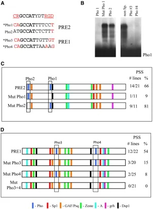
Figure 2 Defining site A and ChIP with aGrh. (A) The sequence of the Z1/A 18-bp annealed oligonucleotide used in the gel mobility shift assays with Schneider cell extract (SL2) shown in B and C and the mutant sites tested: Zeste site (Mut Z1), site A (Mut A), and the Zeste/siteA double mutant (Mut A+Z1). The mutated ba-ses are shown for each site, and altered baba-ses were made in the context of the 18-bp oligo sequence. (B) Gel mobil-ity shift assay using SL2 extract on the Z1/A oligo with 503
activity of this fragment, suggesting that this sequence may play a role in PcG repression (Okulskiet al.2011). There is only one TGTGT sequence in PRE2 that lies in the region of the 181-bp PRE that can be deleted without affecting PSS. There are three sequences in PRE1 that show GT repeats (GTGTGT or GTGTG). These sites have not been tested within the context of PRE1. To date, the protein that binds to GT repeats has not been identified.
PRE1
PRE1 originally referred to a 2-kb region from 22.4 kb to2576 bp upstream of theentranscription start site. This region acts as a PRE, mediates PSS, and contributes to hom-ing (Kassis 1994; DeVido et al. 2008; Cheng et al. 2012). Here we wanted to define the minimal fragment for PRE1. A 441-bp region (21944 to 21503) encompasses a region showing the most clustering of binding sites associated with PSS/PRE activity (DeVidoet al.2008). In a previous study this fragment gave PSS of mini-whitein 3/5 lines, but con-structs with the region 21944 to 21671bp (273 bp,
PRE1.1) or with 21672 to21503 (169 bp, PRE 1.2) did not (Kassis 1994) (Figure 1A). We generated additional lines of PRE1 and PRE1.1 and found that 12/22 PRE1 lines gave PSS while 0/19 PRE1.1 lines gave PSS. From here on we use PRE1 to refer to the 441-bp region from 21944 to21503 bp upstream of theentranscription start site.
PRE1 and PRE2 differ in their requirements for the number of Pho-binding sites for PSS
Mohd-Sarip et al. (2002, 2005, 2006) proposed a model requiring two Pho sites forin vivo chromatin association of Pho and the Polycomb core complex (PCC) with the iab-7
and bxd PREs. They proposed that sequences around the core consensus Pho-binding site contribute on one side to enhancing PHO binding and on the other side to coop-erative binding of Pho and the PRC1 core complex. The consensus sequence that they derived for Pho sites is CRGCCATYDTRGD (R is A or G; D is A, T, or G). CR (under-lined) is called the U sequence and is thought to enhance Pho binding. RGD (underlined) is the consensus for the site
Figure 3 Defining the PHO site requirements of PRE1 and PRE2. (A) Sequences of Pho1-4 lined up with the extended Pho consensus-binding sequence proposed by Mohd-Sarip
et al. (2005). The bases that are part of the extended
that is required for association of PCC. When RGD was mu-tated, both the cooperative binding of Pho with PCC and PSS mediated by theiab-7PRE were abolished (Mohd-Sarip
et al.2005).
We have previously shown that mutation of a Pho-binding site (Pho1 in Figure 3) greatly diminishes PSS by PRE2 (Brownet al.1998). PRE2 has a second potential Pho-binding site (Pho2) on the 59end of the PRE (Figure 3). However, when this site is used as a competitor in a gel shift assay with in vitro-transcribed/translated full-length Pho bound to the Pho1 site, there is very little competition (Fig-ure 3B). Conversely, when the labeled Pho2 oligo was used in the gel mobility shift assay, no specific gel shift was detected (data not shown). Thus, this second Pho site is at best a very weak binding site for Pho compared to the Pho1 site. However, in the context of the PRE, perhaps a weak site can bind Pho in combination with a high-affinity binding site. We mutated the Pho2-binding site in the context of PRE2 in a transformation vector carrying the mini-white
gene and looked for PSS. PSS was unaffected in lines where the Pho1 site was mutated (88% PSS for mutated Pho1 compared to 66% for wild-type PRE2) (Figure 3C). Thus, PRE2 does not require two Pho sites for PSS activity.
There are two predicted Pho sites in PRE1 and both compete as well as the Pho1 site in a gel mobility shift assay usingin vitro-transcribed/translated full-length Pho protein (Figure 3B). We note that Pho1, -3, and -4 all give good matches to the U site, whereas Pho2 does not (Figure 3A).
These data support the view that the U site enhances Pho binding. In contrast, none of the Pho sites contain a perfect match to the RGD sequence required for recruitment of PCC
in vitro(Mohd-Saripet al.2005). We introduced mutations that disrupt Pho binding into the Pho3 and Pho4 sites in-dividually and together in PRE1. Disruption of either the Pho3- or the Pho4-binding sites decreased the PSS activity of the fragment (Figure 3D). When both Pho sites were mutated, we saw no PSS. From this, we conclude that the PRE1 requires two Pho-binding sites for full function. Muta-tion of either site leads to only partial loss of activity. Sim-ilarly, mutation of the single Pho site in PRE2 does not completely abrogate function. We suggest that in PRE2 an-other DNA-binding protein plays the role of the second Pho site for PcG complex recruitment.
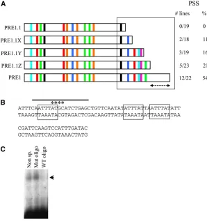
An AT-rich region is required for full activity of PRE1
PRE1.1 contains binding sites for all known factors required for the activity of PRE2, but it has only one of the two Pho sites required for PRE1 activity (Figure 1 and Figure 3). This led to the prediction that adding back the piece of DNA that contains the second Pho site to PRE1.1 might restore full PSS activity to PRE1.1. Surprisingly, adding back the second Pho site only gives PSS in 11% of the lines, which is no better than having a single Pho site (Figure 4A). Clearly, something else is required for the PSS activity of PRE1. We then added back more pieces of DNA from this fragment to see what else was required to recover full PSS activity.
The sequences added back contain consensus-binding sites for a number of factors believed to be involved in PRE activity (Sp1, Site A, Grh, Zeste site) (Figure 4A). As these binding sites are added back, PSS increased from 11% (with the second Pho site) to 21%, but full PSS activity was not restored until we included the entire PRE1 fragment (54%). The last sequence added back did not contain any matches to the binding sites previously identified at theDrosophila
PREs. This region of DNA is extremely AT rich (72%) and is perhaps needed for structural reasons such as the ability of the DNA to bend and facilitate long-range interactions (Fig-ure 4B). The sequence contains three repeats of ATTTAT. Gel mobility shift experiments show that at least one factor spe-cifically binds to this sequence (Figure 4C).
Studies of evolutionary conservation of transcription-factor-binding sites in the 12 sequenced species of Drosoph-ila have proven to be a very valuable tool in the study of enhancers; however, they have not been as useful in the study of PREs. PREs seem to have built-in plasticity in the arrangement of potential binding sites, making them very hard to identify when using a best-fit analysis based on homology over large regions (Dellinoet al.2002; Hauenschild
et al. 2008). Comparison of the en 2.4-kb region encom-passing PRE1 and PRE2 over the 12 Drosophila species showed little conservation between all 12 species. How-ever, when the equivalent region of each species is searched in GenePalette for consensus sequences for the factors that we have discussed here, many of the sites are present. The Pho and GAF/PSQ sites are the most conserved features. Kassis et al.(1989) compared the upstream region of the
en gene betweenD. melanogasterand D. virilis. The corre-sponding region for PRE2 in D. virilis was identified and shown to give PSS (Kassis 1994), implying that this element most likely contains all the protein-binding sites necessary for PRE function. Using GenePalette, we searched the
D. virilis PRE2 sequence for the binding sites that we have studied here. The results are shown in Figure 5A. Between
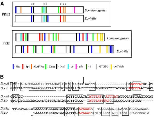
D. melanogaster and D. virilis there is conservation of the Pho and overlapping Dsp1 site, site A, site B, Spps, and GAF/PSQ. There is conservation of a sequence that we have designated site B (GCCGCT), which gives a specific gel shift with Drosophilaembryonic extracts (Americo et al. 2002). Not conserved are the Zeste consensus-binding sites, again implying that Zeste is most likely not the protein that binds to these sites. We extended this analysis to look for conser-vation of PRE1, although we note that this DNA fragment has not been tested for PRE activity. In PRE1, the order and spacing of the predicted binding sites is not conserved, al-though many of the binding sites are present. Furthermore, there is an AT-rich region at the end of the predicted
D. virilis PRE1 including multiple copies of the ATTTAT se-quence (Figure 5B). Without further mutational analysis, it is not possible to distinguish between a requirement for general AT richness and a specific protein-binding DNA sequence.
Activity of PRE1 and PRE2 differs in a bxd-Ubx-lacZ reporter construct
We have previously shown that PRE2 can act as a PRE-maintenance element in an embryonicbxd-Ubx-lacZ P-element reporter construct (Americo et al. 2002). Furthermore, both PRE2 and PRE1 (contained within a 2-kb fragment) acted as PREs in an embryonic en-lacZreporter construct, but PRE1 had greater activity in this context (DeVido et al.
2008). To directly compare the activities of PRE1 and PRE2, they were tested in abxd-Ubx-lacZreporter inserted into the genome using fC31 RMCE (Groth et al. 2004; Bateman
et al.2006) incorporated into the attP insertion site located at 52D (see Bateman et al.2006). The recombinants gen-erated by this system do not carry the mini-white gene;
Figure 5 Comparison of predicted binding sites between
D. melanogasterandD. virilisat PRE1 and PRE2. (A) The
predicted binding site comparison between PRE1 and -2 of
D. melanogasterandD. virilis. See Figure 1 legend for the
consensus-binding sequences used. The asterisks denote sites conserved in PRE2. Note that theD. virilisPRE1 frag-ment has not been tested for PRE activity. (B) Comparison of the AT-rich sequence at the end of PRE1 between
D. melanogaster andD. virilis. The arrows delineate the
AT-rich sequence of PRE1; extra bases on either side are included. Spaces are introduced to get the best lineup of sequence. TheD. virilissequence contains several ATTTAT sequences (highlighted in red), and boldface indicates the most AT-rich part of the sequence: 72% (83/113 bases) and 77% (111/144 bases) AT richness forD. melanogaster
therefore PSS cannot be assessed at this location. This vector and insertion site was previously used to demonstrate the embryonic maintenance activity of the eve PRE (Fujioka
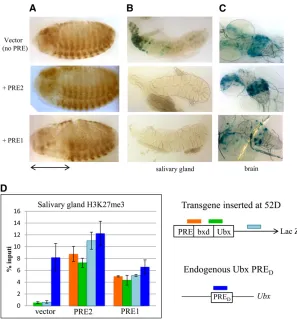
et al. 2008). Based on genome-wide studies of H3K27me3 in embryos (Schuettengruberet al.2009), the 52D insertion site does not lie in an H3K27me3 domain. Embryos with vector alone, PRE1 vector, and PRE2 vector were stained with an antibody againstb-galactosidase (b-gal). In the ab-sence of a PRE,bxd-Ubx-lacZis expressed in parasegments 6–13 early in development, but later becomes derepressed and is expressed throughout the entire embryo (Figure 6A). PRE1, but not PRE2, was able to maintain the restricted expression of bxd-Ubx-lacZ in this chromosomal insertion site (Figure 5A). That PRE2 did not maintain repression was somewhat surprising, since we previously showed that it was able to maintain the restricted pattern of the same
bxd-Ubx-lacZreporter gene at 9 of 12 chromosomal insertion sites in aP-element-based vector (Brownet al.2005). With RCME, constructs were inserted in the genome in either orientation, and similar results were obtained from both orientations. This shows that there is a difference in how PRE1 and PRE2 work in embryos at the 52D insertion site. We next examined thelacZexpression in thebxd-Ubx-lacZ
larvae, with and without the PREs (Figure 6A). In larvae with vector alone,b-gal activity was detected in the salivary glands and in a subset of cells in the brain. This pattern of expression was not due to the insertion site since a similar pattern was observed using the same vector inserted into an attP site at
95E (Fujiokaet al.2008; data not shown). Addition of PRE2 into the vector abrogated expression ofb-gal in the salivary glands but not in the brain of the transgenic larvae. Addition of PRE1 to the vector abrogatedb-gal expression in the sal-ivary gland and greatly reduced the number of cells in the brain that express b-gal. To investigate whether the repres-sion ofb-gal activity was due to the action of the PcG pro-teins, we assayed for the presence of H3K27me3 on the transgene by chromatin immunoprecipitation followed by qPCR. H3K27me3 is a signature of PcG activity, and Ubx
PREDis included as a positive control. In larvae with vector
alone, H3K27me3 is not associated with the lacZ gene in either the brain or the salivary glands. In contrast, salivary glands from flies with vector plus PRE1 or PRE2 have H3K27me3 associated with thelacZgene. The fact that PRE2 can act as a PRE in the salivary glands, but not in the brain or embryo, suggests that either its activity is tissue specific or that the large number of closely aligned transgenes present in the polytene chromosomes potentiated the activity of PRE2, allow-ing it to overcome the activity of the transcriptional activators acting on this construct.
Discussion
PRE architecture
PREs are made up of binding sites for many different proteins, some known and others unknown. Our detailed
Figure 6 Activity of PRE1 and PRE2 in the Ubx-lacZ re-porter construct inserted at 52D in embryos and larval tissues. (A) Anti-b-gal-stained embryos, (B) salivary glands, and (C) brains from transgenic lines carrying a
bxd-Ubx-lacZreporter gene inserted at 52D. (B, C)b-gal activity
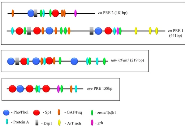
analysis of PRE2 suggests that at least six DNA-binding proteins, including Pho/Phol, Dsp1, Spps, GAF/Psq, Zeste/ Fs(1)h, and Protein A, are required for its PSS activity. PRE1 contains presumptive binding sites for all of those proteins as well as an AT-rich region not found in PRE2. Furthermore, the spacing and order of sites differs between these two PREs (Figure 7). We examined the minimal eve
and iab-7/Fab-7PREs for the presence of these sites (Fig-ure 7). We note that not all sites were found in theeveand
iab-7/Fab-7PREs. For example, we could not find a Dsp1 consensus sequence in theevePRE, and Dsp1 is not bound to this locus in embryos as shown by chromatin immuno-precipitation (Schuettengruberet al.2009). A comparison of the order and arrangement of the confirmed and puta-tive binding sites ofenPRE1,enPRE2,evePRE, andFab7/ iab-7PRE shows a large variation in the order and spacing of the binding sites between the different PREs. This vari-ation may reflect critical differences that are integral to how a specific PRE functions at its endogenous locus; how-ever, this is hard to assess given that even within a given PRE the number, order, and spacing of predicted Dsp1, Pho, GAF, and Zeste sites was found to diverge rapidly within PREs present at conserved positions in various Drosophila
species (Hauenschild et al. 2008). The functional conse-quences of these differences have not been explored, but it does point to an overall plasticity in the system.
This rapid evolution may result in part from redundancy in PRE activity. In the en gene, PRE1 and PRE2 are con-tained within a 1.6-kb region and may act together to re-cruit PcG complexes in their normal context. However, both of these PREs are dispensable for viability in the lab-oratory (Chenget al.2012). Theen/invgene complex has two other major PREs and a number of minor ones that can apparently take over PRE1 and PRE2 functions. This ap-parent redundancy in PRE activity may allow for rapid evolution and increase the diversity of these regulatory elements.
Functional differences between PREs
Both PRE1 and PRE2 mediate PSS of the mini-white gene but behave differently in thebxd-Ubx-lacZreporter complex inserted by RCME at an attPsite at 52D. While both PRE1 and PRE2 could silence the bxd-Ubx-lacZ gene in salivary gland nuclei via the PcG proteins (as measured by H3K27me3 association with thelacZgene), only PRE1 was active in em-bryos and larval brain. Why this difference? We suggest that PREs evolve in the context of other regulatory DNA and that, while retaining overlapping functions, they may adapt to work better with different enhancers. So, while core PRE DNA-binding proteins may remain relatively constant between PREs, some PREs may require additional factors to overcome the activity of specific enhancers.
Differences in PRE activities in transgenes have been previously described (Horard et al. 2000; Okulski et al.
2011; Parket al.2012). In one study, multimerized subfrag-ments of thebxdPRE showed different behaviors in a
mini-whiterepression assay (Horardet al.2000). In another, dif-ferent PREs from thePsc/Su(z)2complex exhibited different silencing activities on an imaginal disk reporter construct (Park et al. 2012). Both these studies were done in P -ele-ment-based vectors, and thus the activities of the reporter constructs were not examined in the same insertion sites. Since PREs are notoriously sensitive to the effects offlanking DNA, it is not easy to determine whether the differences observed were a reflection of intrinsic differences in the activity of the PREs or of the different insertion sites of the reporter genes. To avoid this problem, Okulskiet al.(2011) examined the activity of a PRE from thevggene with activity of the iab-7/Fab-7PRE inserted in the genome at four dif-ferent insertion sites viafC31 integration. This careful study showed that these two PREs exhibited different degrees of mini-whiterepression at different insertion sites and be-haved differently with respect to PSS at the same insertion sites. The problem with this study was that the investigators
Figure 7 Comparison of the order and spatial arrange-ment of consensus-factor-binding sites in en, eve, and
iab-7/Fab7 PREs. The sequences of enPRE1 and PRE2,
used relatively large fragments of DNA (1.6 kb). Thus it is likely that other regulatory sequences were contained within the PRE fragments, and it is not known how additional reg-ulatory sequences modify PRE activity. Even in the 181-bp DNA fragment that we call PRE2, there is another regulatory activity, a promoter-tethering element, which may affect its ability to act as a PRE in some chromosomal locations (Kwon
et al.2009). In previous studies from our group, when a 2.6-kb piece ofenDNA that included PRE1 and PRE2 andfl ank-ing DNA was included in a bxd-Ubx-lacZ P-element-based transgene, the enfragment almost completely silenced the entire lacZ expression pattern in embryos (Americo et al.
2002). We suspect that regulatory DNA within that frag-ment was able to silence the bxd enhancer and that this activity might be separable from the PRE activityper se.
Concluding remarks
From this work and the work of many others we conclude that PREs are a diverse group of related elements that have overlapping but not identical activities. PREs vary in both size and binding-site composition requiring a number of different DNA-binding proteins to function collaboratively to recruit PcG complexes and introduce repressive histone modifications. The diverse PRE architecture could influence exactly which PcG protein complexes are recruited and what types of enhancers they are able to silence or repress.
Acknowledgments
We thank Miki Fujioka for the bxd-Ubx-lacZ RCME vector and for transgenic lines; Bill McGinnis for the generous gift of Grh antibodies; and Emma Hornick for construction of the
bxd-Ubx-lacZ reporter constructs. We also thank Yuzhong Cheng, Payal Ray, and Sandip De for critical reading of this manuscript. This work was supported by the Intramural Research Program of the Eunice Kennedy Shriver National Institute of Child Health and Human Development, National Institutes of Health.
Literature Cited
Almeida, M. S., and S. J. Bray, 2005 Regulation of post-embryonic neuroblasts byDrosophilaGrainy head. Mech. Dev. 122: 1282– 1293.
Americo, J., M. Whiteley, J. L. Brown, M. Fujioka, J. B. Jayneset al., 2002 A complex array of DNA-binding proteins required for pairing-sensitive silencing by a Polycomb group response ele-ment from the Drosophilaengrailedgene. Genetics 160: 1561– 1571.
Bateman, J. R., A. M. Lee, and C.-T. Wu, 2006 Site-specific trans-formation of Drosophila via fC31 Integrase-mediated cassette exchange. Genetics 173: 769–777.
Benson, M., and V. Pirrotta, 1988 TheDrosophila zeste protein binds cooperatively to sites in many gene regulatory regions: implications for transvection and gene regulation. EMBO J. 7: 3907–3915.
Blastyák, A., R. K. Mishra, F. Karch, and H. Gyurkovics, 2006 Efficient and specific targeting of Polycomb group proteins
requires cooperative interaction between Grainy head and Pleio-homeotic. Mol. Cell. Biol. 26: 1434–1444.
Brown, J. L., and J. A. Kassis, 2010 Spps, a Drosophila Sp1/KLF family member binds to PREs and is required for PRE activity late in development. Development 137: 2597–2602.
Brown, J. L., D. Mucci, M. Whiteley, M. L. Dirksen, and J. A. Kassis, 1998 TheDrosophilaPolycomb group genepleiohomeotic enc-odes a DNA binding protein with homology to the transcription factor YY1. Mol. Cell 1: 1057–1064.
Brown, J. L., C. Fritsch, J. Müller, and J. A. Kassis, 2003 The Drosophila pho-like gene encodes a YY1-related DNA binding protein that is redundant withpleiohomeoticin homeotic gene silencing. Development 130: 285–294.
Brown, J. L., D. J. Grau, S. K. DeVido, and J. A. Kassis, 2005 An Sp1/KLF binding site is important for the activity of a Polycomb group response element from the Drosophila engrailed gene. Nucleic Acids Res. 33: 5181–5189.
Busturia, A., A. Lloyd, F. Bejarano, M. Zavortink, H. Xin et al., 2001 The MCP silencer of the Drosophila Abd-B gene requires both Pleiohomeotic and GAGA factor for the maintenance of repression. Development 128: 2163–2173.
Cao, R., L. Wang, H. Wang, L. Xia, H. Erdjument-Bromage et al., 2002 Role of histone H3 lysine 27 methylation in Polycomb-group silencing. Science 298: 1039–1043.
Chang, Y. L., B. King, S. C. Lin, J. A. Kennison, and D. H. Huang, 2007 A double-bromodomain protein, FSH-S, activates the homeotic geneUltrabithoraxthrough a critical promoter-proximal region. Mol. Cell. Biol. 15: 5486–5489.
Cheng, Y., D. Y. Kwon, A. L. Arai, D. Mucci, and J. A. Kassis, 2012 P-element homing is facilitated byengrailed polycomb-group response elements in Drosophila melanogaster. PLoS ONE 7: e30437.
Cunningham, M. D., J. L. Brown, and J. A. Kassis, 2010 Characterization of the Polycomb group response elements of the Drosophila melanogaster invectedlocus. Mol. Cell. Biol. 30: 820–828. Czermin, B., R. Melfi, D. McCabe, V. Seitz, A. Imhof et al.,
2002 Drosophila enhancer of Zeste/ESC complexes have a his-tone H3 methyltransferase activity that marks chromosomal Pol-ycomb sites. Cell 111: 185–196.
Decoville, M., E. Giacomello, M. Leng, and D. Locker, 2001 DSP1, an HMG-like protein, is involved in the regulation of homeotic genes. Genetics 157: 237–244.
Dejardin, J., and G. Cavalli, 2004 Chromatin inheritance upon Zeste-mediated Brahma recruitment at a minimal cellular mem-ory module. EMBO J. 23: 857–868.
Dejardin, J., A. Rappailles, O. Cuvier, C. Grimaud, M. Decoville
et al., 2005 Recruitment of Drosophila Polycomb group pro-teins to chromatin by DSP1. Nature 434: 533–538.
Dellino, G. I., C. Tatout, and V. Pirrotta, 2002 Extensive conser-vation of sequences and chromatin structures in the bxd Poly-comb Response Element amongDrosophilidspecies. Int. J. Dev. Biol. 46: 133–141.
DeVido, S. K., D. Kwon, J. L. Brown, and J. A. Kassis, 2008 The role of Polycomb-group response elements in regulation of en-grailedtranscription in Drosophila. Development 135: 669–676. Fiedler, T., and M. Rehmsmeier, 2006 jPREdictor: a versatile tool for the prediction of cis-regulatory elements. Nucleic Acids Res. 34: W546–W550.
Fritsch, C., J. L. Brown, J. A. Kassis, and J. Müller, 1999 The DNA-binding Polycomb group protein Pleiohomeotic mediates silencing of aDrosophilahomeotic gene. Development 126: 3905–3913. Fritsch, C., D. Beuchle, and J. Müller, 2003 Molecular and genetic
analysis of the Polycomb group gene sex combs extra/Ring in Drosophila. Mech. Dev. 120: 949–954.
both active and repressed transcriptional states through a single site. Development 135: 4131–4139.
Gieni, R. S., and M. J. Hendzel, 2009 Polycomb group protein gene silencing, non-coding RNA, stem cells, and cancer. Bio-chem. Cell Biol. 87: 711–746.
Grimm, C., R. Matos, N. Ly-Hartig, U. Stuerwald, D. Lindneret al., 2009 Molecular recognition of histone lysine methylation by the Polycomb group repressor dSfmbt. EMBO J. 28: 1965–1977. Groth, A. C., M. Fish, R. Nusse, and M. P. Calos, 2004 Construction of transgenic Drosophila by using the site-specific integrase from phagefC31 integrases. Genetics 166: 1775–1782.
Hagstrom, K., M. Müller, and P. Schedl, 1997 A Polycomband GAGA dependent silencer adjoins the Fab-7 boundary in the Drosophila bithorax complex. Genetics 146: 1365–1380. Hauenschild, A., L. Ringrose, C. Altmutter, R. Paro, and M.
Re-hmsmeier, 2008 Evolutionary plasticity of Polycomb/Trithorax response elements in Drosophila species. PLoS Biol. 6: e261. Horard, B., C. Tatout, S. Poux, and V. Pirrotta, 2000 Structure of
a polycomb response element and in vitro binding of Polycomb group complexes containing GAGA factor. Mol. Cell. Biol. 20: 3187–3197.
Hur, M. W., J. D. Laney, S. H. Jeon, J. Ali, and M. D. Biggin, 2002 Zeste maintains repression of Ubxtransgenes: support for a new model of Polycomb repression. Dev. 129: 1339–1343. Kassis, J. A., 1994 Unusual properties of regulatory DNA from the Drosophilaengrailedgene: three“pairing-sensitive”sites within a 1.6-kb region. Genetics 136: 1025–1038.
Kassis, J. A., 2002 Pairing-sensitive silencing, Polycomb group re-sponse elements, and transposon homing in Drosophila. Adv. Genet. 46: 421–438.
Kassis, J. A., and J. L. Brown, 2013 Polycomb group response ele-ments in Drosophila and vertebrates. Adv. Genet. 83: 83–118. Kassis, J. A., C. Desplan, D. K. Wright, and P. H. O’Farrell,
1989 Evolutionary conservation of homeodomain-binding sites and other sequences upstream and within the major tran-scription unit of the Drosophila segmentation gene engrailed. Mol. Cell. Biol. 9: 4304–4311.
Kassis, J. A., E. P. Vansickle, and S. M. Sensabough, 1991 A frag-ment of engrailed regulatory DNA can mediate transvection of the white gene in Drosophila. Genetics 128: 751–761. Kerppola, T. K., 2009 Polycomb group complexes: many
combi-nations, many functions. Trends Cell Biol. 19: 692–704. Klymenko, T., B. Papp, W. Fischle, T. Kocher, M. Schelderet al.,
2006 A Polycomb group protein complex with sequence-specific DNA-binding and selective methyl-lysine-binding activities. Genes Dev. 20: 1110–1122.
Kim, M., and W. McGinnis, 2011 Phosphorylation of Grainy head by ERK is essential for wound-dependent regeneration of an epidermal barrier but dispensable for embryonic barrier devel-opment. Proc. Natl. Acad. Sci. USA 108: 650–655.
Kwon, D., D. Mucci, K. Langlais, J. L. Americo, S. K. DeVidoet al., 2009 Enhancer-promoter communication at theDrosophila en-grailedlocus. Development 136: 3067–3075.
Kwong, C., B. Adryan, I. Bell, L. Meadows, S. Russell et al., 2008 Stability and dynamics of Polycomb target sites in Dro-sophila development. PLoS Genet. 4: e1000178.
Lagarou, A., A. Mohd-Sarip, Y. M. Moshkin, G. E. Chalkley, K. Be-zatarostiet al., 2008 dKDM2 couples histone H2A ubiquitina-tion to histone H3 demethylaubiquitina-tion during Polycomb group silencing. Genes Dev. 22: 2799–2810.
Mishra, R. K., J. Mihaly, S. Barges, A. Spierer, F. Karch et al., 2001 The iab-7 Polycomb response element maps to a nucleo-some-free region of chromatin and requires both GAGA and Plei-ohomeotic for silencing activity. Mol. Cell. Biol. 21: 1311–1318. Moazed, D., and P. H. O’Farrell, 1992 Maintenance of the
en-grailedexpression pattern by Polycomb group genes in Drosoph-ila. Development 116: 805–810.
Mohd-Sarip, A., F. Venturini, G. E. Chalkley, and C. P. Verrijzer, 2002 Pleiohomeotic can link Polycomb to DNA and mediate transcriptional repression. Mol. Cell. Biol. 22: 7473–7483. Mohd-Sarip, A., F. Cleard, R. K. Mishra, F. Karch, and C. P. Verrijzer,
2005 Synergistic recognition of an epigenetic DNA element by Pleiohomeotic and a Polycomb core complex. Genes Dev. 19: 1755–1760.
Mohd-Sarip, A., J. A. Van der Knaap, C. Wyman, R. Kanaar, P. Schedlet al., 2006 Architecture of a polycomb nucleoprotein complex. Mol. Cell 24: 91–100.
Mohrmann, L., A. J. Kal, and C. P. Verrijzer, 2002 Characterization of the extended Myb-like DNA-binding domain of trithorax group protein Zeste. J. Biol. Chem. 277: 47385–47392.
Moses, A. M., D. A. Pollard, D. A. Nix, V. N. Iyer, X.-Y. Liet al., 2006 Large-scale turnover of functional transcription factor binding sites inDrosophila. PLOS Comput. Biol. 2: e130. Mulholland, N. M., I. F. King, and R. E. Kingston, 2003 Regulation
of Polycomb group complexes by the sequence-specific DNA binding proteins zeste and GAGA. Genes Dev. 17: 2741–2746. Müller, J., and M. Bienz, 1991 Long range repression conferring
boundaries of Ultrabithorax expression in the Drosophila em-bryo. EMBO J. 10: 3147–3155.
Müller, J., and J. A. Kassis, 2006 Polycomb response elements and targeting of Polycomb group proteins inDrosophila. Curr. Opin. Genet. Dev. 16: 476–484.
Müller, J., and C. P. Verrijzer, 2009 Biochemical mechanisms of gene regulation by Polycomb group protein complexes. Curr. Opin. Genet. Dev. 19: 150–158.
Müller, J., C. M. Hart, N. J. Francis, M. L. Vargas, A. Senguptaet al., 2002 Histone methyl transferase activity of a Drosophila Poly-comb group repressor complex. Cell 111: 197–208.
Negre, N., J. Hennetin, L. V. Sun, S. Lavrov, M. Bellis et al., 2006 Chromosomal distribution of PcG proteins during Dro-sophiladevelopment. PLoS Biol. 4: 170.
Nekrasov, M., T. Klymenko, S. Fraterman, B. Papp, K. Oktabaet al., 2007 Pcl-PRC2 is needed to generate high levels of H3–K27 trimethylation at Polycomb target genes. EMBO J. 26: 4078– 4088.
Oktaba, K., L. Gutierrez, J. Gagneur, C. Girardot, A. K. Sengupta
et al., 2008 Dynamic regulation of polycomb group protein complexes controls pattern formation and the cell cycle in Dro-sophila. Dev. Cell 15: 877–889.
Okulski, H., B. Druck, S. Bhalerao, and L. Ringrose, 2011 Quantitative analysis of Polycomb response elements (PREs) at identical genomic locations distinguishes contribu-tions of PRE sequence and genomic environment. Epigenetics Chromatin 4: 1–16.
Park, S. Y., Y. B. Schwartz, T. G. Kahn, D. Asker, and V. Pirrotta, 2012 Regulation of Polycomb group genes Psc and Su(z)2 in Drosophila melanogaster. Mech. Dev. 128: 536–547.
Pirrotta, V., 1988 Vectors for P-mediated transformations in Dro-sophila, pp. 437–456 inVectors: A Survey of Molecular Cloning Vectors and Their Uses, edited by R. L. Rodriguez, and D. T. Denhardt. Butterworths, Boston.
Rebeiz, M., and J. W. Posakony, 2004 GenePalette: an unusual software tool for genome sequence visualization and analysis. Dev. Biol. 15: 431–438.
Ringrose, L., M. Rehmsmeier, J. M. Dura, and R. Paro, 2003 Genome-wide prediction of Polycomb/Trithorax response elements inDrosophila melanogaster. Dev. Cell 5: 759–771. Saurin, A. J., Z. Shao, H. Erdjument-Bromage, P. Tempst, and R. E.
Kingston, 2001 A Drosophila Polycomb group complex in-cludes Zeste and dTAFII proteins. Nature 412: 655–660. Scheuermann, J. C., A. G. Alonso, K. Oktaba, N. Ly-Hartig, R. K.
Schuettengruber, B., D. Chourrout, M. Veroort, B. Leblanc, and G. Cavalli, 2007 Genome regulation by polycomb abd trithorax proteins. Cell 128: 735–745.
Schuettengruber, B., M. Ganapathi, B. Leblanc, M. Portoso, and R. Jascheket al., 2009 Functional anatomy of polycomb and tri-thorax chromatin landscapes inDrosophilaembryos. PLoS Biol.
7:0001–0018.
Schwartz, Y. B., T. G. Kahn, D. A. Nix, X. Y. Li, R. Bourgonet al., 2006 Genome-wide analysis of Polycomb targets in Drosophila melanogaster. Nat. Genet. 38: 700–705.
Shao, Z., F. Raible, R. Mollaaghababa, J. R. Guyon, C. T. Wuet al., 1999 Stabilization of chromatin structure by PRC1, a Polycomb complex. Cell 98: 37–46.
Shields, J. M., and V. W. Yang, 1998 Identification of the DNA sequence that interacts with the gut-enriched Krüppel-like fac-tor. Nucleic Acids Res. 26: 796–802.
Shimell, M. J., A. J. Peterson, J. Burr, J. A. Simon, and M. B. O’Connor, 2000 Functional analysis of repressor binding sites in the iab-2 regulatory region of the abdominal-A homeotic gene. Dev. Biol. 218: 38–52.
Simon, J. A., and R. E. Kingston, 2009 Mechanisms of Polycomb gene silencing: knowns and unknowns. Nat. Rev. Biol. 10: 697– 708.
Simon, J., A. Chiang, W. Bender, M. J. Shimell, and M. O’Connor, 1993 Elements of the Drosophila bithorax complex that
mediate repression by Polycomb group products. Dev. Biol. 158: 131–144.
Tolhuis, B., E. de Wit, I. Muijrers, H. Teunissen, and W. Talhout
et al. 2006 Genome-wide profiling of PRC1 and PRC2 Poly-comb chromatin binding in Drosophila melanogaster. Nat. Genet. 38: 694–699.
Tuckfield, A., D. R. Clouston, T. M. Wilanowski, L. L. Zhao, J. M. Cunninghamet al., 2002 Binding of the RING Polycomb pro-teins to specific target genes in a complex with the Grainy head-like family of developmental transcription factors. Mol. Cell. Biol. 22: 1936–1946.
Venkatesan, K., H. R. McManus, C. C. Mello, T. F. Smith, and U. Hansen, 2003 Functional conservation between members of an ancient duplicated transcription factor family, LSF/Grainy head. Nucleic Acids Res. 31: 4304–4316.
Wang, H., and L. Wang, H. Erdjument-Bromage, M. Vidal, P. Tempstet al., 2004 Role of histone H2A ubiquitination in Polycomb silencing. Nature 431: 873–878.
Wang, L., N. Jahren, E. L. Miller, C. S. Ketel, D. R. Mallinet al., 2010 Comparative analysis of chromatin binding by sex comb on midleg (SCM) and other Polycomb group repress-ors at a Drosophila Hox gene. Mol. Cell. Biol. 30: 2584– 2593.