| INVESTIGATION
NATF (Native and Tissue-Speci
fi
c Fluorescence): A
Strategy for Bright, Tissue-Speci
fi
c GFP Labeling of
Native Proteins in
Caenorhabditis elegans
Siwei He,*,†Andrea Cuentas-Condori,†and David M. Miller, III*,†,1
*Program in Neuroscience, Vanderbilt University, Nashville, Tennessee and†Department of Cell and Developmental Biology, Vanderbilt University School of Medicine, Nashville, Tennessee 37240 ORCID IDs: 0000-0002-4847-0031 (A.C.-C.); 0000-0001-9048-873X (D.M.M.)
ABSTRACTGFP labeling by genome editing can reveal the authentic location of a native protein, but is frequently hampered by weak GFP signals and broad expression across a range of tissues that may obscure cell-specific localization. To overcome these problems, we engineered a Native And Tissue-specific Fluorescence (NATF) strategy that combines genome editing and split-GFP to yield bright, cell-specific protein labeling. We use clustered regularly interspaced short palindromic repeats CRISPR/Cas9 to insert a tandem array of seven copies of the GFP11b-strand (gfp11x7) at the genomic locus of each target protein. The resultantgfp11x7knock-in strain is then crossed with separate reporter lines that express the complementing split-GFP fragment (gfp1-10) in specific cell types, thus affording tissue-specific labeling of the target protein at its native level. We show that NATF reveals the otherwise undetectable intracellular location of the immunoglobulin proteinOIG-1and demarcates the receptor auxiliary proteinLEV-10at cell-specific synaptic domains in theCaenorhabditis elegansnervous system.
KEYWORDSC. elegans; cell specificity; genome editing; GFP; NATF; native expression; protein localization
R
ELIABLE localization of a given protein can provide useful clues to its mechanism of action. One way to achieve this goal is to label the protein of interest with tags, such as fluorescent proteins (e.g., GFP) (Chalfie 2009; Remington 2011) or small peptides (e.g., FLAG or HA) (Terpe 2003). Because tagged proteins are typically expressed with heter-ologous promoters or from multicopy transgenic arrays, this approach can result in misleading signals due to overexpres-sion (Praitis et al.2001). This problem can be obviated by using clustered regularly interspaced short palindromic re-peats (CRISPR)/Cas9 for single-copy labeling of the native protein (Hsu et al. 2014; Dickinson et al. 2015), but this genome-editing strategy suffers from two additional limita-tions. First, the endogenous expression level of a target pro-tein may be too low for detection. Second, the propro-tein ofinterest may be expressed in several tissues, thus preventing a clear delineation of cell-specific localization. Previous ef-forts have focused on solving each of these problems sepa-rately. For example, strategies to enhance detection include the use of brighter fluorescent proteins (El Mouridi et al.
2017; Hostettler et al. 2017) and the development of the SunTag label, which uses a scaffold-like structure to recruit multiple copies of GFP (Tanenbaumet al.2014). The“FLP-on” strategy uses cell-specific FLP drivers to activate GFP expression from CRISPR/Cas9-engineered FRT sites but may not yield a visible signal for low-expressing genes (Schwartz and Jorgen-sen 2016). Here, we describe an experimental approach, NATF (Native And Tissue-specific Fluorescence or“Native”), that ex-ploits a combinatorial strategy to achieve both bright and cell-specific labeling of the protein of interest.
Our approach relies on thefinding that the barrel-like GFP structure can be reconstituted by the spontaneous interaction of two separate GFP peptides derived from the highly stable GFP variant, superfolder GFP. The larger of these fragments is comprised of thefirst 10b-strands (GFP1-10). Its smaller complement, a short, 16 amino acid sequence, contains the 11thb-strand (GFP11) (Cabantouset al.2005). The reconstituted Copyright © 2019 by the Genetics Society of America
doi:https://doi.org/10.1534/genetics.119.302063
Manuscript received February 27, 2019; accepted for publication March 28, 2019; published Early Online April 5, 2019.
Available freely online through the author-supported open access option. Supplemental material available athttps://doi.org/10.25386/genetics.7898075.
1Corresponding author: Department of Cell and Developmental Biology, Vanderbilt
split-GFP hybrid produces afluorescent signal that is substantially brighter than weak backgroundfluorescence arising from over-expression of the GFP1-10 fragment (Fenget al.2017). Thus, to enhance the GFP signal, a target protein can be tagged with multiple copies of the short GFP11 peptide and then coex-pressed with excess GFP1-10 (Kamiyamaet al.2016; Feng
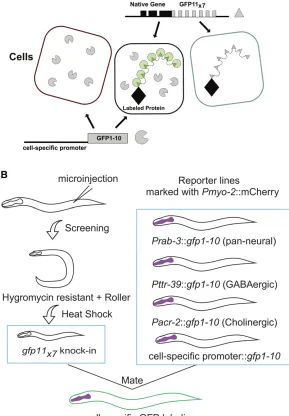
et al. 2017). In addition to labeling the native protein with smaller covalent tags, this combinatorial approach offers the further benefit of limiting the GFP signal to the specific cell type in which GFP1-10 is expressed (Figure 1A).
In this report, we describe a NATF toolbox that combines split-GFP and CRISPR technology for live-cell imaging of labeled Caenorhabditis elegansproteins expressed at native levels. With this approach, a GFP11 multicopy DNA array (GFP11X7) is inserted into the target gene. The resultant knock-in strain can then be crossed with separate reporter lines in which GFP1-10 is expressed in different cell types for tissue-specific visualization of the reconstituted NATF GFP (Figure 1A). We utilized this strategy for effective en-hancement of an otherwise weak signal from single-copy la-beling of a key protein (OIG-1) as well as the cell-specific resolution of a receptor accessory protein (LEV-10) at closely spaced but functionally distinct synapses in the C. elegans
nervous system.
Materials and Methods
C. elegans strains
C. elegansstrains were maintained at room temperature on NGM plates seeded with OP50 (Brenner 1974). Some strains were obtained from the Caenorhabditis Genetics Center (CGC). The N2 Bristol strain was used as the wild-type ref-erence. Transgenic animals were generated using standard microinjection techniques (Evans 2006). Strains used in this study are described in Supplemental Material, Table S1.
Molecular biology
single-guide RNA/Cas9 plasmid design:A 200 bp DNA se-quence that contained the desired cut site was submitted to the optimized CRISPR Design online tool (http://crispr.mit. edu/) to predict single-guide RNA (sgRNA) sequences. To enhance gene-editing efficiency, we selected a 59N18GGNGG sequence (Farboud and Meyer 2015) as an sgRNA targeting site for bothoig-1andlev-10. Foroig-1, 59-GGAGAGAAAGAC GAAAATGG-39was cloned into pDD162 (#47549; Addgene), a plasmid that contains the sgRNA backbone and Cas9 expres-sion system, using Q5 site-directed mutagenesis (New England Biolabs, Beverly, MA). Similarly, for lev-10, 59-ACGAATCGA CTGGTGGCCGG-39was used as the sgRNA target sequence, which is80 bp upstream of thelev-10stop codon.
CRISPR repair template for oig-1 and lev-10: To create the Self-Excising drug selection Cassette (SEC) SEC repair template for oig-1 TagRFP (Tag red fluorescent protein) CRISPR knock-in, flanking500-bp genomic DNA regions
immediately upstream and downstream of the desired inser-tion site were amplified by PCR using the following primers (Primer 1 and Primer 2 for the upstream homology arm, and Primer 3 and Primer 4 for the downstream homology arm) with overlap regions to the target plasmid pDD284 (#66825; Addgene):
OIG-1 Primer 1: 59-GACGTTGTAAAACGACGGCCAGTCGA CCTAACCATTCCAAAAGAT-39.
OIG-1 Primer 2: 59-TGAGCTCCTCTCCCTTGGAGACCATCG CATTTATTCCAACTGATA-39.
OIG-1 Primer 3: 59-TTACAAGGATGACGATGACAAGAGAA AATCTTCGCATATAGAAGA-39.
OIG-1 Primer 4: 59-CAGGAAACAGCTATGACCATGTTATCC AAGTCGGAGTACTGTTCA-39.
The amplified DNA fragments were cloned into plasmid pDD284 using Gibson cloning (New England Biolabs) to create the repair template. The corresponding protospacer adjacent motif (PAM) PAM sequence in the repair template plasmid was mutated from AGG to CCC using Q5 site-directed mutagenesis to produce the final plasmid, pSH30. Correct insertions and mutations were confirmed by sequencing.
To create the SEC repair template (pSH55) for theoig-1 GFP11x7CRISPR knock-in, the GFP11x7coding sequence was amplified from a previously published plasmid (#70224; Addgene) and inserted into pSH30 to replace the TagRFP sequence by In-Fusion cloning (Takara) with the following primers.
Fragment.FOR 221 59-GTTGGAATAAATGCGATGCGTGACC ACATGGTCCTT-39.
Fragment.REV 222 59-AAAGTACAGATTCTCGGTGATACCG GCAGCAT-39.
Vector.FOR 223 59-GAGAATCTGTACTTTCAATCCGGAAA GGTAAG-39.
Vector.REV 224 59-CGCATTTATTCCAACTGATAGAAAGCAT AAAAGTAGT-39.
To create the GFP and GFP11x7knock-in repair template for LEV-10, we used a two-step In-Fusion cloning method. Next,500 bp of DNA sequences upstream and downstream of
thelev-10stop codon were selected forflanking homology arms.
DNA was amplified, and then sequentially cloned into pSH30 or pSH55 to replace the originaloig-1homology arms. The re-sultant plasmids were then used as templates for site-direct mutagenesis to create thefinal repair template plasmid with sgRNA-binding sequences mutated, pSH84 (GFP knock-in), and pSH85 (GFP11x7knock-in). The primers for these cloning steps were designed with a strategy similar that used foroig-1as described above. Primer sequences are available on request.
promoter Pacr-2, and the muscle-specific Pmyo-3promoter were amplified to replace the Prab-3 promoter in pSP1 to create pSH79 (Pttr-39::gfp1-10), pSH88 (Pacr-2::gfp1-10), pSH86 (Pmyo-3::gfp1-10), and pSH87 (Pflp-13::gfp1-10). To create a secreted GFP1-10 construct, In-Fusion cloning was used to add the first 114 bp of the oig-1sequence in-cluding the signal sequence (Philbrooket al.2018) prior to the start codon of GFP1-10 in pSP1. The combined sequence was analyzed using the SignalP 4.1 Server to confirm that the predicted signal peptide was intact. Thefinal plasmid, pSH69 (Prab-3::ssGFP1-10), was confirmed by sequencing.
Confocal microscopy and image processing
Fluorescent images were captured at room temperature using a Nikon (Garden City, NY) A1R confocal microscope. Nema-todes were immobilized with 15 mM levamisole/0.05%
tricaine on a 2% agarose pad in M9 buffer. All images for ACR-12::GFPfluorescence quantification were obtained with the same settings using a 403/1.4 oil objective and Nyquist sampling. Constant laser power was used to compare the LEV-10::GFPfluorescence intensity to that of the NATF GPF signal produced by the combination of LEV-10::GFP11x7with
Pmyo-3::GFP1-10. Images in Figure 4 were three-dimensionally deconvolved with NIS-Elements with the automatic algo-rithm. For other images, ND2 files generated with NIS-Elements were imported into Fiji for analysis. Maximum intensity projections were generated by selecting stacks that had both ventral and dorsal signals. Line scans of dorsal and ventral cords (Figure S2, B–D), and of the nerve ring (Figure 3C), were adjusted by subtraction of background fluorescence measured from an adjacent region for compar-ison offluorescence intensities between samples. To compare the stability of GFP signals inlev-10::gfp vs. lev-10::gfp11x7;
Figure 1 Robust, tissue-specific labeling of a target pro-tein at its native expression level. (A) NATF. CRISPR/Cas9-mediated gene editing is used to label a protein of interest with seven copies of GFP11 (GFP11x7). Transgenic expres-sion of GFP1-10 with cell-specific promoters results in a bright, stable NATF fluorescent signal from multiple, reconstituted GFP molecules in specific tissues. (B) NATF workflow. Worms are injected with sgRNA, repair tem-plate, and co-injection markers.gfp11x7knock-in worms
are recovered after heat shock-induced excision of posi-tive-selection genes. Crossing thegfp11x7knock-in with
Pmyo-3::GFP1-10 strains, a region of interest (ROI) of the same size in each strain was bleached with a 405-nm laser for 15 sec at 50% laser power. Images of the ROI were col-lected and compared, before and after photobleaching. OIG-1::GFP11x7and mCherry::OIG-1 strains were imaged using an A1R Nikon laser confocal to obtain intensity profiles (Fig-ure 2A and Fig(Fig-ure S3). Fiji was used to draw 15-mm long-line scans on the ventral cord and extract intensity profiles, which were exported to Excel. Fluorescent intensities were normal-ized to the maximum intensity value of each line scan. In-tensities were plotted using Prism 6 software. Peaks exceeding a threshold of 75% of the normalized intensity for each sample were counted.
AiryScan imaging
Worms were mounted on 10% agarose pads and immobilized with 15 mM levamisole/0.05% tricaine dissolved in M9. A Zeiss ([Carl Zeiss], Thornwood, NY) LSM880 microscope equipped with an AiryScan detector and a 633/1.40 Plan-Apochromat oil objective lens was used to acquire superreso-lution images of the DD neuron (Figure 4E). Images were acquired as a Z-stack (0.19 mm/step), spanning the total volume of the DD neuron and submitted for AiryScan image processing by ZEN software.
Statistical analysis
For all experiments, sample numbers weren.10. The Stu-dent’st-test was used for comparison between two groups.
P,0.05 was considered significant. Prism 6 was used for statistical analysis.
Immunoblotting
OIG-1CRISPR and transgenic overexpression strains were cul-tured on NGM plates seeded with OP50. Mixed-stage worms were collected into a 1.5-ml tube by washing them off NGM plates with M9 buffer and pelleted by centrifugation. Next, 50ml of 23SDS-PAGE protein sample buffer was added to a 50ml pellet of each genotype and heated to 90°for 15 min. Samples were centrifuged at 8000 rpm for 5 min to remove debris (Milleret al.1983). Then, 30ml of supernatant from each genotype was loaded on a 10% protein gel to run at 110 V for 60 min, before being transferred to a PVDF membrane for immunoblot analysis (Duerr 2006). Immunoblots were treated with monoclonal anti-FLAG M2 antibody (1:500) (Sigma [Sigma Chemical], St. Louis, MO) to the 3XFLAG peptide followed by HRP-conjugated goat anti-mouse IgG antibody (1/2000), and soaked for 2 min in developing solution for chemiluminescent detection (Alegria-Schafferet al.2009).
Data availability
All reagents andC. elegansstrains described in this work are available on request. Plasmids will be deposited at Addgene and C. elegans strains submitted to the CGC stock center. Supplemental figures have been uploaded at the Genetics Society of America Figshare portal. Supplemental material available athttps://doi.org/10.25386/genetics.7898075.
Results
Tool box and strategy for NATF GFP labeling
We used a previously described CRISPR/Cas9 system for genome editing inC. elegans(Dickinsonet al.2015). In this approach, homology armsflank a self-excising cassette that carries positive selection markers (sqt-1) for the identification of transgenic worms (“rollers”) and drug resistance (hygR) for the detection of CRISPR/Cas9-induced integrants. A brief heat-shock treatment induces excision of the marker cassette to restore wild-type movement (“nonroller”) (Figure 1B). For split-GFP experiments, we replaced the fluorescent protein sequence in the original repair template plasmid with a
gfp11x7 insert (Kamiyamaet al. 2016). Homology arms of
500 bp were used for the two genes targeted (oig-1 and
lev-10) in this study (Figure S1A). We also constructed
sep-arate plasmids for expressing GFP1-10 in specific cell types including body muscles, all neurons, cholinergic neurons, and GABAergic neurons (Figure 1B and Figure S1B). In these lines, the GFP1-10 transgenes are carried as extrachromo-somal arrays that are maintained by selecting for a pharyn-geal co-injection marker (Pmyo-2::mCherry) (Figure 1B). Cell-specific drivers are flanked with multiple cloning sites to facilitate the construction of plasmids for GFP1-10 expres-sion in other tissues (Figure S1B). gfp11x7knock-in strains can be confirmed within 2 weeks of the initial injection and then crossed with GFP1-10-expressing lines for characteriza-tion (Figure 1B).
NATF GFP labeling reveals the intracellular localization of OIG-1 in GABAergic motor neurons
oig-1encodes a soluble protein with a single
immunoglobu-lin domain (Figure 2B) that is temporally regulated in GABAergic motor neurons to antagonize a synaptic remodel-ing program; inoig-1mutants, a postsynaptic acetylcholine receptor (AChR) containing the AChR subunit, ACR-12::GFP, is ectopically relocated from dorsal to ventral GABAergic neu-ron processes. OIG-1is secreted when overexpressed from multicopy transgenic arrays to produce bright puncta adja-cent to clusters of ACR-12::GFP (Figure 2, A and B) (Heet al.
2015; Howellet al.2015). To ask ifOIG-1is also secreted when expressed from the native locus, we used CRISPR/Cas9 to engineer a single-copy knock-in of the TagRFP to-gether with a 3XFLAG epitope tag (Figure 2B and Figure S2, A–C). We used immunoblotting to confirm expression of TagRFP::3XFLAG::OIG-1 (Figure 2C) but failed to detect TagRFP expression in vivo either by TagRFP fluorescence (Figure 2, D–G) or by immunostaining against the 3XFLAG epitope (data not shown). To produce a potentially brighter signal, we created agfp11x7::oig-1knock-in (Figure 2B) with a sgRNA that targeted the same 59-N18GGNGG site used for the TagRFP insert (Farboud and Meyer 2015). This strategy was designed to enhance a potential fluorescent signal by attaching seven copies of the GFP11 peptide to the OIG-1
N-terminus (Kamiyamaet al.2016). Successful knock-in of
We have previously shown that ACR-12::GFP in VD-class GABAergic motor neurons mislocalizes to the ventral side in
oig-1mutants, thereby resulting in an asymmetric ACR-12::
GFP signal that is brighter in the ventralvs.dorsal nerve cords (Figure S2) (He et al. 2015). In contrast, in the wildtype, ACR-12::GFP puncta are evenly distributed between dorsal and ventral nerve cords. This symmetry is maintained in the
gfp11X7::oig-1strain, thus arguing that the GFP11x7adduct does not significantly disruptOIG-1function (Figure S2, C and D). We then crossed thegfp11x7::oig-1knock-in with a pan-neural
Prab-3::gfp1-10transgenic line. Consistent with our previous findings, theOIG-1NATF GFP signal can be detected in head neurons, and in both dorsal and ventral nerve cords (Figure 2, H–K) (Heet al.2015). Colocalization ofOIG-1NATF GFP with the nuclear-localized pan-neural marker Prab-3::NLS:: mCherry confirmedOIG-1expression in neurons (Figure 2L). As an independent strategy to validate OIG-1 expression in GABA neurons, we crossed the gfp11x7::oig-1 line with Pttr-39::gfp1-10, which is selectively expressed in DD- and VD-class GABAergic motor neurons (Cinar et al. 2005). In Figure 2 CRISPR knock-in of GFP11x7 in OIG-1 reveals its intracellular localization in GABAergic motor neurons. (A) Overexpression of mCherry::OIG-1 from a transgenic array (Punc-25::mCherry::oig-1) produces bright puncta (magenta) adjacent to ACR-12::GFP-labeled postsynaptic clusters (green) of AChRs in GABAergic motor neu-rons (Punc-47::acr-12::gfp). Merge shows ventral nerve cord. Bar, 10 mm. Line scan of mCherry:: OIG-1 in the ventral nerve cord (bottom) shows punctate signal. Arrowhead points to coelomo-cyte. (B) OIG-1 protein showing N-terminal SP, C-terminal Ig (Im-munoglobulin) domain, and inser-tion site for fluorescent and epitope tags (mCherry, TagRFP, 3xFLAG, and GFP11x7). (C) Im-munoblot stained for the 3XFLAG tag detects expression of single-copy TagRFP::OIG-1 knock-in and confirms over expression of mCherry::3XFLAG::OIG-1 from multicopy transgenic array. (D–G) TagRFP knock-in at theoig-1 lo-cus (top) does not result in detect-able TagRFP::OIG-1fluorescence. Note absence of TagRFP::OIG-1 signal in head neurons (D), body (E), and dorsal (F) and ventral (G) nerve cords. Asterisks mark auto-fluorescent granules. (H–K)OIG-1 expression in the nervous sys-tem. Thegfp11x7::oig-1knock-in
(top) was crossed with the pan-neural transgenic line expressing Prab-3::GFP1-10. A diffuseOIG-1 NATF GFP signal is detected in head neurons (H) and in VD but not DD GABAergic motor neuron cell soma (dashed outlines) in the ventral cord (I). NATF GFP-labeledOIG-1is detectable in both dorsal and ventral nerve cords (J and K). Asterisk marks autofluorescent granules. (L and M)OIG-1expression in the nervous system and in GABAergic motor neurons. (L) Thegfp11x7::oig-1knock-in line was crossed
with the pan-neural markerPrab-3::gfp1-10and all neurons labeled with a nuclear-localized pan-neural label,Prab-3::NLS::mCherry. NoteOIG-1NATF GFP signal specifically in VD but not DD cell soma (dashed outlines), nor in additional ventral cordPrab-3::NLS::mCherry-labeled nuclei corresponding to cholinergic motor neurons. (M) Thegfp11x7::oig-1knock-in was crossed with the GABAergic motor neuron-specific markerPttr-39::gfp1-10and all GABA neurons were labeled
withPunc-47::mCherry. Note theOIG-1NATF GFP signal in VD but not DD motor neurons (dashed outlines)14. GFP1-10 is cytosolically expressed from the
this case, theOIG-1NATF GFP signal is limited to VD neurons with either weak or undetectable expression in DD neurons in L4 larvae (Figure 2M). Thisfinding confirms previous results obtained with a Poig-1::gfptranscriptional reporter that was expressed in VD, but not DD, neurons after the L2 larval stage (Heet al. 2015). Because the GFP1-10 peptide is expressed intracellularly in these strains, the NATF GFP signal likely de-rives from cytoplasmicOIG-1. Notably, theOIG-1NATF GFP signal is visible throughout VD neuron soma and neurites (Figure 2, H–M), and does not show the distinctive highly punctate appearance of OIG-1 when overexpressed from a multicopy array (Figure 2A) (Figure S3) (He et al. 2015; Howellet al., 2015). To test for potential secretion ofOIG-1
from the native locus, thegfp11x7::oig-1knock-in was crossed with a transgenic line in which the GFP1-10 peptide is secreted from neurons (Prab-3::ss::gfp1-10). However, this experiment did not produce a detectable extracellular NATF signal nor GFPfluorescence in coelomocytes in the body cavity, which normally function as macrophage-like cells and thus can be used to detect secreted protein markers (Figure S4A) (Fares and Greenwald 2001). Notably, overexpression of mCherry::
OIG-1from an extrachromosomal array does label coelomo-cytes (Heet al.2015; Howellet al.2015). As a positive control, we showed that the secreted form of GFP1-10 in the Prab-3::ss::gfp1-10strain is functional because it robustly labels a GFP11 peptide fused to the extracellular domain of the syn-aptic membrane proteinNLG-1(Feinberget al.2008) (Figure S4, B–D). As negative controls, we showed that neither GFP11x7nor GFP1-10 by themselves produce visible GFPfl uo-rescence (Figure S2, E and F). Although undetectably low levels of secretedOIG-1could be produced by this experiment, our overall results are consistent with the hypothesis thatOIG-1
is not secreted when expressed at the native level but local-izes intracellularly (S. He, A. Cuentas Condori, D. Miller, un-published data). For example, genetic disruption of theOIG-1
signal peptide blocksOIG-1secretion but does not disruptoig-1 function in vivo (He et al. 2015). Our finding that OIG is intracellularly localized depended on the use of the NATF strategy to reveal low levels of nativeOIG-1 expression and thereby circumvent artifactual extracellular localization due to OIG-1 overexpression from multicopy arrays (He et al.
2015).
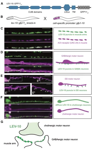
NATF GFP labeling reveals discrete locations for the transmembrane domain protein LEV-10 in different cell types
Having shown that NATF could detect a soluble protein ( OIG-1), we next targetedLEV-10, a CUB domain transmembrane protein that clusters AChRs at postsynaptic sites in body mus-cles (Gally et al. 2004). First, we created a CRISPR/Cas9 knock-in line in which a single copy of GFP was fused to the intracellular C-terminus ofLEV-10(see Figure 4A). We detected LEV-10::GFP in both ventral and dorsal nerve cords, as predicted for a protein that localizes to body muscle syn-apses (Gallyet al.2004). LEV-10::GFP puncta were also de-tected in the head region where motor neurons synapse with
body muscles on the inside surface of the nerve ring (White
et al.1986; Von Stetinaet al.2006) (Figure 3A). For NATF GFP labeling of body muscle synapses, we generated a lev-10::gfp11x7 knock-in and crossed it with a muscle-specific line expressing GFP1-10 (Pmyo-3::gfp1-10) from an extra-chromosomal array. The LEV-10 muscle-specific NATF GFP signal in the head region and axial nerve cords (Figure 3B) mimics that of the single-copy lev-10::gfp knock-in (Figure 3A), but is noticeably brighter. We quantified the GFP signal for each marker at the nerve ring muscle synapses to confirm that theLEV-10NATFfluorescence is brighter (around three times) than the GFP signal from thelev-10::gfpsingle-copy insertion, as predicted from measurements of single-copyvs.
multicopy split-GFP expressed in cultured cells (Figure 3C) (Kamiyamaet al.2016). In addition to determining that the
lev-10::gfp11x7array yields a stronger signal than that of the single-copy lev-10::GFP insert, we also showed that NATF GFP is substantially more resistant to photobleaching, as pre-viously demonstrated for reconstituted split-GFP from mea-surementsin vitro(Kamiyamaet al.2016) (Figure 3D).
In addition to expression in muscle, our independent studies have shown thatLEV-10is also expressed in ventral cord neurons where it colocalizes with AChRs at postsynaptic sites in GABAergic motor neurons (S. He, A. Cuentas Condori, D. Miller, unpulished data). In the motor neuron circuit, cho-linergic motor neurons form dyadic synapses that innervate closely spaced postsynaptic domains in body muscle and GABA neurons (see Figure 4G) (Whiteet al.1986). Both of these postsynaptic regions in the ventral nerve cord region should be labeled in thelev-10::gfpknock-in and, thus, can-not be unambiguously identified (Figure 3A). To resolve this problem, we crossed the lev-10::gfp11x7 knock-in with transgenic lines that express GFP1-10 in either body mus-cles (Pmyo-3::gfp1-10), or in DD and VD GABAergic motor neurons (Pttr-39::gfp1-10). NATF GFP puncta can be readily detected in both cases (Figure 4, C and D), but are brighter in muscles than in GABAergic neurons (data not shown). Expression of a TagRFP-labeled AChR subunit UNC-29
(Gally et al. 2004) in muscle confirms colocalization of UNC-29::TagRFP with LEV-10NATF GFP reconstituted in muscle. (Figure 4C). Expression of GFP1-10 in DD and VD neurons produces LEV-10 NATF GFP puncta that overlap with a cytoplasmic GABA neuron mCherry marker ( Punc-47::mCherry), as predicted for theLEV-10protein that lo-calizes to GABA neuron synapses (Figure 4D). To confirm the postsynaptic location of LEV-10in GABA neurons, we used a DD-specific construct (Pflp-13::gfp1-10) to generate a LEV-10 NATF GFP signal. In this case, superresolution imaging resolves distinctLEV-10NATF GFP puncta at the tips of postsynaptic spine-like projections that have been recently described in the ventral processes of mature DD neurons (Fig-ure 4E) (Philbrooket al.2018). Notably, we have also observed that the AChR marker, ACR-12::GFP, is positioned in the same distal location in DD dendritic spines and that these spines are aligned with presynaptic cholinergic vesicles (Cuentas-Condori
LEV-10localization at distinct postsynaptic locations in muscle
vs. GABA neurons, we also used a cholinergic motor neuron driver (Pacr-2::gfp1-10) to detect a separateLEV-10NATF sig-nal in ventral cord cholinergic neurons. In this case, LEV-10
NATF GFP is diffuse (Figure 4F) and asymmetrically localized to the ventral, but not dorsal, nerve cord (data not shown), a labeling pattern that closely resembles the perisynaptic position of the AChR subunit ACR-12::GFP in cholinergic motor neurons (Petrashet al.2013). BecauseLEV-10is expressed at its native level and retains its AChR clustering function (data not shown) when fused to the GFP11X7adduct, it seems likely that each of the three distinct, cell-specificLEV-10NATF signals (i.e., mus-cle, GABA neurons, and cholinergic neurons) marks authentic subcellular locations for the endogenously expressed LEV-10
protein.
Discussion
We have shown that NATF offers a robust strategy for pro-ducing bright, cell-specific signals for theC. elegansproteins OIG-1 andLEV-10expressed from their native genomic loci. These results suggest that NATF should be especially useful
for marking connections in the compactC. elegans nervous system, where most synapses are located in the densely packed nerve ring and axial nerve cords (Whiteet al.1986). For example, GFP-tagging of a core presynaptic protein (e.g.,
RAB-3) by conventional CRISPR/Cas9 editing should mark synapses throughout the nervous system. In contrast, labeling with the NATF strategy should result in a bright, photostable GFP signal that is limited to the presynaptic domains of specific neurons. Although our results have determined that fusion with the GFP11X7peptide does not result in detectable disrup-tion of thein vivofunction of either OIG-1 (Figure S2, B–D)
orLEV-10(Figure 4C and S. (S. He, A. Cuentas Condori, D. Miller, unpublished data), other proteins may be less tolerant. In that event, smaller adducts with fewer copies of GFP11 could be attempted. In that case, GFP signal augmentation will be diminished but tissue-specific labeling is still possible (Nomaet al.2017). Because thegfp11X7insert is stably inte-grated at the native locus and is thus limiting, the comple-menting GFP1-10 peptide can be provided from multicopy transgenic arrays without risk of inducing overexpression artifacts. Thus, a given GFP11X7split GFP insert can be rapidly tested with multiple tissue-specific GFP1-10 transgenic lines, Figure 3 lev-10::gfp11x7yields a
stronger NATF GFP signal than the single-copylev-10::gfpknock-in at synapses in neurons and muscle cells. (A) Confocal image showing localization of LEV-10::GFP in a sin-gle-copy GFP knock-in at the na-tive lev-10 gene (lev-10::gfp). LEV-10::GFP puncta are visible at the nerve ring (arrow), and in ven-tral and dorsal nerve cords (ar-rowheads). (B) Confocal image of the LEV-10 NATF GFP signal at body muscle synapses arising from the combination of the lev-10::gfp11x7knock-in with
Pmyo-3::gfp1-10. NATF GFP (arrow) is detected at neuromuscular synap-ses near the nerve ring. Bar, 20
mm. Insets (right) shows rotated views of anterior regions of im-ages on left to depict nerve ring labeling. Asterisks mark gut auto-fluorescence. (C) LEV-10 NATF GFP at body muscle synapses in the nerve ring labeled with lev-10::gfp11x7is significantly brighter
(around three times) (34506 441) than the single-copy lev-10::gfp knock-in (12806184).P,0.001, N= 15, Student’st-test. Error bars are SD. (D) TheLEV-10NATF GFP signal at body muscle synapses in the nerve ring labeled withlev-10::gfp11X7is
significantly more stable (0.75 6
which can be readily generated using conventional methods. A similar combinatorial approach should also be useful for tissue-specific protein labeling in other model organisms (Kelliher et al. 2018). We note that NATF can be modified to reduce weak backgroundfluorescence from the GFP1-10 fragment (Fenget al.2017), and for multicolor split-GFP im-aging with cyan (CFP) and yellow (YFP) GFP variants, or with the sfmCherry marker (Kamiyama et al. 2016; Feng et al.
2017).
Acknowledgments
We thank members of the Miller laboratory for critical reading of the manuscript, Sierra Palumbos and Alice Siqi Chen for
plasmid construction, Lakshmi Sundararajan for help with confocal imaging, and Oliver Hobert for sharing strains. Some nematode strains used in this work were provided by the
CaenorhabditisGenetics Center, which is funded by the National Institutes of Health (NIH) National Center for Research Re-sources. Superresolution images were acquired in the Vanderbilt Cell Imaging Shared Resource (1S10 OD-201630-01). This work was supported by NIH grants to D.M.M. (R01 NS-081259 and R01 NS-106951). A.C.-C. is supported by an American Heart Association predoctoral fellowship (18PRE33960581).
Author contributions: S.H., A.C.-C., and D.M.M. conceived the project. S.H. and A.C.-C. performed the experiments and analyzed the data. S.H., A.C.-C., and D.M.M. wrote the manuscript. The authors declare no competing interests.
Figure 4 Visualization ofLEV-10NATF GFP signal at cell-specific synapses. (A) Schematic of LEV-10::GFP11x7 show-ing extracellular complement C1r/C1s, Uegf, Bmp1 (CUB) CUB and low-density lipoprotein receptor domain class A (LDLa) LDLa domains, with TM region and cytoplasmic tail with GFP11x7insert. (B) Cell-specific labeling strategy. The
lev-10::gfp11x7 knock-in strain is crossed with separate
transgenic lines expressing GFP1-10 in specific cell types. (C–F) Representative images (left) and schematics (right) of ventral nerve cord region of L4 larvae showing LEV-10 NATF GFP arising from complementation of thelev-10:: gfp11x7knock-in with cell-specific expression of GFP1-10:
Literature Cited
Alegria-Schaffer, A., A. Lodge, and K. Vattem, 2009 Performing and optimizing Western blots with an emphasis on chemilumi-nescent detection. Methods Enzymol. 463: 573–599. https:// doi.org/10.1016/S0076-6879(09)63033-0
Brenner, S., 1974 The genetics of Caenorhabditis elegans. Genet-ics 77: 71–94.
Cabantous, S., T. C. Terwilliger, and G. S. Waldo, 2005 Protein tagging and detection with engineered self-assembling frag-ments of green fluorescent protein. Nat. Biotechnol. 23: 102– 107.https://doi.org/10.1038/nbt1044
Chalfie, M., 2009 GFP: lighting up life. Proc. Natl. Acad. Sci. USA 106: 10073–10080.https://doi.org/10.1073/pnas.0904061106
Cinar, H., S. Keles, and Y. Jin, 2005 Expression profiling of GA-BAergic motor neurons in Caenorhabditis elegans. Curr. Biol. 15: 340–346.https://doi.org/10.1016/j.cub.2005.02.025
Cuentas-Condori, A., B. Mulcahy, S. He, S. Palumbos, M. Zhenet al., 2019 Dendritic spines on GABAergic neurons respond to cho-linergic signaling in the Caenorhabditis elegans motor cir-cuit. bioRxiv. Available at: https://www.biorxiv.org/content/ 10.1101/598714v2.
Dickinson, D. J., A. M. Pani, J. K. Heppert, C. D. Higgins, and B. Goldstein, 2015 Streamlined genome engineering with a self-excising drug selection cassette. Genetics 200: 1035–1049.
https://doi.org/10.1534/genetics.115.178335
Duerr, J. S., 2006 Immunohistochemistry (June 19, 2006),
WormBooked. TheC. elegansResearch Community, WormBook, doi/10.1895/wormbook.1.105.1, http://www.wormbook.org. El Mouridi, S., C. Lecroisey, P. Tardy, M. Mercier, A. Leclercq-Blondel
et al., 2017 Reliable CRISPR/Cas9 genome engineering in Cae-norhabditis elegans using a single efficient sgRNA and an easily recognizable phenotype. G3 (Bethesda) 7: 1429–1437.https:// doi.org/10.1534/g3.117.040824
Evans, T. C., 2006 Transformation and microinjection (April 6, 2006), WormBook, ed. TheC. elegansResearch Community, WormBook, doi/10.1895/wormbook.1.108.1,http://www.wormbook.org. Farboud, B., and B. J. Meyer, 2015 Dramatic enhancement of
genome editing by CRISPR/Cas9 through improved guide RNA design. Genetics 199: 959–971.https://doi.org/10.1534/ genetics.115.175166
Fares, H., and I. Greenwald, 2001 Genetic analysis of endocytosis in Caenorhabditis elegans: coelomocyte uptake defective mu-tants. Genetics 159: 133–145.
Feinberg, E. H., M. K. Vanhoven, A. Bendesky, G. Wang, R. D. Fetter
et al., 2008 GFP reconstitution across synaptic partners (GRASP) defines cell contacts and synapses in living nervous systems. Neu-ron 57: 353–363.https://doi.org/10.1016/j.neuron.2007.11.030
Feng, S., S. Sekine, V. Pessino, H. Li, M. D. Leonetti et al., 2017 Improved splitfluorescent proteins for endogenous pro-tein labeling. Nat. Commun. 8: 370.https://doi.org/10.1038/ s41467-017-00494-8
Gally, C., S. Eimer, J. E. Richmond, and J.-L. Bessereau, 2004 A transmembrane protein required for acetylcholine receptor clus-tering in Caenorhabditis elegans. Nature 431: 578–582.https:// doi.org/10.1038/nature02893
He, S., A. Philbrook, R. McWhirter, C. V. Gabel, D. G. Taubet al., 2015 Transcriptional control of synaptic remodeling through regulated expression of an immunoglobulin superfamily protein. Curr. Biol. 25: 2541–2548. https://doi.org/10.1016/ j.cub.2015.08.022
Hostettler, L., L. Grundy, S. Käser-Pébernard, C. Wicky, W. R. Schafer
et al., 2017 The brightfluorescent protein mNeonGreen facilitates protein expression analysis in vivo. G3 (Bethesda) 7: 607–615.
https://doi.org/10.1534/g3.116.038133
Howell, K., J. G. White, and O. Hobert, 2015 Spatiotemporal con-trol of a novel synaptic organizer molecule. Nature 523: 83–87.
https://doi.org/10.1038/nature14545
Hsu, P. D., E. S. Lander, and F. Zhang, 2014 Development and applications of CRISPR-Cas9 for genome engineering. Cell 157: 1262–1278.https://doi.org/10.1016/j.cell.2014.05.010
Kamiyama, D., S. Sekine, B. Barsi-Rhyne, J. Hu, B. Chen et al., 2016 Versatile protein tagging in cells with split fluorescent protein. Nat. Commun. 7: 11046. https://doi.org/10.1038/ ncomms11046
Kelliher, M. T., Y. Yue, A. Ng, D. Kamiyama, B. Huang et al., 2018 Autoinhibition of kinesin-1 is essential to the dendrite-specific localization of Golgi outposts. J. Cell Biol. 217: 2531– 2547.https://doi.org/10.1083/jcb.201708096
Miller, D. M., I. Ortiz, G. C. Berliner, and H. F. Epstein, 1983 Differential localization of two myosins within nematode thick filaments. Cell 34: 477–490. https://doi.org/10.1016/ 0092-8674(83)90381-1
Noma, K., A. Goncharov, M. H. Ellisman, and Y. Jin, 2017 Microtubule-dependent ribosome localization in C. ele-gans neurons. Elife 6: e26376. https://doi.org/10.7554/ eLife.26376
Petrash, H. A., A. Philbrook, M. Haburcak, B. Barbagallo, and M. M. Francis, 2013 ACR-12 ionotropic acetylcholine receptor com-plexes regulate inhibitory motor neuron activity in Caenorhab-ditis elegans. J. Neurosci. 33: 5524–5532. https://doi.org/ 10.1523/JNEUROSCI.4384-12.2013
Philbrook, A., S. Ramachandran, C. M. Lambert, D. Oliver, J. Florman
et al., 2018 Neurexin directs partner-specific synaptic connec-tivity in C. elegans. Elife 7: e35692. https://doi.org/10.7554/ eLife.35692
Praitis, V., E. Casey, D. Collar, and J. Austin, 2001 Creation of low-copy integrated transgenic lines in Caenorhabditis elegans. Ge-netics 157: 1217–1226.
Remington, S. J., 2011 Greenfluorescent protein: a perspective. Protein Sci. 20: 1509–1519.https://doi.org/10.1002/pro.684
Schwartz, M. L., and E. M. Jorgensen, 2016 SapTrap, a toolkit for high-throughput CRISPR/Cas9 gene modification in Caenorhab-ditis elegans. Genetics 202: 1277–1288. https://doi.org/ 10.1534/genetics.115.184275
Tanenbaum, M. E., L. A. Gilbert, L. S. Qi, J. S. Weissman, and R. D. Vale, 2014 A protein-tagging system for signal amplification in gene expression andfluorescence imaging. Cell 159: 635–646.
https://doi.org/10.1016/j.cell.2014.09.039
Terpe, K., 2003 Overview of tag protein fusions: from molecular and biochemical fundamentals to commercial systems. Appl. Microbiol. Biotechnol. 60: 523–533. https://doi.org/10.1007/ s00253-002-1158-6
Von Stetina, S. E., M. Treinin, and D. M. Miller, III, 2006 The motor circuit. Int. Rev. Neurobiol. 69: 125–167. https:// doi.org/10.1016/S0074-7742(05)69005-8
White, J. G., E. Southgate, J. N. Thomson, and S. Brenner, 1986 The structure of the nervous system of the nematode Caenorhabditis elegans. Philos. Trans. R. Soc. Lond. B Biol. Sci. 314: 1–340.