BraTS : Brain Tumor Segmentation – Some
Contemporary Approaches
Mahantesh K1, Kanyakumari2
Assistant Professor, Department of Electronics & Communication Engineering, S. J. B Institute of Technology, BGS,
Bengaluru, India1
P.G. Student, Department of Electronics & Communication Engineering, S. J. B Institute of Technology, BGS,
Bengaluru, India2
ABSTRACT: Brain tumor is nothing but the abnormal growth of cancerous cells within Brain. In medical field, segmentation of brain regions & detection of brain tumor is very challenging task because of its complex structure. Magnetic resonance imaging (MRI) provides the detailed information about brain anatomy. Proper brain tumor segmentation using MR brain images helps in identifying exact size & shape of Brain tumor, this intern helps in diagnosis & treatment of brain tumor. Detection of brain tumor from MRI images involves various steps such as MR image pre-processing, segmentation of image & feature extraction. Proposed paper explains about the methods that use Histogram Thresholding, K-means clustering, and Fuzzy C- Means & Support Vector Machine (SVM). Presented method includes several steps such as pre-processing; high frequency components and noise are removed & RGB to gray conversion, global image threshold; converts intensity image into binary image, erosion & dilation of binary image to locate tumor exactly, detecting the stage of the tumor whether Benign or Malignant.
KEYWORDS: Brain tumor, Magnetic resonance imaging (MRI), Histogram Thresholding K-Means clustering, FCM
& SVM, Neural Network.
I. INTRODUCTION
Brain is one of the complex organ in human body as it contains 50-100 billion neurons in it forming a network. Brain tumors are classified as Benign Brain tumor and malignant brain tumor based on the type of tissue present in involved in formation of tumor.
Benign Brain tumor: - Benign Brain tumor normally does not contain any cancer causing cells & it doesn’t affect the nearby healthy tissues. These cells can be removed by surgery.
Malignant Brain tumor: - malignant Brain tumor contain cancer causing cells, these cells grows very rapidly with the time & affect other nearby health brain tissues and cause severe problem, ultimately that may cause the death of the person.
From past few years Brain tumor segmentation becomes a very challenging research area in the medical field. Brain tumor segmentation helps in identifying exact size, shape & location of brain tumor. The segmentation experimented with magnetic resonance imaging (MRI) scanned images in proposed method [1]. Usually MRI scan & CT scan are used to view the anatomy of brain, but MRI scan is more convenient as compare to CT scan for diagnosis. [2], the K-Means clustering & fuzzy C-K-Means algorithms are used to detect the exact location of brain tumor. Using fuzzy C- means algorithm stage of patient’s tumor is determined.
segmentation techniques to locate tumor and fuzzy C-Means algorithm is proposed to segment the brain MR images, this is also used to calculate the tumor area this information helps to differentiate between type of brain tumor.
The MRI of brain is normally diagnosed by physician to locate brain tumor, this detection method does not provides the accurate result. To overcome from this [6], project that produces accurate result & uses computer aided method to detect brain tumor. This intern reduces the consumption of time for diagnosis. Stage of the tumor can be calculated based on the amount of area computed from algorithm.
From past few years many brain tumor segmentation techniques are implemented to locate the brain tumor in MRI. Nowadays Image processing became a very important part of medical field. [7], includes survey on different brain tumor segmentation techniques such as fuzzy C-Means algorithm; this algorithm is used to detect area, size & shape of tumor, knowledge based algorithm; this technique uses multispectral histogram to differ tumor from other healthy parts of the brain, probabilistic neural network (PNN); it gives much accurate & faster result compare to other segmentation techniques. Clustering meant to be dividing the set of data into similar objects groups, groups includes objects of similar characteristics. Objects are grouped depending on some object (pixel) characteristics. K-Means clustering is most useful to divide mass of data quickly & efficiently, intern segmentation became easier. K-means clustering technique for brain tumor segmentation described, [8].
Most of the studies & survey in different countries proved that number of brain tumor patients died because of imprecise tumor detection. Brain tumor may be primary or secondary, it will be primary when tumor at origin stage & will be secondary when tumor cells starts spreading to other healthy tissues of brain. Both Primary &secondary brain tumor can be detected using method that uses K-Means clustering, [9]. [10], proposes histogram Thresholding & artificial neural network. Modification was done on previous segmentation methods to overcome from drawbacks; ROI (region of interest) parameter is used in diagnosis of MRI images. Noise is removed in pre-processing step using Gaussian filter. Edge detection carried out fallowed by ROI & Finally feature extraction is done.
II. RELATED WORK
Most commonly used algorithm is K-Means clustering in image segmentation techniques. Researchers made several efforts to improve efficiency and accuracy of K- Means clustering algorithm. [11], Presented method, Histogram Thresholding to detect and segment brain tumor. Knowledge of area of tumor helps in diagnosis & treatment of tumor. Manual segmentation of MRI is very stimulating & time consuming work. Histogram Thresholding method helps researcher to overcome from these drawbacks.
[12], Describes modified fuzzy c-means method for brain MRI image segmentation, it uses both local & non-local spatial context in order to decrease the noise in segmentation process. The FCM algorithm presented to overcome from the disadvantages of other previous segmentation techniques. In this FCM method both local and non-local information integrates into FCM clustering for better efficiency, method allows cataloguing of one pixel get prejudiced from another pixel and this algorithm suppresses the noise during segmentation procedure.Magnetic resonance imaging provides detailed anatomy of human tissue. Mathematical morphology is used in many applications like object segmentation boundary detection & to decrease noise effect, [13].
III.PROPOSED METHODOLOGY
In this section, we proposed different brain tumor segmentation techniques such as Histogram Thresholding, k-means clustering, fuzzy c- means & support vector machine. This section addresses the difficulties in segmenting the brain tumor image because of its complexity and solution to overcome from those problems. These methods are implemented on set of datasets to check accuracy & efficiency.
A. HISTOGRAM THRESHOLDING
Histogram Thresholding is one of the most commonly used & simple technique in image segmentation field. Thresholding method replaces each pixel to black pixel if ,
< T,
i.e. pixel intensity , less than that of fixed threshold value T and Thresholding replaces pixel with white pixel if , is greater than that of fixed threshold value Histogram means representation of numerical data in graphical form. Image histogram contains peaks & valleys. Histogram Thresholding has many steps such as pre-processing; RGB to grey conversion, Image is partitioned into two equal parts with central axis in histogram Thresholding, plot histogram for those parts & are compared to locate the tumor in MRI brain image, segmentation done using threshold point, Exact dimensions of brain tumor are detected by cropping method; cropping helps in getting the required part of the image, intern makes analysis easier, finally area of tumor is calculated. In Histogram Thresholding method gray image has the intensity value between 0-255, 0 for black pixel & 255 for white pixel.B. K-MEANS CLUSTERING
K-means clustering is one of the unverified algorithms for clusters. Grouping of pixels based on some parameter is called clustering. We have to declare the number of clusters k in this algorithm, declared k clusters centers are selected randomly. Have to calculate the distance between each cluster & each pixel for further use. The distance between cluster & pixel is calculated using simple Euclidian function. First consider a pixel from an image & compare that pixel with all k- cluster centers by distance formula. After comparing, that pixel is shifted to some particular cluster among k- cluster which has shortest distance among all other clusters. This process continues for all other pixels until center meets or joins. Therefore k-means clustering function is used to cluster the data.Calculate Cluster for given image, from below equation (1).
=
∑ ( )(1)
Where k= 1………KDistance between each pixel & cluster given by equation (2).
( )=arg
min
||
–
MK|| (2)
Where i=1………NC. FUZZY C-MEANS ALGORITHM
Dunn introduced the fuzzy c-means (FCM) clustering algorithm & it further improved by Bezdek. In fuzzy c-means algorithm a set of n-vectors are divide into c fuzzy groups & cluster center is found in each group so that dissimilarity is reduced. Fuzzy c-means algorithm is simple to implement & faster as compare to other segmentation techniques. FCM resist noise & can achieve efficient segmentation. In this algorithm partial membership is given to each pixel of the image. The range of membership value is between 0 & 1. Fuzzy c-means algorithm provides intermediate fuzzy values for each pixel i.e.in the same image one fuzzy set may be part of other fuzzy set, hence in FCM part of data is associated with more than one cluster.
In FCM noise is almost vanished by decreasing below objective function, i.e. equation (3).
=
∑
∑
||
–
||
(3)=
measured data of d-dimensional=
center of d-dimensional clusterD. SUPPORT VECTOR MACHINE
Support vector machine is one of the enhanced methods for classification of tumor from brain MR images. [17], oriented Rician noise reduction anisotropic diffusion filter is used in SVM method to remove noise. This method includes Digitization of image, removing noise from image using FCM (skull part is removed from the image), features are extracted using FCM algorithm, using joint entropy & genetic algorithm desired features are selected, and finally classification is done using SVM with kernels. The SVM classifier found more accurate with GRBF (Gaussian radial based functional) kernel. After classification the stage of the tumor is identified whether it is Benign or Malignant. Fig.1. Explains how histogram Thresholding & fuzzy c-means works stepwise. Select input image, pre-processing; converts RGB to grey image & remove noise, segmentation procedure, this flowchart proposes two segmentation methods. Firstly the Histogram Thresholding; morphological operations are carried out & finally image is segmented to locate the tumor in MR images of brain. In FCM clustering is carried out to detect tumor efficiently compare to other clustering methods
Fig.1. Flowchart for Fuzzy C-Means Clustering&Histogram Thresholding.
Input Image
RGB to Grey conversion
Filtering the image
Segmentation
Pre-processing
Preprocessing
B. Histogram Thresholding
Morphological operation
Thresholding A. FCM
IV.EXPERIMENTAL RESULTS& PERFORMANCE ANALYSIS
This section contains result of above explained segmentation methods. We verified set of datasets using histogram Thresholding, k-means, fuzzy c-means & support vector machine and got accurate & efficient results. Fig.2. shows result of Histogram Thresholding. Fig.3. shows result of k-means clustering. Fig.4. shows result of fuzzy c-means algorithm. Fig.5. shows result of SVM method. We compared output of all these methods with manually segmented images to check accuracy & got succeeded also. We can locate tumor very efficiently using these methods
.
V.
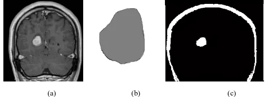
Fig. 2. Result of Histogram Thresholding , (a) Original Input Image , (b) Manually segmented brain MRI, (c)Output of Histogram Thresholding.
Fig.3. Result of k-means algorithm, (a) Original Input Image, (b) Manually segmented brain MRI, (c)k-means algorithm output.
Fig. 4. Result of fuzzy c-means algorithm, (a) Original Input Image , (b) Manually segmented brain MRI, (c)fuzzy c-means algorithm output. (a) (b) (c)
(a) (b) (c)
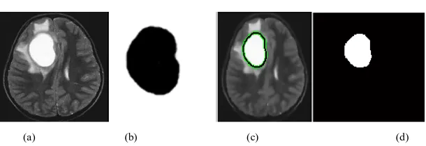
Fig. 5.Result of SVM method, (a) Original Input Image, ( b) Manually segmented brain MRI, (c) SVM classifier, (d) Segmented image.
VI. CONCLUSION
Several methods discussed in the Introduction & related work, failed due to inaccuracy in locating tumor. Based on research & survey, number of brain tumor patients died because of imprecise brain tumor location. Hence it’s very important to locate brain tumor exactly. Using proposed methods in this paper tumor is located & identified accurately, intern helps in differentiating stage of the tumor (benign or malignant). Every method segmented output is compared with manually segmented images and found more accurate. Proposed methods help in proper treatment of tumor by locating tumor accurately.
REFERENCES
[1] Mustaqeem, A., Javed, A., & Fatima, T, “An Efficient Brain Tumor Detection Algorithm Using Watershed &Thresholding Based Segmentation”, International Journal of Image, Graphics and Signal Processing, Vol. 10, no. 3,pp. 34-39, 2012.
[2] Alan Jose1, Sambath, M., & Ravi, S, “Brain Tumor Segmentation Using K-Means Clustering And Fuzzy C-Means Algorithms And Its Area Calculation”, International Journal of Innovative Research in Computer and Communication Engg., Vol. 2, Issue 3, pp. 3496-3501, 2014. [3] Moumen T Melegy and Hashim M Mokhtar, “Tumor segmentation in brain MRI using a fuzzy approach with class center priors”,
El-Melegy, &Mokhtar EURASIP Journal on Image and Video Processing, volume 21, 2014.
[4] Swati Chawla, &NehaGarg, “the Automated Brain Tumor Detection Based On Fuzzy Clustering Segmentation Approach”, International Journal of Emerging Research in Management &Technology ISSN, Volume-3, Issue-7, pp. 107-111, 2014.
[5] Gauri P. Anandgaonkar, &Ganesh.S.Sable, “Detection and Identification of Brain Tumor in Brain MRI Using Fuzzy C-Means Segmentation”, International Journal of Advanced Research in Computer and Communication Engineering, Vol. 2, Issue 10, pp. 3964-3967, 2013.
[6] VarshaKshirsagar, &Prof.JagrutiPanchal, “Segmentation of Brain Tumor and Its Area Calculation”, International Journal of Advanced Research in Computer Science and Software Engineering, Volume 4, Issue 5, pp. 523-530, 2014.
[7] UpasanaGaikwad, KanikaDebbarma&SilkeshaThigale, “Survey Paper on Clustering based Segmentation Approach to Detect Brain Tumor from MRI Scan”, International Journal of Computer Applications (0975 – 8887), Volume 115, no. 14, 2015.
[8] Malathi, R., & Dr. Nadirabanu Kamal, A.R., “Brain Tumor Detection and Identification UsingK-Means Clustering Technique”, Proceedings of the UGC Sponsored National Conference on Advanced Networking and Applications, pp. 14-18, 2015.
[9] Sanghamitra T. Kamble, &Rathod, M.R., “Brain Tumor Segmentation using K-Means Clustering Algorithm”, International Journal of Current Engineering and Technology, Vol.5, no.3, pp. 1521-1524, 2015
[10] Swathi, P. S., DeepaDevassy, Vince Paul, &Sankaranarayanan, P.N., “Brain Tumor Detection and Classification Using Histogram Thresholding and ANN”, International Journal of Computer Science and Information Technologies,Vol. 6, no. 1, pp. 173-176, 2015. [11] Manoj K Kowar, &SourabhYadav, “Brain Tumor Detction and Segmentation Using Histogram Thresholding”, International Journal of
Engineering and Advanced Technology (IJEAT) ISSN: 2249 – 8958, Volume 1, Issue 4, pp. 16-20, 2012.
[12] Jianzhong Wang , Jun Kong , YinghuaLub, Miao Qi , &Baoxue Zhang, “modified FCM algorithm for MRI brain image segmentation using both local and non-local spatial constraints”, Computerized Medical Imaging and Graphics 32, pp. 685–698 , ELSEVIER, 2008.
[13] Pankaj Kr. Saini, &Mohinder Singh, “Brain tumor detection in medical imaging using MATLAB”, International Research Journal of Engineering and Technology (IRJET), Volume 02, Issue 02, pp. 191-196, 2015.
[14] Ganesh Madhikar, &Sunita S. Lokhande, “Brain Tumour Detection and Classification by UsingModified Region Growing Method: A Review”, International Journal of Engineering Research & Technology (IJERT), Vol. 2, Issue 12, pp. 2316-2320, 2013.
[15] CH. Rambabu, &B. Siva AyyappaKumar, “Brain Tumor Classification Using Multi Wavelet Transform and Neural Network”, International Journal of Advanced Research in Computer Science and Software Engineering, Volume 4, Issue 9, pp. 932-935, 2014.
[16] Yash Sharma, &MeghaChhabra, “An Improved Automatic Brain Tumor Detection System”, International Journal of Advanced Research in Computer Science and Software Engineering, Volume 5, Issue 4, pp. 11-15, 2015.
[17] Madheswaran, M., &AntoSahayaDhas, D., “Classification of brain MRI images using support vector machine with various Kernels”, Biomedical Research, vol. 26, no. 3, pp. 505-513, 2015.