Level Set Based Segmentation in Spine CT
Images Using Random Contour Initialization
Jenny Patrick1, Indu M.G.2
PG Scholar [Signal Processing], Dept. of ECE, TKM Institute of Technology, Kollam, Kerala, India1
Assistant Professor, Dept. of ECE, TKM Institute of technology, Kollam, Kerala, India2
ABSTRACT: Segmentation technique plays a crucial role in medical image processing. Due to the advancement in medical imaging techniques, in -depth details of the biological structural can be viewed, which may be helpful for the early and proper diagnosis of many diseases. Segmentation seems to be a highly challenging process in the case of images affected with intensity inhomogeneity. In such cases, level set techniques of segmentation works well and is widely used in the segmentation of image acquired using computer tomography (CT). The recent researches on the medical imaging focus on the segmentation which facilitates the 3-D visualization of medical data. So a framework for vertebrae segmentation in CT slice is put forward, which uses variational level, set technique for vertebrae segmentation by minimizing an energy function formulated using standard k-means criterion. Further, to enhance the inner details, 3-D visualization for segmented vertebrae from a single slice is also done.
KEYWORDS: Bias field, Energy Minimization, CT image, Segmentation, Variational Level set, 3-D Visualization.
I.INTRODUCTION
Diagnostic imaging is a valuable tool in the field of medicine today. X-rays imaging, Computer tomographic (CT) imaging, Magnetic resonance (MR) imaging and other imaging modalities provide an efficient means for the noninvasive mapping of internal structures within the body. After the acquisition of images, for the proper diagnosis of the body parts, these images have to be precisely analyized. For the proper image analysis, image segmentation algorithms play a vital role. Here, the focus is given for vertebrae segmentation in CT image with intensity non-uniformity. Trauma and fractures on specific region of vertebrae (cervical, thoracic, lumbar or sacrum) may occur due to the injuries, accidents, spinal abnormality, ageing etc.. Assessment of the severity of vertebrae deformality will help physicians to determine the most effective treatments for spinal disorders. It is difficult to accurately segment images with intensity variations especially in CT images of spine, because most of the existing segmentation algorithms are region-based that depend on intensity homogeneity of the region of interest. Various techniques such as thresholding, watershed transform and partial differentiation equation based methods have been employed so far for image segmentation problems. Among all these techniques, level set based method which comes under partial differential equation based technique has been proved to be an efficient framework in the past years. Intensity inhomogeneity is caused due to the imperfection in imaging devices or due to the illumination variations that occurs in real-world images, which leads to serious misclassification by intensity-based segmentation algorithms.
I1.RELATED WORKS
affected by intensity inhomogeneity , bias estimation has to be done, for this Li et.al. [10] proposed a novel variational level set method, that uses the weighted K-means clustering method to evaluate the bias field of image intensities in a neighbourhood around each point in the image. With reference to Li et. al. [10] a new segmentation approach is used, which makes use of variational level set with random contour initialization , where the local region difference in the image is identified using K-means clustering with three cluster centres adding to three phase level set technique . So by using this local clustering criterion, the bias field estimation and errors in segmentation results can be effectively corrected in each iteration for different initial contours.The intended method is expected to give accurate segmentation result in single CT slice of vertebra in cross sectional view and the 3-D visualization of the segmented vertebra is done to enhance its details.
III. METHODOLOGY
1. Level Set segmentation technique
The basic idea of the level set method is to represent a contour as the zero level set of higher dimensional function, known as level set function (LSF) which is usually a binary step function. This function is used to formulate an energy function which has to be minimized as the contour evolves to the object boundary. In conventional level set formulation, the level set functions typically develop irregularities during its evolution, which may cause errors and eventually destroy the contour evolution to the object boundary. This problem is rectified by using variational level set formulation. For the level set evolution to be stable and accurate, the LSF has to be maintained in good condition. This condition is well satisfied by signed distance functions because of their unique property │∇∅│ = 1 where ∇∅ is the gradient of level set function ∅ [1], [3], [10].
2. Vertebra Segmentation
Variational level set framework applied in the segmentation stage is a three phase level set. So, there are three regions within the specified contours , , and . Figure 1 shows the details of how level set method is formulated for vertebrae segmentation in CT slice.
The bias field indicates the intensity inhomogeneity and is represented as b. The bias field is slowly varying, which implies that b can be approximated by a constant in a neighbourhood of each point in the image domain. Segmentation of the vertebra is achieved by finding contours which separates the image domain into disjoint regions. For getting different regions that corresponds to vertebra, an energy function has to be minimized within a boundary where the level set evolves. The boundary for level set evolution is obtained with the help of Neumann Boundary condition [10]. Within this specific boundary, the Level Set evolution process takes place. The level set function is obtained by taking the signed function of randomized image and has values 0, 1, and -1. This local clustering criterion is integrated over the neighbourhood centre to define an energy function, which is converted to a level set formulation. Minimization of this energy is achieved by an interleaved process of level set evolution and estimation of the bias field. The above described local intensity clustering property indicates that the intensities in the neighbourhood can be classified into three clusters. This allows us to apply the standard K-means clustering to classify these local intensities, which can be defined in a continuous form as
∑ ( )
│ ( ) │ (1)
where ( − ) is a Gaussian function. During the evolution of level set function, and the bias field b are updated by minimizing the energy function E(∅, c, b) .
(∅, , ) =∫ ∑ ( ) (∅ ) (2)
where (∅ ) is the member ship function which is used as the phase indicator for the regions and level set function represented by ∅. in the above equation is defined by
∫ ( )│ ( ) │ (3)
Energy minimization in the variational level set formulation is given by
∅
=− (4)
where F represents the speed function that controls the evolving level set.
(∅, , ) = + (∅) ∇∅
│∇∅│ + (∅)│∇∅│ (5)
where (∅) represents the dirac function which is the derivative of Heaviside function and , are the regularizing constants.
3. 3-D Visualization
Three dimensional image visualization is used for enhancing the visual content in the image [6]. For decades, several techniques were used to enrich the depth information in images but most of them target at creating information regarding depth using more than one image. Most of these techniques differ among themselves on the basis of attaining depth information. In spite of several techniques developed for three dimensional image formations, the problem with accuracy and pixel information still remains as a challenge for the conversion of two dimensional image into three dimensional image. Thus, a feasible solution can be formulated from a single image for the three dimensional conversion. For this, the segmented image obtained using level set technique, which is a grayscale image, has been converted into indexed one. Then the corresponding contour slice is obtained. The contour slice of different sizes is stacked together to get the three dimensional view of vertebra.
IV. RESULTS AND DISCUSSIONS 1. Input Image
Figure 2: Dicom image of L3 vertebra
CT slice is of cross-sectional view, because this view helps in providing clear visualization of variational level set concepts. The simulation platform used is MATLAB R2010a and the results of each stage are as follows.
2. Level Set Evolution
The intensity range of input is reduced to the range [0,255] and the level set function is calculated using signum function which gives the values 0, 1 and -1. Using the level set function, initial contour is set and figure 3 shows the initial contour initialization.
Figure 3: Initial contour based on signed distance function
Figure 4 shows the different stages of level set evolution. The Gaussian kernel used has a mask size of 17x17 with scale parameter = 4. There are four convolutions to be computed at each time step of level set evolution. The time step ∇ is set to 0.1. The regularization parameters , used in energy minimization has values 1 and 65.025. The heaviside function used to smoothening the level set has a value =1.
(a) (b) (c) (d)
3. Segmented Image
Using the Neumann boundary condition, the boundary for level set evolution is identified. Local clustering criterion function evaluates the classification of intensity within the image. Figure 5 shows the final segmented image with clustered regions shown as black, gray and white.
Figure 5: Segmented L3 vertebra
When the segmented result is compared to the input CT image given, the segmentation seems to efficient, highlighting each and every part of vertebral region. The variational level set approach applied in the cross-sectional view of vertebrae gives good result in CT slice in DICOM format.
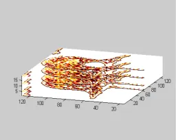
4. 3-D Visualization
Eight duplicated copies of segmented image in figure 5 is concatenated in three dimensions and its singleton values are removed using squeeze function in Matlab to obtain index image. Figure 6 shows the corresponding indexed image.
Figure 6: Segmented L3 vertebra
The contour slice of the indexed image shown in figure 6 is obtained using Matlab command. Then from that contour slice, multiple copies were obtained. The figure 7 displays the contour slice of different size stacked together that will render the 3D visualization of L3 vertebrae from single CT slice. This 3-D visualization can be enhanced if the same procedure is done with multiple CT slices and will help the physician to get the in-depth details of vertebra.
1V.CONCLUSION
Using variational level set technique, a local intensity clustering property is used to define an energy minimization criterion for the level set functions and it is effective for the simultaneous estimation of contour regions and bias field. At present, the 2-D segmentation of vertebra in cross-sectional view is done on single CT slice and the 3-D visualization of the segmented result is achieved. But the segmentation of vertebrae in 2-D image is peripheral, i.e. it does not reflect the actual details and shape of vertebrae, so in future this segmentation has to be extended by using multiple CT slices for 3-D reconstruction of vertebra rather than 3-D Visualization since the 3-D reconstruction helps to give in-depth details of vertebra, which is useful for the diagnosis of several medical conditions affecting spine.
REFERENCES
[1] Kaihua Zhang, Lei Zhang, Kin-Man Lamand David Zhang, “A Level Set Approach to Image Segmentation With Intensity Inhomogeneity” IEEE Transactions On Cybernetics , Vol.46, pp.546-57, 2016 Feb.
[2] Robert Korez, Bulat Ibragimov, Bostjan Likar, Franjo Pernus and Tomaz Vrtovee, “A Framework for Automated Spine and vertebrae Interpolation-Based Detection and Model-Based Segmentation”, IEEE Transactions on Medical Imaging, Vol. 38, No.8, 2015.
[3] Zhang K, Liu Q, Song H, Li X. , “A Variational Approach to Simultaneous Image Segmentation and Bias Correction.” IEEE Transactions On Cybernetics , Vol.45, pp.1426-1437, 2015.
[4] Amandeep Kaur,and Aayushi “Image Segmentation Using Watershed Transform” International Journal of Soft Computing and Engineering (IJSCE) ISSN: 2231-2307, Vol.4, Issue-1, March 2014.
[5] H. Song, “Active contours driven by regularized gradient flux flows for image segmentation,” Electron. Lett., Vol. 50, No. 14, pp. 992–994, 2014.
[6] Lee Sang-Hyun, Park Dae-Won, Jeong Je-Pyong and Moon Kyung, “Conversion 2D Image to 3D Based on Squeeze function and Gradient Map”, International Journal of Software Engineering and Its Applications, Vol.8, No.2, pp: 27-40, 2014.
[7] Mohamed Amine Larhmam, Mohammed Benjelloun and Said Mahmoudi, “Vertebra identification using template matching modelmp and K-means clustering”, Springer-Verlag Berlin Heidelberg, Vol. 60, No.11, 2013.
[8] Poay Hoon Lim, Ulas Bagci and Li Bai, “Introducing Willmore Flow Into Level Set Segmentation of Spinal Vertebrae”, IEEE Transactions On Biomedical Engineering, Vol. 60, No:1, pp: 115-122, January 2013.
[9] Poay Hoon Lim, Ulas Bagci and Li Bai, “A novel Spinal vertebare segmentation framework combining geometric flow and shape prior with level set method”, 9th IEEE International Symposium On Biomedical Engineering (ISBI), pp. 1703-1706 , May 2012.
[10] Chunming Li, Rui Huang, Zhaohua Ding, J. Chris Gatenby and Dimitris N., “A Level Set Method for Image Segmentation in the Presence of Intensity Inhomogeneities With Application to MRI”, IEEE Transactions On Biomedical Engineering , Vol. 20, No.7, July 2011
[11] Melih S. Aslan, Asem Ali, Ham Rara, and Aly A. Farag, “An Automated Vertebra Identification And Segmentation In CT Images”, 17th IEEE International Conference on Image Processing, pp.233-236, September 2010
[12] S. Lankton and A. Tannenbaum, “Localizing region-based active contours,” IEEE Trans. Image Process., Vol. 17, no. 11, pp. 2029–2039, 2008.
[13] C. Li, C.-Y. Kao, J. C. Gore, and Z. Ding, “Implicit active contours driven by local binary fitting energy,” in Proc. IEEE Conf. Comput. Vis. Pattern Recognit., Minneapolis, MN, USA, pp. 1–7, 2007
BIOGRAPHY
Jenny Patrick received her B.Tech degree in Electronics and Communication Engineering from Cochin University of Science and Technology in the year 2014. She is currently pursuing second year M.Tech in Signal Processing at TKM Institute of Technology, Kollam, India. Her areas of interest include image processing and signal processing.