Heart Rate and Respiratory Rate
Measurement Using Image Processing
S.S.Lokhande , Binu K Nair
Dept. of Electronics & Telecommunication, SCOE, Pune, India
ABSTRACT: Respiratory disease is a medical term which affects the organs and tissues which allows gas exchange possible in organism and includes condition of the respiratory tract.Heart rate is the speed of heartbeat measured by the number of contraction of the heart per minute (bpm).According to the physical body of a human being; the heart rate may vary person to person.One of the respiratory diseasescalled severe acute respiratory syndrome (SARS) which spreads around the world in 2003.Therefore many of the quarantine station were affected and a system is launched to detect infected passengers.A method for non contact measurement of multiple vital signs i.e respiratory rate and heart rate based on RGB image processing with CMOS camera is proposed.Monitoring the periodic temperature changes of RGB images at nasal area can calculate respiratory rate.Heart rate is measured by capturing the brightness variations of RGB facial images by fluctuations in skin blood flow.The transmission of disease can be prevented by this non contactable method.
KEYWORDS: CMOS camera; image processing; non contact.
I. INTRODUCTION
Respiratory disease is a medical term which affects the organs in higher organisms.It includes condition of the upper respiratory tract, trachea, bronchi, bronchioles, alveoli, pleura and pleural cavity.Heart rate is the speed of heartbeat measured by the number of contractions of the heart per minute (bpm).According to the body’s physical needs ,the heart rate may vary person to person .One of the respiratory disease known as severe acute respiratory syndrome is caused by coronavirus.An outbreak of SARS in southern china caused an eventual death resulting in 774 deaths during the year of November 2002 and july 2003.No cases of SARS have been reported worldwise since 2004.There is no treatment for SARS which is safe for humans as per 2015.The identification and development of novel vaccines and medicines to treat SARS ia a priority for government and public health agencies arould the world.Some of the international airports adopted the method of screening using thermography.
A. Literature review
The author [1] proposes a system screening of infected individuals by using thermography.This non contact method was assumed to be essential in preventing and controlling the transmission of diseases.In [2] the research work is to measure different parameters that are heart rate, respiratory rate, BP using photoplethysmographic sensors .PPG signal is acquired by PPG sensor, microcontrollerthe acquired PPG signal is displayed in MATLAB. Frequency domain analysis of PPG signal shows two peaks first at around 0.25 to 0.35 Hz and second at around 1 to 1.5 Hz. FFT at 1Hz relates to 60 BPM and FFT at 0.25 Hz relates to 15 respiratory cycles per minute. For BP Measurement, the pulse height of PPG is proportional to the difference between the systolic and the diastolic pressure in the arteries. The standard blood pressure monitoring instrument is used to calculate correlation coefficient. The arterial blood pressure is calculated based on these coefficients. PPG signal is used to detect blood pressure pulsations in a finger and achieved an accuracy of (0.8 ± 7) mmHg and (0.9 ± 6) mmHg for systolic and diastolic pressure, respectively.
receiver's properties, oscillator phase noise, range correlation, and receiver noise.Severe acute respiratory syndrome was first reported in 2003 and very quickly it spreads.Therefore many international airports adopted this technique to detect heart and respiratory disease.Heart rate and respiratory disease can be detected by using thermal and RGB images by using a CMOS IR camera.In this proposed work, a image processing is conducted on thermal and RGB image in a real time .The respiratory rate is determined by the thermal images of the IR camera and heart rate is determined by the RGB images of the CMOS IR camera.Also by the capturing the brightness variation of RGB facial images ,heart rate can be detected.
II. MATERIALSANDMETHODS
The method is divided into two different section .First is to find out the respiratory rate using thermal image and second is to find heart rate using RGB image.
Based on RGB image processing ,we propose a method for non-contact measurement of multiple vital signs, i.e., facial skin temperature and respiratory and heart rates, with a CMOS-IR camera .Respiratory rate is calculated by monitoring the periodic temperature changes of RGB images at nasal area. And by capturing the brightness variations of RGB facial images caused by fluctuations in skin, heart rate is calculated.A CMOS camera is used to evaluate the efficiency of heart rate and respiratory rate.We tested themeasurement of respiratory and heart rates on ten male subjectsunder resting and after exercise conditions with ergometer.We will compare the respiratory rate and heart rate by contact type method in 10 secs.This method can applied in hospitals, airport, station where the chances of getting infection from other people is possible. Hence the risk of infection can be decreased.
A. Real-time RGB Image Processing for Non-contact Vital-sign Measurement
The RGB images were acquired and analyzed inPython using OpencV in real time. In Fig. 1the method tocalculatethe respiratory and heart rates is shown. The TVS-500EXLV is used as a CMOS IR camera that integrates sensor camera and IR camera, and also it provides thermal and RGB fusion mode. Theratio of overlapped thermal and RGB images is adjustable. The thermal/RGB mixed-images were obtained at 30 frames per sec with a 640 × 480 pixelsolution; A camera will capture the image of a person using CMOS camera and each image is send to PC.
Figure 1. General Block Diagram
To calculate the heart rates,the first image of RGB-predominant images is used.The ROI (pixel solution wasapproximately 150 × 180 pixels) is set as to be the center of the
Subject’s face. All images were obtained as those of configuring by only the green plane signals; we calculated the mean brightness value of each image asfollows CMOS camera. We used the obtained mean brightness value to create thewaveform. The heart rates were calculated in thesame analysis program with respiratory rates, except for thesetting of the band-pass filter (0.83–2.0 Hz). The underlyingsignal of interest is a blood volume pulse that propagatesthroughout the body. During the cardiac cycle, volumetricchanges in the facial blood vessels modify the amount ofambient light absorption according to subsequent changes inthe amount of reflected light. These changes indicate the timingof cardiovascular events. By shooting a video of the facialregion with a CMOS camera, the red, green, and blue colorsensors pick up a mixture of the reflected plethysmographicsignals, including other sources of fluctuations in light due toartifacts. We adopted the green signal, which is the mostsuitable color for calculating the heart rate. By capturingthe tiny changes of the green signal, we successfully calculatedthe heart rate.
B. Laboratory Test of CMOS Camera
The respiratory rate and heart rate is calculated using both non-contact method and contactmethod.during measurement the person is asked to remain motionless and keep his/her breathing process spontaneously for 30sec.The CMOS camera captures the subjects face.at last we recorded the measurement value using both contact and non contact type sensors.
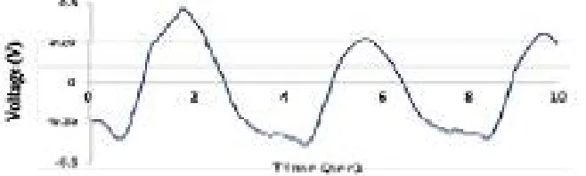
b) The respiratory rate signal obtained from CMOS camera
c) The heart beat signal obtained from ECG signal
d) The heart beat signal obtained from CMOS camera
III. RESULTS
a) b) c)
g) h) i)
Fig 3.a)Original image b) Region of interest (ROI) c)Resized image d) color transformed image e) hue f) saturation g) value h) median filter i) gray image
b)
d)
Figure. 4 a) raw signal b) band pass filter applied to raw signal c) filtered signal d) FFT transform
Output:-
HEART RATE 93.0bpm
RESPIRATORY RATE 18.0bpm
This work involves determining the respiratory rate by the RGB images of the CMOS camera and the heart rate by the RGB images of the CMOS camera
IV. DISCUSSIONANDCONCLUSION
The proposed method is for non-contact measurement of therespiratory and heart rates using a CMOS camera. Our results showed that there was a strong correlation between parameters obtained from the CMOS camera and contacttype sensors (respiratory effort belt: r = 0.99, p <0.01; ECG: r= 0.96, p <0.01) within 10 s.This method enables screening vital signs using only a CMOS camera, indicating that no equipment or measuring hardware is added. Furthermore, thismethod requires no contact with machines, resulting inreduction of secondary infection.
REFERENCES
[1]. BMY. Cheung, LS. Chan, IJ. Lauder, and CR. Kumana, “Detection of body temperature with infrared thermography: accuracy in detection of fever,”Hong Kong, vol. 18(4 Suppl 3), pp. 31–34, 2012.
[2]. NiveditaDaimiwal, M. Sundhararajan and RevatiShriram, “Respiratory Rate, Heart Rate and Continuous Measurement of BP Using PPG”, International Conference on Communication and Signal Processing, April 3-5, 20 14, India
[3]. Ng, GJ. Kaw, and WM. Chang, “Analysis of IR thermal imager for mass blind fever screening”, Microvascular Research, vol. 68(2), pp. 104– 109, 2004.
[4]. G. Sun, S. Abe, Y. Hakozaki, and T. Matsui, “A Novel Noncontact Infection Screening System Based on Self-Organizing Map with K-means Clustering,” Communications in Computer and Information Science, vol. 258, pp. 125–132, 2011.
[5]. T. Matsui, Y. Hakozaki, S. Suzuki,K. Hasegawa, Y. Sugiyama and M. Sugamata,“A novel screening method for influenza patients using a newly developed non-contact screening system,” Journal of Infection, vol. 60 (4), pp. 271–277, 2010.