The Drosophila retained/dead ringer gene and ARID gene
family function during development
TETYANA SHANDALA
1, R. DANIEL KORTSCHAK
1and ROBERT SAINT
1,2,*
1Centre for the Molecular Genetics of Development and Dept. of Molecular Biosciences, Adelaide University, Adelaide, SA, Australia and 2Research School of Biological Sciences, Australian National University, Canberra, ACT, Australia
ABSTRACT The recently discovered ARID family of proteins interact with DNA through a phylo-genetically conserved sequence termed the A/T Interaction Domain (ARID). The retained/dead ringer (retn/dri) gene of Drosophila melanogaster is a founding member of the ARID gene family, and of the eARID subfamily. This subfamily exhibits an extended region of sequence similarity beyond the core ARID motif and a separate conserved domain termed the REKLES domain. retn/ dri is involved in a range of developmental processes, including axis patterning and muscle development. The retn/dri ARID motif has been shown by in vitro studies to exhibit sequence-specific DNA binding activity. Here we demonstrate that the ARID domain is essential for the in vivo function of retn/dri during embryonic development by showing that a mutant form of RETN/DRI, deleted for part of the ARID domain and unable to bind DNA in vitro, cannot rescue the retn/dri mutant phenotype. In the presence of wild-type RETN/DRI this construct acts as a dominant negative, providing additional support for the proposal that RETN/DRI acts in a multiprotein complex. In contrast, we are yet to find an in vivo role for the REKLES domain, despite its clear evolutionary conservation. Finally, we have used germline clone analysis to reveal a requirement for retn/dri in the Drosophila preblastoderm syncytial mitoses.
KEY WORDS:
ARID motif, gene regulation, cell cycle control, DNA binding proteins
0214-6282/2002/$25.00
© UBC Press Printed in Spain
www.ijdb.ehu.es
*Address correspondence to: Prof. Robert Saint. Centre for the Molecular Genetics of Development, Research School of Biological Sciences, Australian National University, Canberra, ACT 0200, Australia. Fax: +61-2-6125-8294. e-mail: robert.saint@anu.edu.au
Abbreviations used in this paper: ARID, AIT-rich interaction domain; EMS,
ethylmethanesulphonate.
Introduction
The development of multicellular organisms requires tight spa-tial and temporal control of switches in the activity of key regulatory genes. Tissues are specified by transcriptional regulatory cas-cades that respond to a variety of extracellular and intracellular cues. It has become clear in recent years that the regulators of tissue-specific gene expression, the transcription factors, are assembled into multi-protein complexes. These complexes in-clude DNA-binding proteins, which recruit other components that are not able to bind DNA directly. The composition of complexes assembled on a specific promoter can be transient, as seen in the cascade of gene regulation in the early Drosophila embryo. Curi-ously, many transcription factors fall into a relatively small number of phylogenetically conserved families, although different family members can assume very different developmental roles in differ-ent organisms (see, for example, Lall and Patel, 2001). As a result, studies of members of conserved protein families very often provide new approaches to understanding mechanisms that regu-late a variety of developmental events.
One of the most recent of the phylogenetically conserved families to have been identified is the ARID family (see Kortschak
et al., 2000). The family was discovered simultaneously by the characterisation of two proteins, mouse Bright (Herrscher et al., 1995; reviewed in Webb, 2001) and Drosophila Dead ringer (Gregory et al., 1996). The dead ringer gene has since been shown to correspond to the retained (retn) gene (Schüpbach and Weischaus, 1991; Ditch, Pitman, Finley, Edeen and McKeown, unpublished observations) and is here referred to as retn/dri. Bright and RETN/DRI were found to contain a conserved sequence-specific DNA binding motif, termed the A/T Interaction Domain (ARID) (Herrscher et al., 1995; Gregory et al., 1996). Members of this family have since been found in Saccharomyces cerevisiae, Caenorhabditis elegans, Danio rerio, mouse and human (Fig. 1A, Kortschak et al., 2000). Sequence-specific DNA binding appears not to be an intrinsic feature of the ARID motif, since the ARID motif proteins human SMARCF1 and Drosophila OSA (orthologs of yeast SWI1) exhibit non-sequence-specific DNA binding (Dallas et al., 2000; Collins et al., 1999).
these, which contains Drosophila RETN/DRI and mouse Bright, exhibits an extended region of similarity either side of the core ARID motif. We have termed this larger region of similarity the extended ARID (eARID) motif (Kortschak et al., 2000). eARID subfamily members also share an independent conserved domain termed the REKLES domain (see below). The second subfamily is defined on the basis of the presence of several C-terminally located conserved domains, including PHD finger domains and a highly conserved PLU-domain. This sub-family includes Ustilago maydis Rum1 (Quadbeck-Seeger et al., 2000), mouse and human SmcX/ Y (Agulnik et al., 1994a; Agulnik et al., 1994b), mouse Jumonji and Desrt (Takeuchi et al., 1995; Lahoud et al., 2001), and human PLU-1, XE169, RBPPLU-1, RBP2, MRF1 and MRF2 (Lu et al., 1999; Wu et al., 1994; Fattaey et al., 1993; Huang et al., 1996). The third subfamily of ARID proteins includes yeast SWI1 (Quinn et al., 1996), Drosophila OSA/ELD (Treisman et al., 1997), a Caenorhabditis elegans ortholog (GeneBank accession no.U80439), mouse Osa1 and human SMARCF1/OSA1/p270/ B120/BAF250 (Takeuchi et al., 1997; Kozmik et al., 2001) proteins. In addition to the ARID motif, these proteins have N-terminal Osa Homology Domains 1 and 2 (OHD1, OHD2) containing four LXXLL motifs, which are implicated in nuclear hormone receptor binding (NR-box).
From the outset, it appeared that members of this gene family would play roles in developmental regulation. For example, retn/dri exhibits a highly regulated temporal and spatial pattern of expression (Fig. 2, Gregory et al., 1996; Shandala et al., 1999), while Bright, a B cell regulator of IgH transcription also exhibits tissue-restricted patterns of expression (Herrscher et al.,1995; Webb et al., 1998).
Evidence for a developmental role for ARID family genes has also come from ge-netic analysis. The SWI1 gene of Saccharo-myces cerevisiae is part of the Swi/Snf com-plex required for the regulation of many genes involved in simple developmental pro-cesses such as mating type switching (see Sudarsanam and Winston, 2000, for a re-view). More recently, the regulator U. maydis 1 (rum1) gene has been shown to be essen-tial for normal development of Ustilago maydis spores (Quadbeck-Seeger et al., 2000). The effect on sporulation appears to be mediated by downregulation of a very defined set of genes, including egl1, dik1, lga2, hum2, that are normally induced by two homeobox genes, bE and bW, encoded by the U. maydis mating type locus.
A role for the Drosophila retn/dri gene in embryonic development was revealed by characterisation of retn/dri EMS-induced al-leles (Shandala et al., 1999). retn/dri was found to be essential for several aspects of embryogenesis, including anterior/posterior and dorsal/ventral patterning. One of the targets of retn/dri transcriptional regulation in the blastoderm embryo appears to be the dorsal-specific gene zerknüllt (zen). zen is normally expressed in a narrow dorsally located stripe along the embryo. In retn/dri
A
B
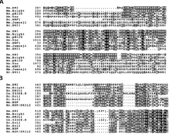
Fig. 1. Sequence comparison of the ARID and REKLES motifs of selected ARID family proteins. (A) eARID and ARID alignment showing the extended region of homology of RETN/DRI and Bright, both N and C-terminal to the core ARID which is conserved across all family members. (B) Alignment of amino acid sequences of the REKLES region from all known eARID proteins.
mutant embryos, zen expression fails to refine to its normal amnioserosa-specific expression (Valentine et al., 1998). The activity of RETN/DRI in this case is to convert the normal transcrip-tional activator Dorsal (DL) into a repressor by binding adjacent to DL and creating a surface to which the Groucho (GRO) WD-40 repeat co-repressor protein binds (Valentine et al., 1998). GRO has been found to interact with the N-terminal tail of histone H3 and with the histone deacetylase Rpd3, an enzyme involved in chroma-tin condensation and gene inactivation (Palaparti et al., 1997; Chen et al., 1999).
Repression of huckebein along the ventral side of the embryo trunk is also mediated by RETN/DRI and DL, through recruitment of GRO to the ventral repression element of hkb (Hader et al., 2000). huckebein is a Drosophila terminal gap gene expressed in the anterior and posterior caps. It is activated along the ventral side of the embryo by DL. For both zen and hkb, there is a difference between the behaviour of the endogenous genes and of the minimal repression element derived from them, the minimal ele-ments exhibiting much greater sensitivity to retn/dri regulation. This is presumably the result of a level of redundancy in the repression of the wild-type gene that is lost in the reduction to the minimal regulatory element.
In addition to its role as a repressor, evidence suggests that RETN/DRI can act as an activator. retn/dri is required for activation of the argos gene in two near-terminal domains of expression in the blastoderm embryo, accounting for the head skeleton phenotype observed in retn/dri mutant embryos (Shandala et al., 1999).
A
C
D
E
B
of the protein. This issue is made more significant by theobserva-tion that the ARID motif is dispensable for the in vivo funcobserva-tion of the S. cerevisiae SWI1 protein (C. Peterson, personal communica-tion). Here we provide evidence that the ARID domain is essential for the in vivo function of retn/dri. In addition, we explore the function of the separate REKLES domain within retn/dri. Finally, we show that retn/dri is required for normal mitosis during the syncytial cleavage stage of Drosophila development.
Results
The ARID Domain is Necessary for DNA Binding
To assess the importance of the RETN/DRI ARID motif, in vitro mutagenesis was used to generate a retn/dri construct, termed ARIDδH5, in which the third α-helix of the ARID motif was mutated. This helix was selected on the basis that it is highly conserved and includes an invariant tryptophan residue found in all ARID do-mains, indicating that it should be essential for ARID function. The RETN/DRI eARID, spanning the sequence from amino acid 258 to 410, has previously been shown to be sufficient for sequence-specific DNA binding to an oligonucleotide consisting of repeats of a consensus Engrailed binding site termed NP (Gregory et al., 1996). To determine whether helix 5 of the RETN/DRI ARID is necessary for binding to the NP sequence, electrophoretic mobility shift assays were conducted using both the wild-type ARID (GST-RETN/DRIARID), as has been previously described (Gregory et al., 1996), and the mutant ARID (GST- RETN/DRIARIDδH5) lacking the helix 5 sequence. Using elevated protein levels to detect any potential binding by the mutant, GST- RETN/DRIARID was able to retard the available NP6 while GST-RETN/DRIARIDδH5 was not able to retard the NP6 DNA (Fig. 3A). We conclude that the RETN/DRI ARID is necessary for DNA binding in vitro.
The ARID Motif is Essential for RETN/DRI Function In Vivo
To test the in vivo importance of the ARID motif, the ARIDδH5 construct was placed downstream of a yeast GAL4 upstream activator sequence (UAS) and transformed into the Drosophila germline. Genetic crosses were used to place this construct under the transcriptional control of endogenous retn/dri enhancers via a retn/dri::GAL4 enhancer-trap P-element insertion line (Shandala et al., 1999). When a wild-type retn/dri construct is expressed in this way, embryonic lethality of retn/dri1 homozygotes is rescued
(Shandala et al., 1999). However, when the ARIDδH5 construct was tested in the same assay, no rescue was observed. We conclude that the ARID domain is essential for RETN/DRI function in vivo.
RETN/DRIARIDδH5 Acts as a Dominant Negative
Failure of the retn/dri ARID deletion mutant to rescue the mutant phenotype demonstrated the in vivo requirement for the ARID domain. The RETN/DRIARIDδH5 construct used in this experiment had the capacity to interact with co-factors involved in RETN/DRI func-tion. If so, the construct should act in an antimorphic, or dominant negative, way in the presence of the wild-type gene. To test for the antimorphic potential of this construct, wild-type RETN/DRI and RETN/DRIARIDδH5 lacking DNA-binding activity was expressed in the wild-type retn/dri pattern as described above, but now in the pres-ence of the wild-type retn/dri gene. No flies were observed to carry both the UAS::retn/driARIDδH5 transgene and the retn/dri::GAL4 driver, indicating that these genes are synthetically lethal, consistent with an antimorphic function.
In order to confirm the antimorphic activity of retn/driARIDδH5, we examined flies heterozygous for an amorphic retn/dri allele which were also expressing retn/driARIDδH5 in the wing, a non-essential retn/dri-expressing tissue. Examination of larval retn/dri expression
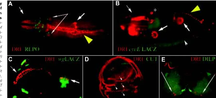
Fig 2.Developmentally regulated pattern of retn/dri expression in Drosophila embryos and larvae. Immu-nochemical staining of whole mount embryos (A-C) or larval tissues (D,E) with rat-anti-DRI (red) (A-E). Images were captured using epifluorescence mi-croscopy, except (D) which is a confocal im-age. Samples are coun-terstained with rabbit anti-REPO (green) (A), rabbit anti-βgal (green)
(B,C), mouse anti-CUT (green) (D) and rabbit anti-DILP (green) (E). Embry-onic tissues that express
UAS:: retn/driARIDδH5 expression was induced in developing wing cells using the GAL471B enhancer trap, which expresses GAL4 in the wing-blade anlagen of the wing imaginal disk (Brand and Perrimon 1993). Expression of UAS::retn/driARIDδH5 under GAL471B control resulted in variable wing vein defects and losses of campaniform sensilla (Fig. 4 A-C), in regions consistent with the endogenous expression pattern of RETN/DRI (see Fig. 2D).
To establish whether retn/driARIDδH5 acts antimorphically, the dose of endogenous retn/dri was halved by placing GAL471B and P[UAS::retn/driARIDδH5] in a retn/dri heterozygous mutant back-ground. Enhancement of the severity of the phenotype in retn/dri1
heterozygotes was observed (Fig. 4, compare D with E), confirming that the retn/driARIDδH5 construct was acting as an antimorphic form of the gene.
Other Effector Domains in eARID-Containing Proteins: the REKLES Domain
The eARID family proteins contain two additional conserved motifs, the REKLESα and REKLESβ domains (Fig. 1B, Kortschak et al., 2000). The REKLES motif is located immediately carboxy-terminal to the eARID. It has a size of a little over 100 amino acids in the C. elegans and vertebrate RETN/DRI homologs, but in Drosophila there is a non-conserved insertion of 237 amino acids within the domain, dividing it into the α and β subdomains. A role for the REKLES domain in eARID protein function has been suggested by the observation that C-terminal deletions of the Bright protein, removing regions that include the REKLES do-main, fail to tetramerise in gel retardation assays (Herrscher et al., 1995).
The REKLESβ Region is not Required for Self-Association
To test whether the most conserved region of the REKLESβ domain was responsible for the homomerisation of RETN/DRI, the most highly conserved region of the RETN/DRI REKLES domain (amino acids 792 to 807), which includes the invariant residues of the REKLESβ region, was deleted by in vitro site-directed mutagenesis. A western blot of wild-type REKLES (GST–RETN/DRIREKLES) and mutant REKLES (GST– RETN/DRIREKLESδβ), was probed with in vitro
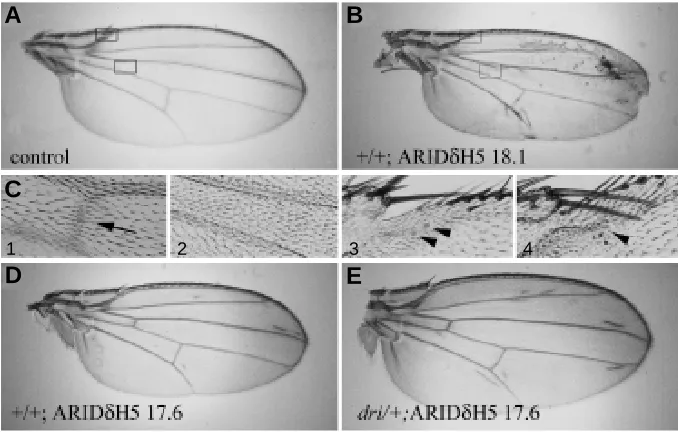
Fig. 4. Effects of mutant and wild-type retn/ dri expression under GAL471B control in the wing-blade anlagen of the wing imaginal disk.
(A-F) Photomicrographs of wings from females of the following genotypes: (A) w1118 (full wing). (B) P[UAS::retn/driARIDδH518.1]/ w1118;+;GAL471B/+ (full wing), (C1) and (C2)
higher magnifications of (A) and (B) respectively (bottom boxed area), showing the region around the anterior cross vein (black arrowhead in C1),
(C3) and (C4) higher magnifications of (A) and (B) respectively (top boxed area), showing the re-gion around the twin campaniform sensilla of the margin (black arrow heads indicate the presence of campaniform sensilla), (D) w1118; +; GAL471B P[UAS::retn/driARIDδH517.6]/+ and (E) w1118; retn/dri1/+; GAL471B P[UAS:: retn/driARIDδH5 17.6]/+, showing an enhanced phenotype when the retn/dri gene dosage was halved.
Fig. 3. In vitro analysis of eARID deletions. (A) Gel electrophoretic mobility shift assay using GST, GST-RETN/DRIARID and GST-RETN/DRIARIDδH5 proteins incubated with labelled NP6 oligonucleotide. (B) Protein blot assay for RETN/DRI REKLES domain self-association. GST-RETN/DRIREKLES and GST-RETN/DRIREKLESδβ were electrophoresed and transferred to nitro-cellulose membranes. Membranes were probed with either anti-DRI antibody or radiolabelled full-length RETN/DRI protein, produced by in vitro transcription and translation. The 29kDa GST protein unfused to RETN/DRI and probed with radiolabelled RETN/DRI protein is also shown. Note that the GST-RETN/DRIREKLES and GST-RETN/DRIREKLESδβ proteins appear as
two bands, both of which are less than the size predicted for a GST-RETN/ DRIREKLES protein. The reason for this has not been established.
patterns revealed that it is expressed in a subset of wing imaginal disk cells that express the homeodomain protein Cut (Fig. 2D). These cut and retn/dri-expressing cells are known to be the precursors of a group of sense organs, known as the campaniform sensilla, located on the wing (Huang et al., 1991; Blochlinger et al., 1993). Additionally, retn/dri appears to be expressed at low levels throughout the wing imaginal disk. Thus the wing was chosen as a target tissue for confirmation of retn/driARIDδΗ5 antimorphism.
A
B
A
B
C
D
E
shock promoter was used to induce recombination between non-sister chromatids of the two homologs. This resulted in the generation of retn/dri mutant cells that no longer carried the dominant female sterile allele ovoD1 and could therefore produce eggs.
Many early embryos derived from germline retn/dri- clones, and
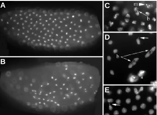
therefore lacking maternally-derived RETN/DRI, exhibited prolif-eration defects. The most pronounced of these defects was the loss of synchrony of cell divisions across the early syncytial embryo (Fig. 6 A-F, 7A-E). Affected embryos exhibited numerous anaphase bridges (Fig. 7 C,D) leading to the formation of aberrant nuclei, including micronuclei and the fusion of nuclei (Fig. 7 C-E). Cell cycle defects of this nature lead to a loss of cells during the syncytial divisions (Sullivan et al., 1993), so it is not surprising that some embryos had fewer cells while still undergoing mitosis (Fig. 6 D-F, 7B,D). However, as previously observed for the zygotic retn/dri phenotypes, the germline clone embryonic proliferation defects were highly variable, with approximately 65% of blastoderm stage embryos exhibiting a normal distribution of nuclei during embryo-genesis.
Discussion
The ARID family of genes are involved in a wide variety of transcriptional regulatory mechanisms and in a diversity of bio-logical processes. The retn/dri gene of Drosophila melanogaster is perhaps the best characterised ARID family gene with respect to its role in development. In this report we extend this characterisation by describing studies of the in vitro and in vivo functions of conserved domains in the RETN/DRI protein. Gel mobility shift assays using the Engrailed consensus binding NP6 DNA sequence revealed that disruption of the ARID motif by deletion of the fifth α-helix disrupts DNA-binding. Similarly,
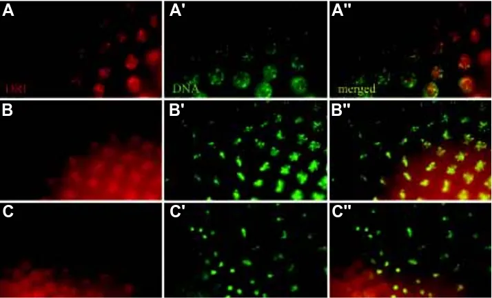
dele-Fig. 5. RETN/DRI distribution during mitosis RETN/DRI distribution, detected using polyclonal rat anti-RETN/DRI (red, A-C) during a wave of mitoses occurring in a syncytial embryo. (A) Speckles of RETN/DRI are observed in interphase nuclei. (B) At prophase/metaphase RETN/DRI can be visualised both on the chromosomes and diffusely at a low level in cytoplasm. (C) During anaphase, RETN/DRI appears to have degraded, before a high level of protein re-accumulates in the telophase nuclei. (A’-C’). The same sections stained forDNA with Hoechst 33258. (A’’-C’’) merged images.
transcription/translated, 35S-methionine-labelled RETN/DRI (Fig. 3B,
arrow). No difference in signal intensity was observed between the wild-type and mutant forms, indicating that the REKLESβ region is not necessary for self-association.
retn/dri Lacking the REKLESβ Region is able to Rescue retn/dri Function In Vivo
In order to examine the in vivo role of the REKLESβ region of RETN/DRI, rescue experiments similar to those described for full length retn/dri and the ARID mutant form of retn/dri were performed. Unlike the ARID motif mutation, constructs lacking the REKLESβ region could still rescue the mutant phenotype. This shows that although it represents the most conserved part of the REKLES motif, the REKLESβ region of RETN/DRI is not essential for function during stages of embryonic and imaginal development rescued by the transgene. However, in vivo expression of retn/dri::GAL4>UAS::retn/ driREKLESδβ in a heterozygous background caused a small but significant reduction in viability (data not shown), suggesting that this construct may have acted as a mild dominant negative form of the protein.
ARID Family Genes and Cell Cycle Regulation: A Role for retn/ dri in Drosophila Preblastoderm Mitoses
retn/dri maternal products are uniformly distributed in the early Drosophila embryo during the preblastoderm cleavage stage, where nuclei are undergoing mitoses in a syncytium (Fig. 5, Gregory et al., 1996). At interphase, the RETN/DRI protein is present in discrete apically located nuclear foci not completely overlapping with the highly condensed DNA foci stained preferentially by Hoechst 33258 dye (Fig. 5A). At metaphase, RETN/DRI can be visualised both on the chromosomes and at a low level in the cytoplasm (Fig. 5B). The cytoplasmic RETN/DRI gradually disappears during the anaphase/
telophase transition, after which high levels of protein accumulate in the newly re-formed nuclei (Fig. 5C). After this stage, retn/dri is expressed in a range of terminally differen-tiating cells that have ceased proliferation (Fig. 2). Analysis of the zygotic retn/dri phenotype showed that retn/dri plays roles in many and perhaps all of the differentiat-ing cells in which it is expressed (Shandala et al., 1999; Shandala, Sibbons and Saint, unpublished observations).
To determine a function for retn/dri dur-ing the syncytial divisions, it is necessary to eliminate the maternal retn/dri product from the egg. This was achieved using mitotic recombination to produce retn/dri mutant clones of cells in the developing oocytes, using the method of the FLP-FRT-ovoD1
system (Chou and Perrimon, 1996). retn/ dri mutant alleles were recombined onto chromosomes carrying FRT recombina-tion sites at the base of chromosome arm 2R and placed in trans to a chromosome carrying a dominant female sterile muta-tion, ovoD1,that prevents the production of
eggs. A transgene expressing the FLP recombinase under the control of the heat
A
A'
A''
B
B'
B''
A
B
C
D
E
F
A'
B'
C'
D'
E'
F'
A''
B''
C''
D''
E''
F''
tions of human SMARCF1 ARID and of helix 1 of mouse Bright dramatically abrogate DNA-binding activity of the resultant pep-tides (Herrscher et al., 1995; Webb, 2001; Dallas et al.,2000). An in vivo requirement for ARID domain function was demonstrated by showing that the deletion of this same helix eliminated the ability of a retn/dri transgene to rescue the mutant phenotype. The in vivo requirement for ARID sequences has also been demon-strated for the mouse Desrt protein. Targeted deletion of the ARID-containing exon of Desrt in mice results in multiple develop-mental defects, including reduced embryonic viability, growth retardation, disruption of spermatogenesis and transient immune abnormalities (Lahoud et al., 2001).
Examination of the phenotype of the deletion transgene in the presence of wild-type retn/dri in the present study showed that
RETN/DRIARIDδH5 acted as a dominant nega-tive. This suggests that RETN/DRI acts as part of a protein complex, consistent with the ob-servation that Bright tetramerises (Herrscher et al., 1995) and that RETN/DRI forms protein complexes with the co-repressor Groucho (Val-entine et al., 1998).
eARID family members exhibit an indepen-dent, less well conserved bipartite domain termed the REKLES domain (Kortschak et al., 2000). Western analysis of the REKLES do-main using radio-labelled RETN/DRI as a probe showed that the REKLES domain is capable of mediating self-association, consistent with the finding of Herrscher et al. (1995) that the region of Bright corresponding to the REKLES domain confers tetramerisation activity. A re-quirement for the REKLESβ domain for RETN/ DRI function in vivo was assayed by expres-sion of RETN/DRI lacking the most conserved part of this subdomain. This construct rescued the retn/dri mutant phenotype as effectively as the full length retn/dri construct. This indicates that the REKLESβ deletion does not result in a significant loss of zygotic retn/dri activity, leav-ing us yet to demonstrate a function for this domain.
In the last of the studies reported here, we used germline clones to examine the role of maternal retn/dri products during the para-synchronous syncytial cleavage divisions that follow fertilisation of the Drosophila embryo. Embryos deprived of their maternally-derived retn/dri products in this way exhibit variable but frequent mitotic aberrations, including the loss of synchrony, the presence of incomplete sis-ter chromatid separation and a reduction in the number of nuclei. RETN/DRI is present in cleavage stage nuclei, but appears to de-crease in level during mitosis to accumulate in the nucleus after nuclear envelope formation. It is possible, therefore, that the mitotic pheno-types observed reflect a role for RETN/DRI in chromosome condensation in cleavage em-bryos, rather than mitosis itself.
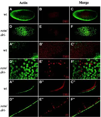
Fig. 6. Loss of nuclei in embryos lacking maternal RETN/DRI protein (A-C) Wild-type embryos. Nuclei were labeled with anti-RETN/DRI (red), actin was detected with monoclonal mouse anti-Actin antibody (green). (D-F) retn/dri mutants. Nuclei were labeled with propidium iodine (PI, propidium iodide, red), anti-Actin staining is green. (A’- C’) Apical veiw of the wild-type embryos in (A-C). (D’-F’) Apical view of the retn/dri mutant embryos in (D-F), showing less cells and disruption of the normal relationship between F-actin and nuclear position in the syncytium.
(A’’-C’’) Transverse optical section through the same wild-type embryo shown in (A-C). (D’’-F’’)
Transverse optical section through the mutant embryo shown in (D-F), showing nuclei sinking into the interior of the embryo.
A
C
B
D
E
in the heart, liver, neural tube, spleen and thymus (Takeuchi et al., 1995; Kitajima et al., 2001). Detailed analysis of jumonji mutant tissues revealed that cardiac trabecular myocytes in the heart and lymphoid cells (megakaryocytes) in all hematopoetic tissues showed over-proliferation and overgrowth (Kitajima et al., 2001). retn/dri appears not to be expressed in all dividing cells, so it is unlikely that it is required in all mitotic divisions in the same way that RBP1 may be generally required. Furthermore, analysis of the phenotypes later in development has not revealed any re-quirement for RETN/DRI in later divisions, nor has it revealed any overgrowth phenotype that would indicate a negative regulatory role of the type inferred for mouse jumonji. However, the cleavage divisions in the Drosophila embryo are highly modified divisions, being syncytial and occurring every approximately 10 minutes. It is possible that RETN/DRI has been recruited to facilitate these extraordinarily rapid early embryonic mitotic cycles.
These studies confirm that the ARID motif is essential for retn/ dri in vivo function, but the function of the more poorly conserved REKLES domain remains to be elucidated. The involvement of retn/dri in a variety of developmental processes, from the syncy-tial cleavage mitoses described here to the early axis patterning and later roles in muscle development (Shandala et al., 1999) and neural development (T. Shandala, J. Sibbons and R.Saint, un-published observation), may mean that the REKLES domain has a stage or role-specific function. Identification of other factors that bind RETN/ARID could provide a way of further exploring the function of the REKLES domain and of the RETN/DRI protein in general.
Materials and Methods
Generation of Domain-Specific Mutant retn/dri Constructs
To analyse the in vitro function of the ARID and REKLES domains, PCR amplification was used to generate pGEX::retn/dri constructs. Accuracy of PCR, and orientation and frame of insertion of the fragment were confirmed Fig. 7. Proliferation defects in embryos with no maternal retn/dri expression. Whole mount embryos stained with Hoechst 33258. (A) Loss of synchrony of cell divisions across the early syncytial embryo. (B) Some embryos had fewer cells while still attempting mitosis. (C-E) Various mitotic defects observed in retn/dri mutant embryos: anaphase bridges (b, thin arrows), incomplete or aberrant nuclear reformation (short arrows) and formation of micronuclei (m, arrowhead).
by sequence analysis. A deletion construct, pGEX::retn/driARIDδH5, lacking the putative eARID helix 5 was generated from pGEX::retn/driARID using the QuikChange™ Site-Directed Mutagenesis Kit (Stratagene, California, USA). Mutant clones were confirmed by sequence analysis. Similarly, wild-type and mutant REKLES constructs were generated; fragments generated by PCR amplification were cloned into pGEX1 to generate pGEX::retn/driREKLES (amino acids 400-901) and pGEX::retn/driREKLESδβ (as above, but lacking the REKLESβ region) respectively. Orientation and frame of insertion of the fragments were confirmed by sequence analysis.
For analysis of the in vivo function, a full length cDNA retn/dri clone (Gregory et al., 1996) was mutagenised using the QuikChange™ Site-Directed Mutagenesis Kit (Stratagene, California, USA) to generate the retn/ dri mutant plasmids pretn/driARIDδH5 and pretn/driREKLESδβ respectively. Mutated sites were confirmed by sequence analysis. Mutant retn/dri cDNAs were cloned into pP[UAST] for Drosophila germline transformation (Spradling and Rubin, 1982) and GAL4-induced expression (Brand and Perrimon, 1993). Correct orientation of the cDNA was confirmed by restriction analysis. In vivo GAL4-induced expression of wild-type and mutant forms of RETN/ DRI was confirmed by western analysis of eye imaginal disks expressing RETN/DRI under the control of the GMR::GAL4 transgene.
Generation of Germline retn/dri Mutant Clones
To abolish the maternal retn/dri contribution, embryos derived from retn/ dri1 and retn/dri2 germline clones were generated using the FLP-FRT-ovoD1
system developed by Chou and Perrimon (1996).
Immunochistochemistry
The following antibodies were used in these studies: polyclonal rat anti-DRI (Gregory et al., 1996), polyclonal anti rabbit-βgal (Rockland Immunochemicals, California, USA), monoclonal anti-CUT, anti-actin (De-velopmental Studies Hybridoma Bank, University of Iowa, USA), rabbit anti-DILP (a gift from Mark R.Brown, Department of Entomology, University of Georgia, Athens, USA) and anti-REPO (a gift from Andrew Travers, Labora-tory of Molecular Biology, Medical Research Council, Cambridge, UK).
Acknowledgements
This work was supported by the Australian Research Council and an Australian Postgraduate Research Award (to RDK). We thank Michelle Coombe for her technical assistance. The monoclonal antibody anti-CUT developed by G.M.Rubin was obtained from the Developmental Studies Hybridoma Bank developed under the auspices of the NICHD and main-tained by The University of Iowa, Department of Biological Sciences, Iowa City, IA 52242.
References
ADNANE, J., SHAO, Z. and ROBBINS, P.D. (1995) The retinoblastoma susceptibility gene product represses transcription when directly bound to the promoter. J Biol Chem 270: 8837-8843.
AGULNIK, A.I., MITCHELL, M.J., MATTEI, M.G., BORSANI, G., AVNER, P.A., LERNER, J.L. and BISHOP, C.E. (1994a) A novel X gene with a widely transcribed Y-linked homologue escapes X-inactivation in mouse and human. Hum Mol Genet 3: 879-884.
AGULNIK, A.I., MITCHELL, M.J., LERNER, J.L., WOODS, D.R. and BISHOP, C.E. (1994b) A mouse Y chromosome gene encoded by a region essential for spermatogenesis and expression of male-specific minor histocompatibility anti-gens. Hum Mol Genet 3: 873-878.
BLOCHLINGER, K., JAN, L.Y. and JAN, Y.N. (1993) Postembryonic patterns of expression of cut, a locus regulating sensory organ identity in Drosophila. Development 117: 441-450.
BRAND, A.H. and PERRIMON, N. (1993) Targeted gene expression as a means of altering cell fates and generating dominant phenotypes. Development 118: 401-415.
CHEN, G., FERNANDEZ, J., MISCHE, S. and COUREY, A.J. (1999) A functional interaction between the histone deacetylase Rpd3 and the corepressor groucho in Drosophila development. Genes Dev 13: 2218-2230.
CHOU, T.B. and PERRIMON, N. (1996) The autosomal FLP-DFS technique for generating germline mosaics in Drosophila melanogaster. Genetics 144: 1673-1679.
COLLINS, R.T., FURUKAWA, T., TANESE, N. and TREISMAN, J.E. (1999) Osa associates with the Brahma chromatin remodelingcomplex and promotes the activation of some target genes. Embo J 18: 7029-7040.
DALLAS, P.B., PACCHIONE, S., WILSKER, D., BOWRIN, V., KOBAYASHI, R. and MORAN, E. (2000) The human SWI-SNF complex protein p270 is an ARID family member with non-sequence-specific DNA binding activity. Mol Cell Biol 20: 3137-3146.
FATTAEY, A.R., HELIN, K., DEMBSKI, M.S., DYSON, N., HARLOW, E., VUOCOLO, G.A., HANOBIK, M.G., HASKELL, K.M., OLIFF, A., DEFEO-JONES, D. et al. (1993) Characterization of the retinoblastoma binding proteins RBP1 and RBP2. Oncogene 8: 3149-3156.
GREGORY, S.L., KORTSCHAK, R.D., KALIONIS, B. and SAINT, R. (1996) Characterization of the dead ringer gene identifies a novel, highly conserved family of sequence-specific DNA-binding proteins. Mol Cell Biol 16: 792-799.
HADER, T., WAINWRIGHT, D., SHANDALA, T., SAINT, R., TAUBERT, H., BRONNER, G. and JACKLE, H. (2000) Receptor tyrosine kinase signaling regulates different modes of Groucho-dependent control of Dorsal. Curr Biol 10: 51-54.
HERRSCHER, R.F., KAPLAN, M.H., LELSZ, D.L., DAS, C., SCHEUERMANN, R. and TUCKER, P.W. (1995) The immunoglobulin heavy-chain matrix-associat-ing regions are bound by Bright: a B cell-specific trans-activator that describes a new DNA-binding protein family. Genes Dev 9: 3067-3082.
HUANG, F., DAMBLY-CHAUDIERE, C. and GHYSEN, A. (1991) The emergence of sense organs in the wing disc of Drosophila. Development 111: 1087-1095.
HUANG, T.H., OKA, T., ASAI, T., OKADA, T., MERRILLS, B.W., GERTSON, P.N., WHITSON, R.H. and ITAKURA, K. (1996) Repression by a differentiation-specific factor of the human cytomegalovirus enhancer. Nucleic Acids Res 24: 1695-1701.
KITAJIMA, K., KOJIMA, M., KONDO, S. and TAKEUCHI, T. (2001) A role of jumonji gene in proliferation but not differentiation of megakaryocyte lineage cells. Exp Hematol 29: 507-514.
KOONIN, E.V., ZHOU, S. and LUCCHESI, J.C. (1995) The chromo superfamily: new members, duplication of the chromo domain and possible role in delivering transcription regulators to chromatin. Nucleic Acids Res 23: 4229-4233.
KORTSCHAK, R.D., TUCKER, P.W. and SAINT, R. (2000) ARID proteins come in from the desert. Trends Biochem Sci 25: 294-299.
KOZMIK, Z., MACHON, O., KRALOVA, J., KRESLOVA, J., PACES, J. and VLCEK, C. (2001) Characterization of mammalian orthologues of the Drosophila osa gene: cDNA cloning, expression, chromosomal localization, and direct physical interac-tion with Brahma chromatin-remodeling complex. Genomics 73: 140-148.
LAHOUD, M.H., RISTEVSKI, S., VENTER, D.J., JERMIIN, L.S., BERTONCELLO, I., ZAVARSEK, S., HASTHORPE, S., DRAGO, J., DE KRETSER, D., HERTZOG, P.J. and KOLA, I. (2001) Gene targeting of Desrt, a novel ARID class DNA-binding protein, causes growth retardation and abnormal development of reproductive organs. Genome Res 11: 1327-1334.
LAI, A., KENNEDY, B.K., BARBIE, D.A., BERTOS, N.R., YANG, X.J., THEBERGE, M.C., TSAI, S.C., SETO, E., ZHANG, Y., KUZMICHEV, A., LANE, W.S., REINBERG, D., HARLOW, E. and BRANTON, P.E. (2001) RBP1 recruits the mSIN3-histone deacetylase complex to the pocket of retinoblastoma tumor suppressor family proteins found in limited discrete regions of the nucleus at growth arrest. Mol Cell Biol 21: 2918-2932.
LALL, S. and PATEL, N.H. (2001) Conservation and divergence in molecular mechanisms of axis formation. Annu Rev Genet 35: 407-437.
LEE, Y., SONG, A.J., BAKER, R., MICALES, B., CONWAY, S.J. and LYONS, G.E. (2000) Jumonji, a nuclear protein that is necessary for normal heart development. Circ Res 86: 932-938.
LU, P.J., SUNDQUIST, K., BAECKSTROM, D., POULSOM, R., HANBY, A., MEIER-EWERT, S., JONES, T., MITCHELL, M., PITHA-ROWE, P., FREEMONT, P. and TAYLOR-PAPADIMITRIOU, J. (1999) A novel gene (PLU-1) containing highly conserved putative DNA/chromatin binding motifs is specifically up-regulated in breast cancer. J Biol Chem 274: 15633-15645.
NEVINS, J.R. (1998) Toward an understanding of the functional complexity of the E2F and retinoblastoma families. Cell Growth Differ 9: 585-593.
NUMATA, S., CLAUDIO, P.P., DEAN, C., GIORDANO, A. and CROCE, C.M.(1998) Bdp, a new member of a family of DNA binding proteins, associates with the retinoblastoma gene product. Cancer Res. 59: 3741-3747.
PALAPARTI, A., BARATZ, A. and STIFANI, S. (1997) The Groucho/transducin-like enhancer of split transcriptional repressors interact with the genetically defined amino-terminal silencing domain of histone H3. J Biol Chem 272: 26604-26610.
QUADBECK-SEEGER, C., WANNER, G., HUBER, S., KAHMANN, R. and KAMPER, J. (2000) A protein with similarity to the human retinoblastoma binding protein 2 acts specifically as a repressor for genes regulated by the b mating type locus in Ustilago maydis. Mol Microbiol 38: 154-166.
QUINN, J., FYRBERG, A.M., GANSTER, R.W., SCHMIDT, M.C. and PETERSON, C.L. (1996) DNA-binding properties of the yeast SWI/SNF complex. Nature 379: 844-847.
SCHUPBACH, T. and WIESCHAUS, E. (1991) Female sterile mutations on the second chromosome of Drosophila melanogaster. II. Mutations blocking oogenesis or altering egg morphology. Genetics 129: 1119-1136.
SHANDALA, T., KORTSCHAK, R.D., GREGORY, S. and SAINT, R. (1999) The Drosophila dead ringer gene is required for early embryonic patterning through regulation of argos and buttonhead expression. Development 126: 4341-4349.
SPRADLING, A.C. and RUBIN, G.M. (1982) Transposition of cloned P elements into Drosophila germ line chromosomes. Science 218: 341-347.
SUDARSANAM, P. and WINSTON, F. (2000) The Swi/Snf family nucleosome-remod-eling complexes and transcriptional control.Trends Genet 16: 345-351.
SULLIVAN, W., DAILY, D.R., FOGARTY, P., YOOK, K.J. and PIMPINELLI, S. (1993) Delays in anaphase initiation occur in individual nuclei of the syncytial Drosophila embryo. Mol Biol Cell 4: 885-896.
TAKEUCHI, T., YAMAZAKI, Y., KATOH-FUKUI, Y., TSUCHIYA, R., KONDO, S., MOTOYAMA, J. and HIGASHINAKAGAWA, T. (1995) Gene trap capture of a novel mouse gene, jumonji, required for neural tube formation. Genes Dev 9: 1211-1222.
TAKEUCHI, T., CHEN, B.K., QIU, Y., SONOBE, H. and OHTSUKI, Y. (1997) Molecular cloning and expression of a novel human cDNA containing CAG repeats. Gene 204: 71-77.
TREISMAN, J.E., LUK, A., RUBIN, G.M. and HEBERLEIN, U. (1997) eyelid antago-nizes wingless signaling during Drosophila development and has homology to the Bright family of DNA-binding proteins. Genes Dev 11: 1949-1962.
VALENTINE, S.A., CHEN, G., SHANDALA, T., FERNANDEZ, J., MISCHE, S., SAINT, R. and COUREY, A.J. (1998) Dorsal-mediated repression requires the formation of a multiprotein repression complex at the ventral silencer. Mol Cell Biol 18: 6584-6594.
WEBB, C.F., SMITH, E.A., MEDINA, K.L., BUCHANAN, K.L., SMITHSON, G. and DOU, S. (1998) Expression of bright at two distinct stages of B lymphocyte development. J Immunol 160: 4747-4754.
WEBB, C.F. (2001) The transcription factor, Bright, and immunoglobulin heavy chain expression. Immunol Res 24: 149-161.