Original Article
Role of cell division in branching morphogenesis and
differentiation of the embryonic pancreas
LORI DAWN HORB and JONATHAN M.W. SLACK*
Developmental Biology Programme, Department of Biology and Biochemistry, University of Bath, U.K.
ABSTRACT A new culture system for the embryonic pancreas enables the formation of a branched organ in vitro. In such cultures, each terminal branch originates as a small bud and the number of buds and of terminal branches increases progressively with the expansion of the culture. However buds can also be resorbed during growth. The normal labelling index of cells in incipient buds (“tips”) is greater than between buds (“dips”) suggesting that budding may be driven by a local increase of cell division. Consistent with this, treatments that reduce cell division repress the formation of buds and branches. It is not possible to initiate budding in isolated endodermal epithelium by treatment with fibroblast growth factor, although this does increase the degree of differentiation of exocrine cells. Cultures in which cell division is completely inhibited by aphidicolin treatment will produce more endocrine cells than usual and inhibit the differentiation of exocrine cells. Consistent with this it is found that in untreated cultures the division of endocrine precursors cannot be detected by BrdU labelling whereas the division of exocrine precursors is frequent. It is concluded that cell division is necessary for bud formation in the embryonic pancreas and that the growth factors required for this normally come from the mesenchyme. Cell division is also necessary for exocrine differentiation. Endocrine cells, however, can arise from undifferentiated progenitors without cell division.
KEY WORDS:
pancreas, amylase, insulin, glucagon, branching morphogenesis, aphidicolin,
dexametha-sone, bromodeoxyuridine.
0214-6282/2000/$20.00
© UBC Press Printed in Spain
www.ehu.es/ijdb
*Address for reprints: J.M.W. Slack. Developmental Biology Programme, Department of Biology and Biochemistry, University of Bath, Bath BA2 7AY, UK. FAX: +44-1225-826-779. e-mail j.m.w.slack@bath.ac.uk
Abbreviations used in this paper: BrdU, bromodeoxyuridine; FGF, fibroblast growth factor; DAPI, 4,6-diamidino-2-phenyl indole.
Introduction
Most glandular organs consist of an epithelium, which forms the functional tissue of the gland, and a mesenchyme that forms the interstitial connective tissue. During embryonic development the epithelial bud forms by invagination and grows in response to permissive inductive signals from the mesenchyme (Grobstein, 1966). In the course of its growth the epithelium undergoes repeated budding to produce a complex branched structure. For exocrine glands, the original site of invagination is retained as a secretory duct, while for endocrine glands the epithelium becomes detached from its original parent tissue (Le Gros Clark, 1975). The appearance of the branched structure is specific to the organ and so clearly distinct in salivary gland, mammary gland, lung and kidney, which are the four organs that have previously been studied in some detail.
Branching morphogenesis has attracted attention in recent years and considerable progress has been made in understanding this phenomenon in the kidney (Sariola and Sainio, 1997), the lungs (Hogan et al., 1997; Warburton et al., 2000) and the tracheal system of Drosophila (Shilo et al., 1997). The branching behaviour of the embryonic pancreas has been well described in normal
The main question that is examined in the present paper is: is budding driven by differential cell division? It is normally assumed that budding must be due to growth, and there are well understood examples of branching morphogenesis where this is the case, for example the lung, where FGF10 from the mesenchyme promotes growth and branching behaviour (Bellusci et al., 1997). But there are also examples where branching morphogenesis can proceed in the absence of cell division, for example in the tracheal system of Drosophila the formation of buds is also controlled by FGF signalling but the morphogenesis is achieved entirely by cell migration (Shilo et al., 1997). Likewise, in the salivary gland, branching can occur after doses of X-irradiation that suppress most cell division (Nakanishi et al., 1987). In this case the branching is sensitive to treatment with cytochalasin B (Spooner and Wessells, 1972) suggesting a primary role for cell movement and rearrange-ment dependent on microfilarearrange-ments.
Here we have examined the labelling index in regions of incipient budding and find that there is an excess of dividing cells. We show that inhibition of cell division using either dexametha-sone, or aphidicolin, two agents with completely different modes of action, prevents branching. It is not possible effectively to augment
duced growth/branching is associated with endocrine differentia-tion. This suggests that there is an antagonism during the develop-ment of the pancreas between continued cell division and the formation of endocrine cells.
Results
Formation of branches and cell division
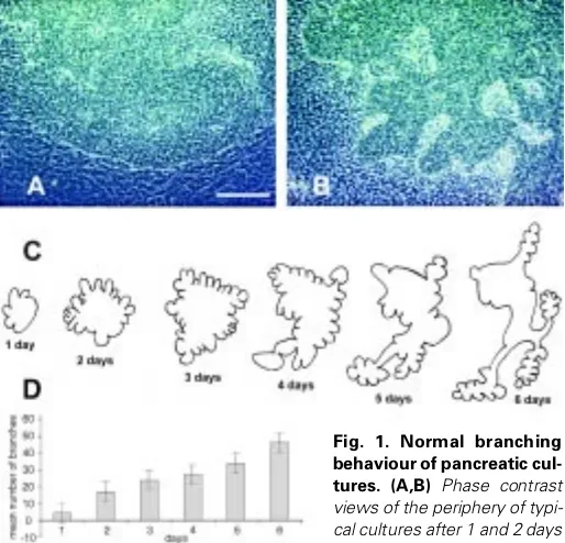
A single E11.5 dorsal pancreas in culture starts out as a simple hemisphere and on the first day of culture it spreads out to become a flattened mound (Fig. 1A). The mesenchyme migrates out rapidly to become a cell monolayer surrounding the epithelium. On the second day the epithelium develops a number of buds at its edge
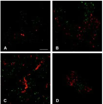
Fig. 2. BrdU incorporation into cultures. These panels all show experi-ments in which the cultures are labelled with BrdU for 4 hours, then fixed and processed for immunostaining of the BrdU. (A) Low power view of an intact culture, showing high labelling index in the epithelium. Scale bar, 400
µm. (B) A peripheral area of mesenchyme, showing moderate labelling index. Scale bar, 100 µm. (C,D) High power views of a tip and a dip, showing higher labelling index in the tip. Scale bars, 20 µm. (E) Labelling pattern of a dexamethasone treated culture. The index is reduced relative to the normal epithelium and there is little branch formation. (F) Labelling of an aphidicolin treated culture, showing no BrdU incorporation. Scale bars for E,F, 400 µm.
TABLE 1
BRDU LABELLING INDICES FOR BUD REGIONS (“TIPS”) AND INTERBUD REGIONS (“DIPS”)
Tips Dips
labelled unlabelled labelled unlabelled chi squared
day 2 111 83 79 88 7.89*
3 173 93 118 135 17.28**
4 383 88 296 163 33.3**
*p<0.005; **p<0.001
(Fig. 1B). These spread out and produce further buds so that the entire structure becomes highly ramified and branched, in a similar way to the pancreas in vivo. Typically about 45 buds are present by 6 days of culture. The behaviour is illustrated in Fig. 1C, which shows camera lucida drawings of a single specimen on successive days, and in Fig. 1D which shows the increase in bud number for a set of cultures. It is apparent that not all buds grow out to become large branches, sometimes they regress leaving a significant unbranched part of the perimeter. This fact shows that the mecha-nism underlying branching does not involve an inhibition from existing buds, as in such a case new buds would always appear at a maximum distance from the existing ones.
During the growth of these cultures the epithelial cell number increases. This was estimated by staining cultures with DAPI and counting the nuclei. Mean values for 2 cultures were 1354 on day 2, 2679 on day 3 and 3543 on day 6. The mesenchymal area, which is a cell monolayer, also increased in proportion to the epithelial cell numbers. This growth rate is not as fast as in vivo, but it does resemble the in vivo pattern insofar as growth is very rapid at the mid-gestation embryo stage and slows down later.
We were interested to know whether buds arose because of increased local cell division. This is not necessarily the case as buds disappear as well as appear and so there must also be an element of cell movement involved in the morphogenesis of the culture. Cultures were labelled with BrdU for 4 hours and then fixed and immunostained to give a labelling index. Cultures were stained with DAPI which stains all nuclei and then the number of DAPI-labelled and BrdU-DAPI-labelled nuclei was counted in a standard size square in regions of incipient budding (“tips”), and in between such regions (“dips”). Typical results from the BrdU labelling are shown in Fig. 2 A-D and results from 8-10 pooled data sets are given in Table 1. The absolute values of the labelling indices are not so important, as the labelling conditions may differ slightly between experiments. However the conditions will be the same in different parts of one culture, so it is the difference between the proportion of labelled cells at tips versus dips that is significant. The results show clearly that there is more cell division at “tips” than at “dips”, and that the effect becomes stronger with successive days in culture.
However, it does not follow from this that the localised cell division is necessarily a driving force for budding. So we then sought to find whether treatments that affected the rate of cell division would affect the branching behaviour. Treatment with the
synthetic glucocorticoid dexamethasone (1 µM) was found to reduce the cell labelling index and also to reduce the formation of branches very considerably. In 4 day cultures, the 4 hour labelling index was 70.7% in controls and 41.8% in the Dex treated cultures (compare Fig. 2 A,E). In Fig. 3 is shown camera lucida drawings of a typical culture, together with phase contrast pictures of the epithelial margin and it can be seen that there is hardly any bud formation in dexamethasone.
A more extreme inhibition of morphogenesis was achieved by treatment with aphidicolin (0.5 µg/ml). This is an inhibitor of DNA synthesis and so should be expected completely to inhibit BrdU incorporation. Indeed the labelling index is reduced to 0% (Fig. 2F). The epithelium spreads out and becomes somewhat flatter than usual with a less distinct margin (Fig. 3). It shows no branching at all
Fig. 3. Camera lucida drawings of typical cultures on successive days, and phase contrast views of the epithelial margin at the end of the culture period. (A) A dexam-ethasone treated culture. The phase view shows only limited incipient branching. (B) An aphidicolin treated culture. The phase view shows no branching and an indistinct epithe-lial boundary.
and sometimes breaks up into more than one domain, as shown in Fig. 3. Despite the substantial change in cell behaviour the cultures are fully viable (see below), and the mesenchyme spreads out to form the usual extended cell monolayer around the epithelium.
As previous work had shown a mitogenic effect of FGFs (Miralles et al., 1999) we attempted to induce the formation of supernumer-ary buds by treatment with fibroblast growth factors. FGF1 was used because it is capable of stimulating all known FGF receptors (Ornitz et al., 1996) and should therefore be able to mimic the effect of any of the 22 known FGFs. For these experiments it is not necessary to know which particular FGFs are active in vivo in order to establish FGF responsiveness. But beads soaked in 10 µg/ml or 100 µg/ml did not show any effect on the formation of branches. When we examined the labelling index it became clear that is not easy to serum-starve the cultures. Even after 2 days in serum free medium they are still growing, although they do retract from the substrate after this time. This is presumably because the mesen-chyme itself secretes various growth factors. So we felt it important to remove the mesenchyme and culture the epithelium alone. This does survive in the culture system and form a small monolayer, but it does not undergo the typical morphogenetic behaviour of the intact cultures. Addition of FGFs does promote differentiation (see below), but has little effect on the labelling index and does not provoke morphogenetic behaviour such as the formation of cell aggregates or buds. So it seems probable that the mesenchyme is necessary for branching morphogenesis for reasons in addition to a supply of mitogenic factors.
Effects on differentiation
We needed to establish that the treatments employed in this study were not toxic and so studied the differentiation patterns of cultures in the various regimes by immunostaining with specific antibodies to three proteins. Amylase is an abundant secretory product of exocrine cells. Insulin and glucagon are hormones
starved, and cultured with and without FGF (100 ng/ml). At least 4 replicates were performed for each of these experiments and typical specimens are shown. In the absence of FGF these cultures show little or no exocrine differentiation (Fig. 4A), but in the presence of FGF there is significantly more (Fig. 4B). The quantity of endocrine differentiation is not much changed although in the presence of FGF the insulin cells become more bunched together into islet-like structures (Fig. 4 C,D). The BrdU labelling is very limited in all cases.
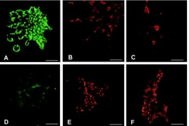
In Fig. 5 are shown normal cultures and cultures treated with aphidicolin to suppress cell division. Again, at least 4 replicates were performed with very similar results. In the normal cultures there is amylase expression around the periphery of most of the branch tips (Fig. 5A) and a sprinkling of insulin and glucagon-positive cells (Fig. 5 B,C). In the aphidicolin-treated cultures there is no exocrine differentiation at all, and the proportion of insulin and glucagon cells is considerably increased (Fig. 5 D-F). However they remain as individual cells and do not aggregate into clumps as in the controls. These results confirm that there is no obvious toxicity of any of the treatments, in that some form of differentiation occurs successfully in all cases. It also confirms the finding of Miralles et al., (Miralles et al., 1999) that FGF can promote exocrine differentiation from isolated epithelium. The most interesting
re-Fig. 5. Differentiation in aphidicolin treated cultures, showing an increase in endocrine and decrease in exocrine differentiation. (A-C) Controls, showing immunostaining for amylase, insulin and glucagon. (D-F) Aphidicolin treated, showing amylase, insulin and glucagon. Amylase is in green, insulin and glucagon in red. Scale bars, 400 µm except B,C, 200 µm.
that the normal cultures showed abundant exo-crine and endoexo-crine differentiation. Some type of differentiation occurred in all the cultures show-ing that none of the treatments were toxic over the time course of the experiment. But there were significant effects on the patterns of differentia-tion; in particular aphidicolin promoted endocrine differentiation, while FGF treatment of isolated epithelia promoted exocrine differentiation. A summary of the effects is given in Table 2, and individual cases are shown in Figs. 4, 5. In Table 2 the proportions of cells immunopositive for amylase, insulin or glucagon are shown coded as -, +, ++ or +++. On this qualitative scale, ++ is normal for amylase and insulin, - means none, + means substantially less than usual and +++ means substantially more than usual. For gluca-gon, + represents the control level.
In Fig. 4 are shown isolated epithelia,
serum-TABLE 2
CELL DIFFERENTIATION PATTERNS
amylase insulin glucagon
control ++ ++ +
dexamethasone ++
aphidicolin - +++ +++
isolated epithelium + ++
sults are those for the aphidicolin treated cultures which show a very substantial shift from exocrine to endocrine differentiation.
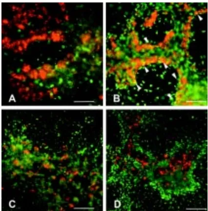
Aphidicolin is an inhibitor of DNA replication. This result raised the possibility that the exocrine cells would need to go through some cycles of DNA replication as an obligatory requirement for their terminal differentiation, while the endocrine cells do not (Satoh and Ikegami, 1981; Temple and Raff, 1986). We attempted to find when the cell types normally underwent their final S-phase by labelling cultures with BrdU on different days, and then finding whether exocrine or endocrine cells had become labelled at the end of the culture period. In fact it is difficult to obtain good BrdU labelling over the early period of the cultures (0-1 or 1-2 days) perhaps because at this stage they are rather compact and do not allow ready penetration of the reagent. Very few amylase-positive and no insulin-positive cells at 4 days were labelled during this period (Fig. 6 A,C). On the other hand, BrdU labelling on days 2-3 or 2-3-4 was very effective and some amylase positive cells were double labelled (visible as the yellow cells in Fig. 6B). No insulin-positive cells were double labelled with BrdU administered at any stage. This suggests that all the insulin-positive cells differentiating over the culture period are cells that were already present when the cultures were set up. It is not possible to prove this because of the difficulty of effective BrdU labelling at the early stages, but certainly there is a tendency for the endocrine cells not to have divided for a long time, compared with exocrine cells whose progenitors may divide within 24 hours of terminal differentiation. The time at which most of the insulin-positive cells actually differentiate is between days 2 and 3 of culture. Before this, only a very few very faintly stained insulin cells are present.
Discussion
The two extreme conceptions of morphogenetic behaviour would involve growth alone or cell movement alone (Bard, 1990). The former case resembles a plant in which the entire morphology arises from growth of rigid and immotile parts (Steeves and Sussex 1989). The latter case resembles a slime mould culmination in which all morphological events are due to cell movement and rearrangement, in the absence of cell division (Bonner 1947). Studies of branching morphogenesis in epithelial organs have suggested a role for cell division in some cases but not in others. We have taken advantage of a new culture system for the developing pancreas, which displays branching morphogenesis and allows visualisation of events by wholemount immunostaining. We find that new buds are associated with elevated cell division, and that treatments which reduce or abolish cell division reduce or abolish branching morphogenesis. However this does not prove that a primary role for local cell division as a driving force in new bud formation. In normal cultures, some new buds arise but then resorb into a smooth perimeter showing that buds may disappear even though the tissue as a whole is growing. The aphidicolin-treated cultures, in which there is no cell division, can form a few buds at early stages that are later resorbed. Moreover, treatment with FGF, although it increases exocrine differentiation, does not itself pro-voke the initiation of new buds. It is very striking that the isolated epithelium does not undergo branching morphogenesis even when treated with FGF suggesting that other factors, either chemi-cal or mechanichemi-cal, are required from the mesenchyme.
The immunostaining for differentiation markers was originally carried out in order to check that the cultures were not adversely
affected by the treatments. But this also revealed that when cell division is prevented, the differentiation of exocrine cells is re-duced while that of endocrine cells is increased. The study of the labelling of differentiated cells with BrdU administered at different times during the culture shows that very few endocrine cells have divided at all during the culture period, whereas the exocrine cells may do so. Due to the relatively low level of double labelling in all experiments it seems unlikely that there is an obligatory sequence of cell divisions preceding exocrine differentiation, as has been found in some other systems (Satoh and Ikegami, 1981; Temple and Raff, 1986). It may be that the intracellular pathways activated by mitogens are antagonistic to endocrine differentiation, and that when growth is prevented then more endocrine cells differentiate as a result. This would also be consistent with the previous results showing that endocrine differentiation represents the main de-fault pathway of differentiation of the pancreatic epithelium in the absence of mesenchyme (Gittes et al., 1996; Miralles et al., 1998).
The overall conclusion is that cell division is probably necessary for branching morphogenesis of the pancreas although it is not the only driving force. Other interactions with the mesenchyme, both mechanical and chemical, are also likely to be important.
Materials and Methods
Cultures
Cultures were set up from E11.5 mouse embryos as described in Percival and Slack (1999). Briefly the dorsal bud is placed cut surface down on a coverslip coated with 50 µg/ml fibronectin. The coverslips are sub-merged in culture medium in 3 cm Petri plates. The medium is BME with Earle’s salts, 10% foetal bovine serum, 50 µg/ml gentamycin. Cultures are maintained for up to 6 days at 37°C in 5% CO2, with a change of medium every 2 days. The day of plating is called day 0.
It was made up in ethanol and used at 1 µM. The medium, containing fresh dexamethasone, was changed each day. Aphidicolin is a tetracyclic diterpene mycotoxin that is a specific inhibitor of DNA polymerase α, not affecting RNA or protein synthesis. Aphidicolin (Sigma cat A0781) was dissolved in DMSO at 10 mg/ml and used at 0.5 µg/ml. The medium, containing fresh aphidicolin, was changed each day. Bromo-2-deoxyuridine (Sigma cat B-9285) was made to 10 mM in water. Labelling of cultures was carried out either overnight or for 4 hours at a concentration of 10 µM.
Immunostaining
Cultures were fixed in MEMFA (10% formalin, 0.1M MOPS pH 7.4, 2 mM EGTA) for 40 min at room temperature. They remain attached to the coverslips throughout the procedures. For BrdU detection they were treated with 2M HCl for 30 min at 37°C to improve accessibility of the DNA. All cultures were permeabilised using 1% Triton-X-100 in PBS for 1 hour. Before the first antibody they were treated with 6% BSA +10% heat-inactivated foetal bovine serum in PBS, 1 hour, to block non-specific sites. The first antibody was applied overnight in the blocking buffer at 4°C. Coverslips were washed with PBS 4 x 20 min. The second antibody was applied in blocking buffer at room temperature for 2 hours, followed by another 4 x 20 min washes in PBS. For double staining the procedure was then repeated for the second pair of antibodies. After the immunostaining stages were completed the coverslips were mounted on slides in gelvatol medium (20% polyvinyl alcohol in 10 mM Tris HCl, pH 8.6). The primary antibodies used, with dilutions, were: Mouse monoclonal anti-BrdU (Sigma B-2531), 1/1000; Guinea pig anti-bovine insulin (Sigma I-6136), 1/200; Mouse monoclonal anti-porcine glucagon (Sigma G2654), 1/200; Rabbit anti-human amylase (Sigma A8273), 1/200. Secondary antibodies, with dilutions: FITC goat anti-mouse (ICN 67228), 1/200; TRITC anti-guinea pig (Sigma T-7153), 1/200; TRITC anti-rabbit (Sigma T-6778), 1/200. Negative controls in which the first antibody was omitted are not shown, but were carried out in all experimental series. No cells were labelled in any of the negative controls. Specimens were viewed with a Leica DMRB fluorescent microscope. Images were captured using Hamamatsu C4880 cooled CCD camera or a JVC colour CCD camera and processed using Adobe Photoshop.
Acknowledgements
This work was supported by the Medical Research Council, grant number G9520375. We should like to thank David Tosh and Andrew Ward for comments on the manuscript.
References
BARD, J.B.L. (1990). Morphogenesis. The Cellular and Molecular Processes of Developmental Anatomy. Cambridge University Press, Cambridge UK.
BELLUSCI, S., GRINDLEY, J., EMOTO, H., ITOH, N. and HOGAN, B. (1997). Fibroblast Growth Factor 10(FGF10) and branching morphogenesis in the embry-onic mouse lung. Development 124: 4867-4878.
BONNER, J.T. (1947). Evidence for the formation of cell aggregates by chemotaxis in the development of the slime mold Dictyostelium discoideum. J. Exp. Zool. 106: 1-26.
HOGAN, B.L.M., GRINDLEY, J., BELLUSCI, S., DUNN, N.R., EMOTO, H. and ITOH, N. (1997). Branching morphogenesis of the lung: new models for a classical problem. Cold Spring Harbor Symposia on Quantitative Biology 62: 249-256.
LE GROS CLARK, W.E.(1975). The Tissues of the Body 6th edn. Clarendon Press, Oxford.
MIRALLES, F., CZERNICHOW, P. and SCHARFMAN, R. (1998). Follistatin regu-lates the relative proportions of endocrine versus exocrine tissue during pancre-atic development. Development 125: 1017-1024.
MIRALLES, F., CZERNICHOW, P., OZAKI, K., ITOH, N. and SCHARFMANN, R. (1999). Signaling through fibroblast growth factor receptor 2b plays a key role in the development of the exocrine pancreas. Proc. Natl. Acad. Sci USA 96: 6267-6272.
NAKANISHI, Y., MORITA, T. and NOGAWA, H. (1987). Cell proliferation is not required for the initiation of early cleft formation in mouse embryonic submandibu-lar epithelium in vivo. Development 99: 429-437.
ORNITZ, D.M., XU, J., COLVIN, J.S., MCEWEN, D.G., MACARTHUR, C.A., COULIER, F., GAO, G. and GOLDFARB, M. (1996). Receptor specificity of the fibroblast growth factor family. J. Biol. Chem. 271: 15292-15297.
PERCIVAL, A.C., and SLACK, J.M.W. (1999). Analysis of pancreatic development using a cell lineage label. Exp. Cell Res. 247: 123-132.
PICTET, R., and RUTTER, W. J. (1972). Development of the embryonic endocrine pancreas. Chapt. 2 Handbook of Physiology, Section 7. American Physiological Society. Eds. Steiner, DF & Frenkel, N, Williams & Wilkins, Washington DC 1: 25-66.
SARIOLA, H., and SAINIO, K. (1997). The tip top branching ureter. Curr. Biol. 9: 877-884.
SATOH, N. and IKEGAMI, S. (1981). A definite number of aphidicolin sensitive cell cycle events are required for acetyl cholinesterase development in the presump-tive muscle cells of the ascidian embryo. J. Embryol. Exp. Morph. 61: 1-13.
SHILO, B., GABAY, L., GLAZER, L., REICHMAN-FRIED, M., WAPPNER, P., WILK, R. and ZELZER, E. (1997). Branching morphogenesis in the Drosophila tracheal system. Cold Spring Harbor Symposia on Quantitative Biology 62: 241-247.
SPOONER, B.S., and WESSELLS, N. (1972). An analysis of salivary gland morpho-genesis: role of cytoplasmic microfilaments and microtubules. Dev. Biol. 27: 38-54.
STEEVES, T.A. and SUSSEX, I.M. (1989). Patterns in Plant Development. Cam-bridge University Press, CamCam-bridge UK.
TEITELMAN, G., ALPERT, S., POLAK, J.M., MARTINEZ, A. and HANAHAN, D. (1993). Precursor cells of mouse endocrine pancreas coexpress insulin, gluca-gon, and the neuronal proteins tyrosine hydroxylase and neuropeptide Y but not pancreatic polypeptide. Development 118: 1031-1039.
TEMPLE, S. and RAFF, M.C. (1986). Clonal analysis of oligodendrocyte development in culture - evidence for a developmental clock that counts cell divisions. Cell 44: 773-779.
UPCHURCH, B.H., APONTE, G.W. and LEITER, A.B. (1994). Expression of peptide YY in all four islet cell types in the developing mouse pancreas suggests a common peptide YY-producing progenitor. Development 120: 245-252.
WARBURTON, D., SCHWARZ, M., TEFFT, D., FLORES-DELGADO, G., ANDER-SON, K.D. and CARDOSO, W.V. (2000). The molecular basis of lung morphogen-esis. Mech. Dev. 92: 55-81.