Scholarship@Western
Scholarship@Western
Electronic Thesis and Dissertation Repository
7-29-2019 1:00 PM
Low Birth Weight and Post-Natal Diet Induced Alterations in
Low Birth Weight and Post-Natal Diet Induced Alterations in
Skeletal Muscle Oxygen Consumption and Fiber Type
Skeletal Muscle Oxygen Consumption and Fiber Type
Composition
Composition
Megan Cedrone
The University of Western Ontario
Supervisor Staples, James F.
The University of Western Ontario Co-Supervisor Regnault, Timothy R.H.
The University of Western Ontario Graduate Program in Biology
A thesis submitted in partial fulfillment of the requirements for the degree in Master of Science © Megan Cedrone 2019
Follow this and additional works at: https://ir.lib.uwo.ca/etd
Part of the Medical Physiology Commons, and the Musculoskeletal, Neural, and Ocular Physiology Commons
Recommended Citation Recommended Citation
Cedrone, Megan, "Low Birth Weight and Post-Natal Diet Induced Alterations in Skeletal Muscle Oxygen Consumption and Fiber Type Composition" (2019). Electronic Thesis and Dissertation Repository. 6446.
https://ir.lib.uwo.ca/etd/6446
This Dissertation/Thesis is brought to you for free and open access by Scholarship@Western. It has been accepted for inclusion in Electronic Thesis and Dissertation Repository by an authorized administrator of
i
Adverse in utero and postnatal conditions can increase susceptibility to metabolic syndrome
(MS). Altered muscle respiration contributes to MS, but the effects of restricted oxygen and
nutrients in utero on skeletal muscle mitochondria remain unknown. In this study guinea pig
sows underwent uterine artery ablations mid-gestation, producing fetuses with low birth
weight (LBW). Soleus muscle was collected near term or at four months of age, from LBW
and control fetuses and offspring, where the offspring were fed either a Western Diet (WD)
or a control diet (CD). Soleus muscles from LBW fetuses exhibit lower maximal respiration
rates than normal birth weight (NBW) sham-surgery controls. Additionally, LBW/CD,
NBW/WD and LBW/WD adult guinea pigs displayed reduced respiration compared with
NBW/CD. Cultured C2C12 cells were utilized to better understand independent effects of
hypoxia and fatty acid saturation upon cellular respiration. Both chronic (5 days) hypoxia and
palmitate (16:0) reduced respiration compared with normoxia.
Lay Summary
The environment in which we develop during pregnancy can determine certain aspects of our
health as adults. Previous studies have identified a correlation between low birth weight, an
outcome related to intrauterine growth restriction and the development of metabolic diseases
such as type 2 diabetes and cardiovascular disease later in life. The present study aimed to
identify a link between an environment low in oxygen and nutrients during pregnancy, which
is observed in intrauterine growth restriction and metabolic disorders in adulthood. As there
is a reduced skeletal muscle mass in cases of intrauterine growth restriction and skeletal
muscle plays an important role in insulin sensing, an important contributor to metabolic
health, it was chosen as the focus of this study. An intrauterine growth restricted fetal guinea
pig model was used to determine a link between a poor environment during pregnancy and
skeletal muscle oxygen consumption. A second study was undertaken where growth
restricted animals were given either a control or Western diet to determine if a post-natal diet
ii
unsaturated fatty acid palmitoleate on skeletal muscle oxygen consumption. Growth
restricted animals had reduced skeletal muscle oxygen consumption; however, the addition of
the Western diet did not further reduce oxygen consumption but did reduce oxygen
consumption in normal birth weight animals. Similarly, hypoxic cultured cells had reduced
oxygen consumption and the saturated fatty acid reduced oxygen consumption of only cells
cultured under normal oxygen levels. The monounsaturated fatty acid had no effect on
oxygen consumption. Discovering this link allows for further studies to be conducted with
potential treatments.
Keywords
Skeletal Muscle, Intrauterine Growth Restriction, Low Birth Weight, Oxygen Consumption,
iii
Acknowledgments
First and foremost, I would like to thank my supervisors, Drs. Jim Staples and Tim Regnault
for the opportunity to do this work and for all of your support. Without you, none of this
would have been possible. I never thought my fourth-year project would have turned into
such an exciting project and that I would have the chance to learn so many different
techniques and skills. All of your support and hard work is very much appreciated! Your
guidance has been invaluable both regarding my research and personal goals, thank you.
Next, I would like to thank my committee: Drs. Chris Guglielmo and Peter Merrifield. You
have helped shape my project and guide my research to a full finished product. Your
feedback is always appreciated and you have helped me expand my knowledge and grow as a
student. To Dr. Brent Sinclair, thank you for helping me return to my studies after an
extended leave, your guidance and persistence is the reason I am here to complete my degree.
Thank you for all your help.
Perhaps the most enjoyable part of this process was interacting and creating strong
friendships with other students, volunteers and lab personnel. The support and feeling of not
being in this alone really helped me push through when working was the last thing on my
mind. Jacky, I feel as though we were in this together, start to finish, you have been there to
give advice, listen to my various complaints, for coffee runs, lab lunches and comedic relief.
Alex, as my favorite science magician, I thank you most for providing motivation both in lab
and while writing, your positive attitude makes everything more enjoyable. Dr. Lin Zhao, I
think I could be your apprentice for life if you would allow it; you have taught me (almost)
everything I know in terms of lab techniques and various other life lessons. I already miss
our daily chats about anything and everything; you have a great perspective on life. To Katie
Belgrave, without whom none of this would have been possible, I’ve practiced your teaching
for the past years and hope to continue practicing safe science for the rest of my life. Noelle,
thank you for lunch time online shopping distractions, it was always easier to go back to
work after some good old-fashioned retail therapy (even if you did most of the buying!). To
the rest of the DDT lab and Staples lab members, there are too many of you to name, but
each of you have contributed to my success and helped push me along, you deserve the most
iv
do what makes me happy, and supported me through thick and thin, I’m so grateful to have
such amazing and supportive parents. Dad, although you are no longer here to see me
complete my degree, I know you would be so proud. I am so grateful that we were able to
spend so much time together, and although we were both battling our medical conditions,
your support has given me strength and determination. And mom, I am eternally grateful for
your unwavering help, you have been patient, kind and loving. You are my pillar of support
(sometimes quite literally) and I would be lots without you. To my brother, J.C. though our
differences are plentiful, our sibling rivalry has always pushed me and given me a
competitive edge. Finally, thank you Scott for your unconditional love and respect. Thank
you all for your support, emotional and financial, this would not have been possible without
v
Table of Contents
Abstract ... i
Acknowledgments... iii
Table of Contents ... v
List of Tables ... viii
List of Abbreviations ... ix
List of Figures ... xi
1 Introduction ... 1
1.1 Developmental Origins of Health and Disease ... 1
1.2 Intrauterine Growth Restriction ... 4
1.3 Hypoxia-induced alterations in skeletal muscle metabolism ... 6
1.3.1 Electron Transport Chain ... 7
1.3.2 Myogenesis and adverse in utero environments ... 9
1.4 Skeletal Muscle Fiber Type Composition and adverse in utero environments .... 11
1.4.1 Skeletal muscle fiber composition ... 12
1.5 The Western Diet ... 14
1.5.1 Western Diet origin and composition and the development of metabolic disease ... 14
1.5.2 Mechanisms of sugar and saturated fats’ negative impacts and increased risk of metabolic disease ... 16
1.6 Thesis Rationale and Objectives ... 17
1.6.1 Rationale ... 17
1.6.2 Objectives and Hypotheses ... 19
2 Materials and Methods ... 20
vi
2.1.3 Tissue Collection ... 23
2.2 Soleus Respirometry ... 24
2.3 Citrate Synthase Activity ... 25
2.4 Molecular Analysis ... 26
2.4.1 Rt-qPCR for Myosin Heavy Chain isoform expression ... 26
2.4.2 Immunoblotting of Electron Transport Chain Complex Subunits ... 28
2.5 Hypoxic C2C12 Cells and Fatty Acid Supplementation ... 29
2.5.1 Time points for collection ... 30
2.5.2 Rate of Oxygen Consumption in Cell Culture ... 31
2.5.3 Extraction and Quantification of Mitochondrial DNA from Cultured Cells ... 32
2.6 Statistics ... 33
3 Results ... 34
3.1 Growth Restriction Induced Alterations in Fetal Soleus Muscle Respiration ... 34
3.2 Low Birth Weight and Western Diet Effects on Soleus Muscle Respiration ... 37
3.2.1 Birth Weights and Thinness ... 37
3.2.2 Soleus Muscle Oxygen Consumption of Young Adult Guinea Pigs ... 40
3.2.3 ETC Protein Expression and Citrate Synthase Activity ... 42
3.2.4 Fiber Type Compositions ... 44
3.3 Hypoxia and Fatty Acid Induced Alterations in Mitochondrial Function in C2C12 Cells ... 46
3.3.1 Hypoxia reduced oxygen consumption which persisted after normoxic recovery... 46
3.3.2 Mitochondrial DNA Was Not Altered by Hypoxia ... 49
4 Discussion ... 50
vii
4.2 IUGR and Diet Independently Reduce Soleus Oxygen Consumption ... 51
4.3 Soleus Myosin Heavy Chain Expression Altered by Birth Weight and Diet ... 52
4.4 Hypoxic Regulation of C2C12 Oxygen Consumption ... 53
4.5 Impact of Fatty Acids on Oxygen Consumption of Hypoxic Cultured Cells ... 54
4.6 Potential Limitations ... 55
4.7 Future Studies ... 56
4.8 Conclusions ... 57
References ... 59
Appendix ... 81
viii
List of Tables
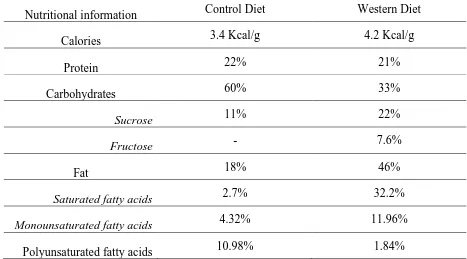
Table 1.6-2 Guinea Pig Diet Comparison (percent calories) ... 23
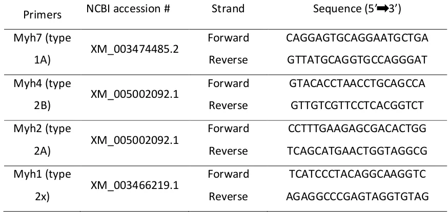
Table 2.4-1 Guinea Pig Myosin Heavy Chain primer pairs... 28
ix
List of Abbreviations
CS Citrate Synthase
CVD Cardiovascular disease
DOHaD Developmental origins of health and disease
ETC Electron transport chain
FA Fatty acid
HIF1-α Hypoxia inducible factor 1-alpha
IMM Inner mitochondrial membrane
IUGR Intrauterine growth restriction
LBW Low birth weight
MEF Myocyte enhancer factors
MHC Myosin heavy chain
MHC Myosin heavy chain
MRF Myogenic regulatory factors
MUFA Monounsaturated fatty acid
NAFLD Non-alcoholic fatty liver disease
NBW Normal birth weight
OXPHOS Oxidative phosphorylation
x
PGC-1α PPAR-γ coactivator-1α
PI Placental insufficiency
PPAR Peroxisome proliferator-activated receptor
PUFA Polyunsaturated fatty acid
SFA Saturated fatty acid
SGA Small for gestational age
SM Skeletal muscle
TCA Tricarboxylic acid
UAA Uterine artery ablation
UCP Uncoupling protein
xi
List of Figures
Figure 1.3-1 Overview of the Electron Transport Chain. Each of the embedded complexes
are depicted within the inner mitochondrial membrane (IMM), and the dissociable carriers’
coenzyme Q and cytochrome C are represented as (Q) and (c) respectively. Electron flow is
indicated by solid arrows and proton pumping by dashed arrows. ... 8
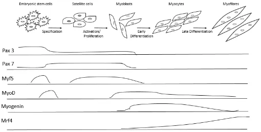
Figure 1.3-2 Embryonic Myogenesis. Determination and differentiation of muscle cells
regulated by Pax3/Pax7and MRFs MyoD, Myogenin, Mrf4 and Myf5. Transcription factor
expression is shown as area under the curve. Modified from Benzinger et al, 2012. ... 10
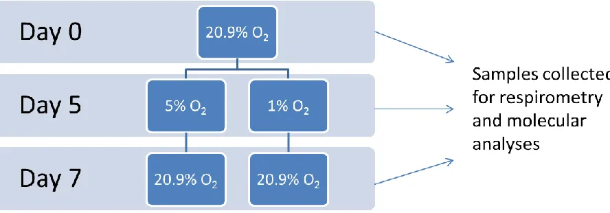
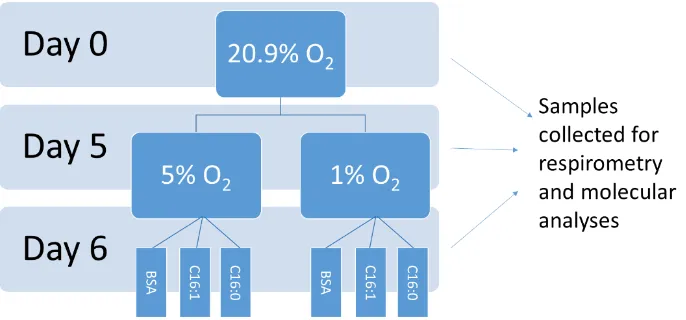
Figure 2.5-1 Diagram of cell culture experiments. C2C12 mouse myoblasts were cultured at
20.9% oxygen until they reached ~ 80% confluence. ... 30
Figure 2.5-2 Diagram of hypoxic cells treated with fatty acids. C2C12 mouse myoblast were
cultured at 20.9% oxygen until they reached ~ 80% confluence. ... 31
Figure 3.1-1 Fetal body weights at collection. ... 34
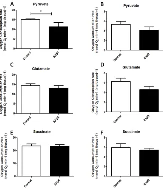
Figure 3.1-2 State 3 (A, C, E) and State 4 (B, D, F) respiration of fetal Guinea Pig soleus
muscle with either (A, B) pyruvate, (C, D) glutamate or (E, F) succinate as oxidative
substrate. ... 36
Figure 3.2-1 Birth weights of 150-day old animals used for respirometry and molecular
analyses. ... 38
Figure 3.2-2 Birth weight to birth length ratio of 150-day old guinea pigs ... 39
Figure 3.2-3 State 3 (A,C, E) and State 4 (B, D, E) respiration of permeabilized soleus from
150 day old Guinea Pig with (A, B) pyruvate, (C, D) glutamate and (D, E) succinate as
oxidative substrate. . ... 41
Figure 3.2-4 Relative protein expression of subunits of electron transport chain complex I
subunit NDUFB8 (A), complex II subunit SDH8(B), complex III subunit UQCRC2 (C),
complex IV subunit MTCO1 (D), complex V subunit ATP5A (E) in NBW or LBW 150 day
xii
Figure 3.2-6 Relative mRNA expression of myosin heavy chain genes (MHC) in NBW or
LBW 4 month old Guinea Pigs fed a Control or Western Diet (normalized to βactin). ... 45
Figure 3.3-1 Rate of oxygen consumption in C2C12 cells cultured under hypoxia using the
Oxygraph-2k. ... 47
Figure 3.3-2 Rates of oxygen consumption of cells cultured in 5% and 1% O2 after 24 hours
0.5mM fatty acid exposure (SFA palmitate C16:0, UFA palmitoleate C16:1 or BSA control)
at 20.9% O2. Rates were measured with 50µM (5% O2; A) and 10uM (1% O2; B) within the
Oxygraph chamber ... 48
Figure 3.3-3 Mitochondrial DNA unaltered after 2 and 5 days of differentiation or after
recovery in hypoxic cultured cells ... 49
1
Introduction
1.1
Developmental Origins of Health and Disease
The in utero environment in which a eutherian mammal fetus develops can determine
pre-dispositions to metabolic diseases later in life. The maternal uterine lining connects to
the developing fetus’ umbilical cord via the placenta. This organ regulates fetal gas
exchange, macronutrient supply and carbon dioxide elimination. Oxygen concentration
and macronutrient supply are two in utero environmental conditions that can affect
immediate fetal and later life post-partum metabolism. In fact, the association between a
poor in utero environment and later life development of metabolic diseases has recently
emerged as a global health concern for humans (Hales and Barker, 2001). The notion that
the in utero environment could play a role in adult-onset diseases was first entertained by
David Barker. His hypothesis (deemed the Barker hypothesis) stated that low oxygen and
nutrient transfer across the placenta can result in increased risk of metabolic syndrome, a
cluster of risk factors for metabolic diseases such as cardiovascular disease (CVD), type 2
diabetes and non-alcoholic fatty liver disease (NAFLD), in later life (Barker, 2004). This
hypothesis led to the investigation of in utero hypoxia and under nutrition as possible
contributors to such diseases (De Boo and Harding, 2006).
Many in-depth human population studies, covering a diversity of race and
socio-economic factors, now provide additional support for the advancement of research into
developmental origins of health and disease (DOHaD) (Villar and Belizán, 1982; Strauss,
2000; De Boo and Harding, 2006). A poor in utero environment, resulting from poor
maternal nutrition may lead to reduced oxygen and nutrient transfer across the placenta,
priming the offspring with a metabolism that is prepared for a similar, nutrient deprived
environment outside the womb (Glickman, Hanson and Beedle, 2007). Additionally,
(Valsamakis et al., 2006). Decreased expression of homeobox genes HLX1 and ESX1L
(Murthi et al., 2006; Murthi et al., 2006)involved in embryonic proliferation, migration
and invasion as well as increased placental a1-antichymotrypsin mRNA (a protease
inhibitor involved in inflammation) have been linked to IUGR (Chelbi et al., 2012).
Independent of these factors, however a nutrient and oxygen deficient in utero
environment is associated with a low birth weight, and is considered crucial to
adult-onset metabolic disorders (Villar and Belizán, 1982; Chatelain, 2000; Cox and Marton,
2009).
Negative early-life environmental experiences, such as in utero hypoxia, can cause
lasting epigenetic perturbations that may underly DOHaD observations(Hales and
Barker, 2001; De Boo and Harding, 2006; Valsamakis et al., 2006). These chemical
modifications of the DNA resulting in alterations in gene expression and regulation are
associated with an increased risk of later life metabolic disorders (Lal et al., 2003;
Gluckman et al., 2005). Such disorders may not emerge until early adulthood, or until a
second challenge, such as poor diet or even exercise, is imposed(Valsamakis et al., 2006;
Reyes et al., 2015), suggesting that an in utero perturbation may impact gene expression
both during development and after birth. It is not yet clear, however, whether in utero
reprogramming events are reversible, and specific genes affected in a variety of tissues
and animal models are topics of current research efforts (Zeng et al., 2013; Iglesias-Platas
et al., 2014; Venhoranta et al., 2014). Interventions such as N‐acetylcysteine a precursor
of the antioxidant glutathione, (Herrera et al., 2017) folic acid, (Ding and Cui, 2018) and
gene therapy (David, 2017) are currently being explored.
Although the mechanisms underlying the altered regulation of specific genes related to an
adverse in utero environment are still being identified, the general pattern of low birth
weight, prioritizing of blood flow to organs such as the brain, heart and adrenal glands,
and resulting changes in expression of specific genes, is termed the “thrifty phenotype”
(Neel, 1999; Hales and Barker, 2001). Thrifty phenotype offspring are not simply
brain to liver mass ratio and reduced muscle mass (Hales and Barker, 2001). During
human evolution, it is speculated that “thrifty genes” were selected for when food was
scarce, and may have primed offspring for a post-natal life where nutrient availability
was poor(Neel, 1999; Hales and Barker, 2001; Valsamakis et al., 2006). Today,
however, maternal and post-natal nutrient deficiency are rare in Western societies, and
offspring that display a thrifty phenotype, often result from a placental
insufficiency-induced oxygen deficient in utero environment (Godfrey and Robinson, 1994). The
thrifty phenotype is assumed to increase the probability of in utero survival, but
establishes a mismatch between the nutrient and oxygen ‘poor’ in utero environment, for
which the fetus was primed, and the nutrient and oxygen rich environment experienced
after birth. As a result, the offspring is born with a metabolic profile programmed to
survive in an energy- and oxygen-deficient (hypoxia) environment, but is exposed to an
environment with sufficient oxygen that is energy dense, especially when exposed to a
Western Diet (WD). In these instances human post-partum growth rates above average
(Boersma and Wit, 2013) are often observed (Bol et al., 2009; Coupé et al., 2009; Crume
et al., 2014). This rapid weight gain in infancy increases the risk of adult-onset obesity
(Adair et al., 2013). There is; however, a critical period of approximately 1000 days
where the thrifty phenotype appears to remain plastic (Victora et al., 2010) which
1.2
Intrauterine Growth Restriction
Placental nutrient and oxygen transfer, genetic growth potential and the maternal
environment all contribute to mammalian fetal development. Intrauterine growth
restriction (IUGR) results in a pregnancy where one or more of these factors are
negatively altered, creating a sub-optimal developmental environment. IUGR is a
condition in which a fetus’ growth is restricted and is associated with low birth weight
(LBW) and in some cases, evidence of asymmetrical growth e.g. high brain to liver
weight ratio (Mitchell, 2001; Bauer et al., 2003; Rosenberg, 2008). Fetuses which fall
below the 10th percentile for gestational age and sex are deemed small for gestational age
(SGA), an outcome related to IUGR. Of these births the majority of pathological cases–
i.e. those related to adult onset metabolic disorders - involve low abdominal
circumference but normal brain circumference (Peleg et al., 1998) . A human birth
weight threshold of 2,500g for IUGR, however, is a crude and somewhat arbitrary
criterion that does not account for fetuses outside these parameters who have failed to
reach their genetic growth potential and were perhaps exposed to a suboptimal in utero
environment (Gale et al., 2006; Mayer and Joseph, 2013; Melamed, 2014). Metabolic
remodeling as a result of a sub-optimal in utero environment, and associated with later
life metabolic disorders may therefore affect individuals across all birth weights (Kuzawa
and Adair, 2003; Gluckman et al., 2009) and there is emerging evidence of long-term
metabolic dysfunction independent of birth weight (Moore and Davies, 2005; Walsh and
McAuliffe, 2015). We must then consider additional parameters for classifying growth
restriction, such as the mentioned brain and abdominal circumferences, blood
inflammatory markers, differential microRNA expression, as well as ponderal index
(mass/height3), a measure of thinness, (Peleg, Kennedy and Hunter, 1998; Hales and Barker, 2001; Lausten-Thomsen et al., 2014; Thamotharan et al., 2017) in order to
include those who are a normal birth weight, but may have undergone a reprogramming
event due to poor in utero conditions. Low birth weight classifications alone fail to
include offspring of average birth weight who may have been larger under more
A well characterized classification of a sub-optimal in utero environment is placental
insufficiency (PI), which is an idiopathic condition characterized by a reduction in
maternal blood flow through the placenta. The resultant hypoxia and poor nutrient
exchange can lead to IUGR (Gagnon et al., 1996; Trudinger and Giles, 1996). PI occurs
when the placental fails to develop or implant properly, often resulting in a smaller
surface area for nutrient and oxygen exchange (Salafia, 1997) leading to a hypoxic,
nutrient-deprived in utero environment (Pardi et al., 2002). Experimentally PI can be
mimicked using partial ablation of the uterine artery in guinea pigs(Briscoe et al., 2004;
Turner and Trudinger, 2009). Larger animal models, such as sheep, which are more
easily probed with oxygen sensors, are excellent models for studying changes in oxygen
flow in IUGR (Gagnon et al., 1996; Morrison, 2008; Thompson et al., 2011). Other
animals, such as swine and rats have also been studied to reveal differences in gene
expression between control and IUGR offspring, which exhibited reduced insulin
sensitivity (Kind et al., 2003; Ying et al., 2019). Moreover, culturing muscle precursor
cells, such as C2C12 mouse myoblast cells, under hypoxia can mimic the effects of PI on
muscle metabolism(Regnault et al., 2010; Yu et al., 2011).
The pathological outcome associated with PI is IUGR, which can be categorized as either
symmetrical or asymmetrical. Symmetrical IUGR is typically a result of maternal or fetal
genetic abnormalities and results in fetuses which are proportionally small relative to the
average fetus. In asymmetrical IUGR certain organs (brain, heart, adrenal gland) are
disproportionately large (Peleg et al., 1998). Asymmetrical IUGR may result from a
number of causes including maternal hypertension, maternal under nutrition and, most
commonly, placental insufficiency (Pollack and Divon, 1992; Monk and Moore, 2004;
Valsamakis et al., 2006). As a result of placental insufficiency, fetal blood flow to the
heart, brain and adrenal glands increases, but blood flow to other organs including the
liver, kidneys and skeletal muscle (SM) decreases. This pattern results in a small
abdomen to head size, i.e. the asymmetrical phenotype. The reduced SM mass has also
been linked to altered insulin sensitivity in adulthood (Phillips et al., 1994; Desai et al.,
1.3
Hypoxia-induced alterations in skeletal muscle
metabolism
Independent of deficient nutrient transfer, chronic hypoxia in utero is associated with
IUGR (Kajimura et al., 2006; Rueda-Clausen et al., 2011); pregnancies at high altitudes
result in higher proportions of IUGR births in human populations that are not adapted to
such environments (Murphy et al., 1986; Cogswell and Yip, 1995; Semenza, 2000).
Further studies in chick embryos isolate hypoxia’s modulating role in IUGR, independent
of the placenta (Giussani et al., 2007; Mehta and Mehta, 2008; Tintu et al., 2009).
In prolonged hypoxia, hypoxia inducible factor 1-alpha (HIF1-α) accumulates, leading to
altered tissue metabolism. HIF1-α is a powerful transcriptional regulator of energy
metabolism (Lee et al., 2004; J. W. Kim et al., 2006; Denko, 2008) and is degraded under
normoxic conditions but allowed to dimerize with HIF1-β under cellular hypoxia (Wiener
et al., 1996; Ladoux and Frelin, 1997; Wenger et al., 1997). Accumulation of HIF1-α
attenuates glycolytic input to the tricarboxylic acid (TCA) cycle through increased
transcription of pyruvate dehydrogenase kinase 1 (PDK1), which inactivates pyruvate
dehydrogenase, halting the conversion of pyruvate to acetyl-CoA (Kim et al.,
2006).Overexpression of HIF1-α in otherwise normoxic pregnant mice led to IUGR and
reduced placental weights compared to controls (Tal et al., 2010), indicating hypoxia as a
contributor to IUGR, independent of reduced nutrient transfer. HIF1 is also involved in
up-regulating the transcription of glycolysis enzymes and glucose transporters (J. W. Kim
et al., 2006; Papandreou et al., 2006; Aragonés et al., 2008) facilitating anaerobic ATP
production. Further supporting the observed transcriptional effects of HIF1, functional
assays in human RCC4 cells show reduced oxygen consumption following normoxic
recovery from chronic hypoxia (Soares et al., 2005; Papandreou et al., 2006). Yet another
study has identified electron transport chain (ETC) complex I as a more specific target for
inhibition under hypoxic conditions; HIF-1α induced downregulation of NDUFA4L2, a
subunit of complex I, lowers oxygen consumption and suppresses complex I activity
1.3.1
Electron Transport Chain
A number of cellular processes, critical to energy metabolism, including angiogenesis
(the formation of new blood vessels) glucose metabolism, and oxidative phosphorylation
are regulated by hypoxia (Mu et al., 2001; Pugh and Ratcliffe, 2003; Magalhães et al.,
2005). As oxygen is central to oxidative phosphorylation (OXPHOS), the role of hypoxia
in IUGR has been chosen as the focus of this study. OXPHOS is an aerobic metabolic
pathway occurring in the inner mitochondrial membrane. In this pathway exergonic
reactions of the ETC, including the donating and accepting of electrons, are coupled with,
and help drive endergonic reactions such as ATP production. The ETC is comprised of
five main enzyme complexes and a variety of electron donors with oxygen as the terminal
electron acceptor. The energy released by the donation of two electrons is used to
transport two protons across the inner mitochondrial membrane, creating an
electrochemical gradient which can drive the phosphorylation of ADP, producing ATP.
The five principal enzyme complexes involved in electron transport are NADH
dehydrogenase (complex I), succinate dehydrogenase (complex II), cytochrome c
oxidoreductase (complex III), cytochrome c oxidase (complex IV), and ATP synthase
(complex V; Figure 1.3-1). Each enzyme complex is comprised of a great number of
proteins that are encoded by both nuclear and mitochondrial DNA.
Complex I oxidizes NADH to NAD+ while reducing coenzyme Q10, or ubiquinone, a coenzyme within the inner mitochondrial membrane. Complex II is the site of succinate
and FADH2 oxidation and is the secondary entry point of the electron transport chain.
While complex II oxidizes succinate to fumarate and reduces ubiquinone, unlike complex
I, the energy released by this reaction is insufficient to transfer of protons into the
Following entry into the ETC from either complex I or complex II, the electrons are
transferred to complex III by ubiquinone. From complex III, cytochrome C transports the
electrons to complex IV. When stimulated by sufficient concentrations of ADP and
inorganic phosphate (Pi), complex V uses the proton gradient created by the ETC to
generate ATP (Figure 1.3-1).
Under acute hypoxic conditions, glucose metabolism is altered through what is termed
the Pasteur Effect. With less oxygen available to act as an electron acceptor at ETC
complex IV, flux through the ETC, as well as ATP synthesis by complex V, slows. As a
result, fewer NADH molecules are oxidized by complex I. The accumulated NADH
would decrease glycolytic flux pyruvate (key TCA metabolite), but it can be oxidized by
lactate dehydrogenase, while converting pyruvate to lactate. As a result, in acute hypoxia,
glycolysis can continue at high rates without the accumulation of pyruvate, permitting
some anaerobic ATP production by way of substrate level phosphorylation. Figure 1.3-1 Overview of the Electron Transport Chain. Each of the embedded
complexes are depicted within the inner mitochondrial membrane (IMM), and the
dissociable carriers’ coenzyme Q and cytochrome C are represented as (Q) and (c)
respectively. Electron flow is indicated by solid arrows and proton pumping by dashed
1.3.2
Myogenesis and adverse
in utero
environments
As previously mentioned, many fetal organ systems are impacted in situations of PI
hypoxia. Skeletal muscle mass is greatly reduced in IUGR offspring (Padoan et al., 2004;
Larciprete et al., 2005), as a result of hypoxic induced cardiac redistribution, suggesting
that myogenesis likely occurs in a sub-optimal oxygen environment. Hypoxia is strongly
linked to the IUGR in utero environment and may contribute to the reduction in overall
terminal differentiation (Di Carlo et al., 2004; Yun et al., 2005) and decreased muscle
mass thereby linking hypoxia to the thinness component of the thrifty phenotype.
Skeletal muscle development during embryogenesis can be affected by the in utero
environment, and in adulthood. Successful myogenesis requires a precise coordination of
spatial and temporal events. Myogenesis involves the synchronization of a number of
embryonic and fetal progenitor cells specified by paired box transcription factors Pax3
and Pax7 respectively 63,64 which results in determination of myofibers. Following
activation by Pax3/Pax7, fetal and embryonic muscle growth is orchestrated by the
myogenic regulatory factors (MRF) myogenin, MyoD, Mrf4, and Myf5 which induce
expression of genes which encode contractile proteins unique to muscle cells (Figure
1.3).
Pax 3 and Pax 7 specify embryonic progenitor cells resulting in satellite cells. These are
later activated through the continued expression of Pax7 with some regulation by Myf5.
Satellite cell proliferation begins with the expression of MyoD in addition to Pax7 and
Myf5 regulation, resulting in myoblast formation. Early differentiation into myocytes is
marked by declined Pax7 and Myf5 expression with increased myogenin expression and
slowly increasing Mrf4 expression. Finally, the transition from myocytes to myofibers, or
late differentiation, is regulated by minimal MyoD expression, tapering myogenin
Downstream targets of myogenin and MyoD include the transcription factors known as
myocyte enhancer factors (MEFs): Mef2A, Mef2B and Mef2C. Together, MRFs and
MEFs activate the transcription of contractile and structural proteins found in myoblasts
(Blais et al., 2005). Under IUGR conditions expression of MEF2a decreases but MEF2d
expression increases, with no change in MEF2c (Raychaudhuri et al., 2008). Because
MEF2 isoform binding with MyoD relates to GLUT 4 transcription (Raychaudhuri et al.,
2008; Zheng, Rollet and Pan, 2012) in addition to fiber type determination (Potthoff et
al., 2007), IUGR changes in MEF2 expression potentially contribute to increased risk of
metabolic syndrome. Additionally, post-natal myogenesis is impaired in IUGR offspring
(Wirfalt et al., 2001; De Blasio et al., 2007) and lean mass and muscle strength remains Figure 1.3-2 Embryonic Myogenesis. Determination and differentiation of muscle cells
regulated by Pax3/Pax7and MRFs MyoD, Myogenin, Mrf4 and Myf5. Transcription
decreased compared to controls (Aihie Sayer et al., 2004; Inskip et al., 2007; Ylihärsilä et
al., 2007).
Moreover, there have been extensive reports studying in vitro differentiation in the C2C12
Mus musculus cell line under hypoxia (Yun, et al., 2005; Cicchillitti et al., 2012; Slot et
al., 2014). Although the cells are derived from adult satellite cells, in vitro differentiation
closely mimics the in utero fetal differentiation process (Messina et al., 2010). Important
markers of differentiation and cell signaling, necessary for the initiation of determination
and differentiation, are expressed similarly in utero and in this cell line (Burattini et al.,
2004) making this cell line a useful and tractable experimental model.
1.4
Skeletal Muscle Fiber Type Composition and
adverse
in utero
environments
Fiber type composition determines contractile characteristics, oxidative function and
metabolic capacity of skeletal muscle. There are two ways to classify fiber types: by their
myosin ATPase type (affecting twitch speed) and their principle mode of generating ATP
(oxidative, glycolytic or oxidative/glycolytic). Oxidative and glycolytic fibers express
different isoforms of myosin heavy chain (MHC) and preferentially metabolize different
substrates in order to produce ATP. Oxidative fibers have a higher concentration of
mitochondria and use predominantly oxidative phosphorylation to yield ATP, whereas
glycolytic fibers primarily use anaerobic glycolysis to yield ATP and have fewer
mitochondria. Muscle fibers can be alternatively categorized as fast-twitch (Type I) or
slow-twitch (Type II), referring to the speed of which myosin can hydrolyze ATP.
Slow-twitch fibers are primarily oxidative and in humans are referred to as type Ia fibers,
whereas Type II fibers can be either oxidative-glycolytic (Type IIa) or glycolytic (Type
1.4.1
Skeletal muscle fiber composition
Slow and fast muscle fibers originate during fetal development as populations of primary
(type I MHC) and secondary (type IIa, IIb and IIx MHC) myotubes respectively. Fiber
type fate is considered plastic; fiber type switching can occur with exercise or diet
(Simoneau et al., 1985; Schrauwen et al., 2001; Handschin et al., 2007). In disease states,
for example individuals with type 2 diabetes and insulin resistance, there is a notable shift
towards more glycolytic fibers (Tanner et al., 2002; Ten Broek, Grefte and Von den Hoff,
2010; Tello et al., 2011), which is associated with reduced muscle oxygen consumption
(He et al., 2001; Oberbach et al., 2006). The mammalian target of rapamycin,
peroxisome proliferator-activated receptor (PPAR)s and PPAR-γ coactivator-1α
(1α) together regulate fiber type composition and, by extension oxidative capacity.
PGC-1α increases insulin sensitivity by maintaining a larger number of functional
mitochondria by inducing fiber type switching towards type I and IIa expression.
The importance of PPARs in normal fetal development is supported by the fact that
PPARγ knock-out mice are growth-restricted at birth (Gokina et al., 2013). Furthermore,
the targeted expression of an activated form of PPARβ/δ in the skeletal muscles makes
mice resistant to obesity by increasing the numbers of oxidative (Type I and IIa) muscle
fibers (Slot et al., 2014), while the selective ablation of PPARβ/δ induces obesity by
reducing the oxidative capacity of the muscles (Schuler et al., 2006). PPARβ/δ also
directly and indirectly regulates genes involved in fatty acid transport and oxidation
through binding to specific peroxisome proliferator response elements (PPREs) on these
genes (Narravula and Colgan, 2001). Examples of these genes include Cluster of
Differentiation 36 or Fatty acid translocase which is involved in fatty acid uptake
(Schuler et al., 2006; Jans et al., 2012), carnitine palmitoyl transferase 1, involved in the
mitochondrial transfer of long-chain fatty acids, and the uncoupling protein (UCP-1, -2
and -3) family (St-Pierre et al., 2003; Cantó et al., 2012). Altered expression of these
genes directly correlates with insulin resistance, altered fatty acid oxidation,
intramyocellular lipid accumulation, and mitochondrial damage(St-Pierre et al., 2003;
Studies of pig and sheep, which have similar muscle differentiation to humans, show that
differentiation occurs near mid-gestation and fiber type ratios are set before birth. As
fiber type determines oxidative capacity, the ratio of oxidative to glycolytic fibers is an
important factor in predisposition to metabolic syndrome. In human studies, reduced
PGC-1α mRNA in adult skeletal muscle has been linked to the development of insulin
resistance and Type II diabetes, in conjunction with inactivity and nutrient excess
(Handschin et al., 2007; Holmstrom et al., 2012; Kristensen et al., 2014). Further, in
IUGR rats, fetal PGC-1α promoter activation and expression is impaired, through
epigenetic modifications(Zeng et al., 2013). These changes are related to the
development of insulin resistance later in postnatal life and intriguingly also in later
generations, supportive of the concept of in utero inter-generational programmed PPAR
modifications(Martins et al., 2012; Shen et al., 2018). These data suggest that adverse
PGC-1α/PPAR control promotes altered muscle fiber determination and oxidative
1.5
The Western Diet
1.5.1
Western Diet origin and composition and the development of
metabolic disease
Metabolic disorders, many of which exhibit reduced skeletal muscle oxygen consumption
and altered fiber type composition, are associated with long term consumption of the
high-fat, high-sugar Western Diet (WD) in adults (Wirfalt et al., 2001). Though diet has
long been the focus of research in metabolic disorders, emerging data suggest the in utero
environment plays a role as well (Valsamakis et al., 2006; Nevin et al., 2018). On an
evolutionary time-scale, agriculture and industrialization are recent developments, which
have caused extreme changes in the human diet over a short time span. Modern
technology and urbanization have drastically altered not only nutrient composition but
also food availability, changing quality and quantity of the human diet. Pre-agricultural
human populations, or hunter-gatherers, did not have a consistent diet but rather one
which varied by climate, location and ecological surroundings (Cordain et al., 2000a).
While their diets varied by location and food availability, many similarities among the
diets of different pre-agricultural populations existed, which provide insight regarding the
Western Diet, as it relates to development of chronic, metabolic disorders.
The World Health Organization recommends humans intake 10% of our daily energy
from sugar (The World Health Organization, 2003); however in 2004 Canadians
consumed, on average, 21% of calories from sugar (Health Canada, 2005). Although it is
difficult to determine sugar consumption of early humans, it is estimated to have
accounted for a much smaller percentage of total energy (Cordain et al., 2000b;
Brand-Miller et al., 2002). Furthermore, there has been a marked shift in fatty acid consumption
between pre- and post-industrial humans (Cordain et al., 2000b). Additional changes in
fatty acid composition between pre- and post-agricultural populations stem from meat
sources. Wild mammals exhibit seasonal changes in fat storage, where excess energy is
(SFAs) (Mercer, 1998). Fatty acids found in muscle and other tissues however are mainly
polyunsaturated fatty acids (PUFAs) and monounsaturated fatty acids (MUFAs) (Rule et
al., 2002) thus hunters eating skeletal muscle as meat would have had a diet rich in
PUFAs and MUFAs for the majority of the year (Cordain and Watkins, 2002). In
contrast, post-agricultural populations consume higher SFA content from meat for two
main reasons: animal husbandry and technological advancements in corn harvesting.
First, animal husbandry has allowed for slaughter at peak fatty acid storage year round
compared to the seasonal peak fatty acid found in wild mammals (Whitaker, 1975).
Second, animals are now kept in feed lots and fed grain (mostly corn) and exhibit
“marbled meat”, i.e. intramuscular adipose deposits resulting from excess triacylglycerols
accumulation (Whitaker, 1975). The triacylglycerol in these deposits is comprised largely
of SFA (Cordain et al., 2005). Wild meat does not exhibit this “marbling” (Cordain and
Watkins, 2002).
On an evolutionary time-scale, 5,000 to 10,000 years is a relatively short period in which
to undergo such drastic environmental changes. Environmental pressures can either
positively or negatively select for genetic traits, and a changing environment may be
discordant with widely favorable genetic traits from a previous environmental pressure
(Gould, 2002; Caldararo, 2003). The current human genome has been primed for the diet
of our ancestors and is discordant to the present day “Western Diet” (Eaton et al., 1988;
Caldararo, 2003). Increased sugar consumption and a shift in the fatty acid composition
from mainly PUFAs and MUFAs to SFAs are staples of the Western Diet (Cordain et al.,
2005).
An emerging body of evidence suggests that high fat, high sugar diets are closely linked
to metabolic disorders (Hu et al., 2001; Cordain et al., 2005; Kuo et al., 2008; Lottenberg
et al., 2012). High sugar diets increase blood glucose levels, leading to increased insulin
secretion (Ludwig, 2002). Over time cells become insulin resistant, even compared to
isocaloric diets with low sugar (Miller, 1994). Promotion of insulin resistance by a high
sugar diet has adverse metabolic effects and is an early indicator for type II diabetes and
cardiovascular disease (Ludwig, 2002; Cordain et al., 2003). Additionally, following a 16
synthase activity (a marker of functional mitochondrial abundance) and decreased
oxidative fiber expression in conjunction with insulin resistance (Bonnard et al., 2008),
indicating an association between mitochondrial dysfunction, diet and development of
metabolic disorders.
1.5.2
Mechanisms of sugar and saturated fats’ negative impacts
and increased risk of metabolic disease
Metabolic dysfunction with respect to FAs occurs due to insulin resistance, increased
plasma cholesterol and adiposity (Hokanson and Austin, 1996; Turco et al., 2014).
Although the total amount of FAs consumed can contribute to metabolic perturbations,
recent studies show that the FA quality (PUFA, MUFA or SFA) can also influence
disease development (Hokanson and Austin, 1996; Turco et al., 2014) likely because of
their different biochemical properties.
Saturated fatty acids are classified as such because each carbon of the acyl chain is
saturated with hydrogen molecules, and there are no double bonded carbons. Mono and
polyunsaturated fatty acids are those with one or multiple double bonds, respectively.
These double bonds create bends in the hydrocarbon chain and therefore unsaturated fats
have less dense overall structures. Dietary intake of SFAs, particularly palmitate, the
most abundant dietary SFA, increases total plasma cholesterol and decreases low density
lipoprotein (LDL) receptor expression, thereby indirectly increasing plasma LDL levels
(Chappell et al., 1993; Rioux and Legrand, 2007). Plasma cholesterol, specifically in the
form of LDL or VLDL are important risk factors in metabolic syndrome, CVD and type
II diabetes (Hokanson and Austin, 1996). Intracerebroventricular injections of palmitate
simulate insulin resistance in the brain (Posey et al., 2009) and decrease PGC-1α
expression in skeletal muscle (Coll et al., 2008), providing further support for palmitate’s
A recent review noted that adolescents in half of the 30 studied countries are ingesting
more SFAs than recommended by World Health Organization, and failing to reach the
recommended intake of PUFAs (Harika et al., 2011). PUFAs are important in membrane
fluidity but can influence lipogenesis by modulation of PPARα, similar to MUFAs
(Lichtenstein and Ausman, 1994; Hannah, Ou and Luong, 2001; Assy and Nassar, 2009;
X. Yang et al., 2011). Studies demonstrate that SFA induced insulin resistance is
associated with impaired glucose transport or phosphorylation(Holland et al., 2011;
Martins et al., 2012). Skeletal muscle specific studies show reduced IRS (insulin receptor
substrates)-1 tyrosine phosphorylation (Yu et al., 2002) following SFA infusion.
Tyrosine phosphorylation of IRS-1 leads to a phosphorylation cascade which results in
increased glucose uptake. Conversely, dietary intake of MUFAs ameliorate insulin
resistance in skeletal muscle (Yang et al., 2011).
1.6
Thesis Rationale and Objectives
1.6.1
Rationale
Metabolic disorders are linked to poor in utero environments and can develop
independent of post-natal diets (Barker, 2004; de Boo and Harding, 2006). A poor in
utero environment, characterized by decreased oxygen and nutrient transfer across the
placenta, is associated with LBW and IUGR outcomes (Byberg et al., 2000; Pardi et al.,
2002; Burke et al., 2006). These outcomes are observable phenotypes linked to an
underlying physiological perturbation: a reprograming event (Neel, 1999; Barker, 2004).
Fetal reprograming occurs in low oxygen and low nutrient environments, and consists
primarily of a redirection of blood flow, sparing some organs (brain, heart and
adrenals)(de Boo and Harding, 2006). Meanwhile, other organs and tissues such as the
liver, kidneys and skeletal muscle experience a reduction in blood flow (Peleg, Kennedy
expression leading to the observed predisposition of IUGR offspring to metabolic
disorders (Barker, 2004; de Boo and Harding, 2006).
In addition to their associations with a poor in utero environment, increased incidence of
metabolic disorders such as insulin resistance and type II diabetes have been linked to the
increased prevalence of a high-fat, high-sugar WD (Cordain et al., 2005). Specifically,
SFAs, and processed sugars, present in higher proportion in the WD compared to other
diets (Cordain et al., 2000a), have been identified as key components in the development
of metabolic disorders (Holland et al., 2011).
Underlying in utero reprogrammed changes in IUGR mitochondrial function and how
these changes may interact with an adverse postnatal environment, such as a WD are
currently ill-defined. Skeletal muscle biopsied from insulin resistant individuals have
reduced rates of oxygen consumption (Gosker et al., 2002; Mogensen et al., 2007).
Oxygen consumption may be altered due to the changes in fiber type composition
because of differences in preferred metabolic pathways of the different fiber types.
Oxidative fibers (type Ia, IIa and IIx) have a high density of mitochondria and capillaries
and therefore respire at higher rates than their glycolytic (type IIb) counterparts (Lillioja
et al., 1987; Schiaffino and Reggiani, 2011). IUGR offspring show impaired
mitochondrial function manifesting as reduced oxygen consumption in skeletal muscle
attributed to reduced electron transport chain complex I activity, therefore skeletal muscle
oxygen consumption is proposed as an early predictor for such metabolic diseases.
I therefore postulate that in utero hypoxia reprograming reduces oxygen consumption,
and that a WD, or palmitate alone, will aggravate mitochondrial dysfunction whereas
palmitoleate (a MUFA) will mitigate hypoxia-induced reductions in oxygen
1.6.2
Objectives and Hypotheses
My first objective was to determine the rate of oxygen consumption in LBW fetal guinea
pig soleus muscle compared to normal birth weight (NBW) animals, ex vivo. I
hypothesized that hypoxia (through placental insufficiency) will alter muscle oxygen
consumption during development. I predicted that LBW animals would have reduced
soleus oxygen consumption.
My second objective was to determine the effects of growth restriction on oxygen
consumption rate and skeletal muscle fiber type composition in young adult LBW guinea
pigs independently and in conjunction with a post-natal WD. I hypothesized that WD
interacts with IUGR metabolic reprogramming and predicted that LBW/control diet-fed
animals and NBW/western diet fed animals will exhibit increased glycolytic fiber ratios
and reduced oxygen consumption while LBW/western diet-fed animals will have even
further increases in glycolytic fiber ratios compared to NBW/control-diet fed animals as
well as either treatment independently. I tested these two hypotheses using a
whole-animal model of IUGR leading to a LBW outcome.
My final objective was to isolate hypoxia-specific effects of IUGR on skeletal muscle
differentiation, metabolism and mitochondrial function and evaluate the effects of
saturated fatty acids and monounsaturated fatty acids on oxygen consumption in a
cell-based model system. It would be difficult to achieve this objective in a whole-animal
model, so I used a skeletal muscle cell culture model where I could control oxygen as
well as nutrient quantity and quality. It was postulated that hypoxia and fatty acid type
alter oxidative metabolism through changes in fiber type. Specifically, I predicted that
hypoxic cultured cells would consume less oxygen than normoxic cultured cells and that
SFAs will exacerbate this while MUFAs would have a mitigating effect through changes
2
Materials and Methods
2.1.1
Surgical Induction of Growth Restriction
All animal procedures were approved by the University of Western Ontario Animal Use
Subcommittee in accordance with the Canadian Council on Animal Care guidelines.
Pregnant Dunkin-Hartley guinea pigs (Charles River Laboratories, Wilmington, MA,
USA) were housed with a 12/12 hour light/dark cycle and allowed access to standard
guinea pig chow (LabDiet diet 5025: 27% protein, 13% fat, 60% carbohydrates) ad
libitium. Fetal samples were obtained from sows that underwent uterine artery ablations
(UAA) performed mid-term (approximately day 35 of a 68-day gestation) to induce
IUGR by a veterinary technician. Following anesthesia (4-5% Isoflurane with 2L/min O2
for induction, followed by 2.5-3% Isoflurane with 1L/min O2 for maintenance), a
subcutaneous injection of Robinul (Glycopyrrolate, 0.01mg/kg, Sandoz Can Inc.,
Montreal QC) was administered. To expose the bicornate uterus, a midline incision was
made below the umbilicus and subsequently the UAA was performed by cauterization of
50% of uterine artery branches using an Aaron 2250 electrosurgical generator (Bovie
Medical, Clearwater, FL)(Turner and Trudinger, 2009)
Guinea pig studies were divided into two experiments, one examined the direct impact of
the in utero insult and a second focused upon the long-term effects of an adverse in utero
environment combined with a second insult of a poor Western diet during a post-natal
growth phase. In the first study, fetal soleus muscle was collected near term
(approximately gestational day 65 of a 68-day term). These muscles were used for
respirometry experiments and came from fetuses that were categorized as either control
(fetuses weighing more than 90 grams) or IUGR (fetuses weighing less than 85 grams;
Figure 2.1-1).
In the second study both UAA and sham-operated controls were carried to term and
below). Animals whose birth weight fell below the 25th percentile of all pups born were
categorized as LBW and those whose birth weight fell between the 25th and the 75th
percentile were categorized as normal birth weight (Thompson et al., 2011). In
accordance with previous data (Kind et al., 2003; Briscoe et al., 2004) and meeting these
criteria, LBW animals were below 85g at birth and normal birth weight animals were
above 90g at birth. Four months after birth soleus muscle was collected from these
animals for respirometry and molecular analysis (further described in section 2.4)
2.1.2
Post-Natal Life and Feeding
Guinea pigs that carried to term were weaned at 15 days of age and housed in individual
cages maintained at 20°C ± 2°C, 30-40% relative humidity with a 12-hour light/dark
cycle. At weaning, offspring were randomly assigned to either a control diet (CD, 3.4
kcal/g; product number TD: 110240, Harlan Laboratories, Madison WI) or a Western diet
(WD, 4.2 kcal/g; product number TD: 110239, Harlan Laboratories, Madison WI; Figure
2.1-1). The control diet was comprised of a high complex carbohydrate content, but low
fructose and sucrose with the majority of fats being PUFA, and very low SFA content. In
contrast, the WD has less overall carbohydrate composition, but higher fructose and
sucrose content, and a larger percentage of SFA (Table 2.2-1). Once weaned, pups were
then classified into four experimental groups: normal birth weight on a control diet
(NBW/CD), low birth weight on a control diet (LBW/CD), normal birth weight on a
Western diet (NBW/WD), and low birth weight on a Western diet (LBW/WD). Animals
were maintained on these diets and sacrificed at ~150 days, corresponding to young
Figure 2.1-1 Overview of Guinea Pig animal model for uterine artery ablation and
post-natal diet. Uterine artery ablations (UAA) or sham control surgeries were
performed on pregnant sows. Pups born from UAA sows weighing less than 85g were
considered LBW, weaned and then given either a control or Western diet. Pups born from
sham surgeries weighing more than 90g were considered NBW, weaned and given either
a control or Western diet. Animals were sacrificed and soleus muscle was collected at
Table 1.6-1 Guinea Pig Diet Comparison (percent calories)
Nutritional information Control Diet Western Diet
Calories 3.4 Kcal/g 4.2 Kcal/g
Protein 22% 21%
Carbohydrates 60% 33%
Sucrose 11% 22%
Fructose - 7.6%
Fat 18% 46%
Saturated fatty acids 2.7% 32.2%
Monounsaturated fatty acids 4.32% 11.96%
Polyunsaturated fatty acids 10.98% 1.84%
2.1.3
Tissue Collection
In the first study, which compared control and IUGR fetuses, pregnant sows near term
(gestational day 65) were sacrificed by CO2 asphyxiation following an overnight fast
(Greulich et al., 2011) and fetuses were removed. In the second study of post-natal diet
effects on LBW and NBW offspring, animals born to both UAA and sham operated sows
were sacrificed at postnatal day 150, corresponding to young adulthood (Briscoe et al.,
2004), by CO2 asphyxiation following an overnight fast (Greulich et al., 2011). From
every animal (fetus and offspring) one soleus muscle was removed and used immediately
for respirometry (described in section 2.2, below). and the contralateral muscle, flash
frozen in liquid nitrogen for CS activity. In addition, for the 150-day old NBW and LBW
animals only, soleus muscle was also collected and frozen for fiber typing mRNA
2.2
Soleus Respirometry
The soleus muscle dissected from control and IUGR fetuses, as well as 150 day old NBW
and LBW animals was stored on ice in BIOPS buffer (50mM K+-MES, 20mM taurine, 0.5mM dithiothreitol, 6.56mM MgCl2, 5.77mM ATP, 15mM phosphocreatine, 20mM
imidazole pH 7.1 adjusted with 5N KOH at 0°C, containing a Ca-EGTA buffer (2.77mM
CaK2EGTA + 7.2mM K2EGTA; 0.1μM free calcium) until permeabilized. The soleus
muscle tissue was kept ice-cold and teased apart using sharp forceps to isolate muscle
fiber bundles 3-4mm long and 1mm in diameter, exposing a fairly large surface area. The
fibers were incubated in BIOPS supplemented with saponin (50μg/ml) for 30 minutes
(optimal for skeletal muscle; Kuznetsov et al., 2008), permeabilizing the cells by binding
to cholesterol in the sarcolemma, thereby allowing access of oxidative substrates and
ADP to the mitochondria without further disrupting the tissue. In contrast to sarcolemma,
mitochondrial membranes contain little cholesterol, so are relatively unaffected by
saponin incubation. Following permeabilization, tissue was washed 3 times in MiR05
buffer (110mM sucrose, 60mM K-lactobionate, 0.5mM EGTA, 3mM MgCl2, 20mM
taurine, 10mM KH2PO4, 20mM HEPES adjusted to pH 7.1 with KOH at 37°C; and
1g/litre BSA fatty acid free) for 2 minutes.
Rates of oxygen consumption, with oxidative substrates and ADP (state 3) and following
ATP synthesis (state 4), were determined using the Oxygraph-2k (Oroboros), with
Clark-type O2 electrodes in airtight 2mL chambers at 37°C. Glutamate (10mM final
concentration) and pyruvate (5mM) were injected into the chamber along with malate
(2mM) as substrates for respiration through ETC complex I. In separate incubations
succinate (10mM) was solely used as an ETC complex II substrate, with rotenone
(0.5μM) added to inhibit ETC complex I arising from endogenous substrate metabolism.
Once respiration rate with only substrates stabilized, ADP (2mM) was added, stimulating
state 3 (maximal physiological) respiration. After recording stable state 3 rates
oligomycin (5mM) was injected into the Oxygraph chamber to inhibit complex V
2.3
Citrate Synthase Activity
Soleus muscle from control and IUGR fetuses, as well as 150 day old NBW and LBW
animals was dissected and frozen in liquid N2 and tissue stored at -80°C. Samples were
then homogenized in radioimmunoprecipitation assay buffer (RIPA; 50mM Tris-HCl,
NP-40 1%, Na-deoxycholate 0.25%, 1mM EDTA, 150mM NaCl, 50mM NAF, 1mM
NaV, 25mM β-glycerophosphate, pH 7.4) with protease and phosphatase inhibitors. One
aliquot was used for western blots (see section 2.4) and another frozen at -80°C until it
was used for citrate synthase (CS) activity. Thawed homogenate was sonicated then
centrifuged at 2000g for 5 min at 4°C.
The CS reaction was followed in 96-well plates in triplicate by measuring absorbance at
412nm using a spectrophotometer. Wells were loaded with 220μl reaction buffer
(consisting of 25mM HEPES, 0.1% Triton X-100 and 2mM EDTA) 30μl of 0.15mM
5,5’-dithio-bis(2-nitrobenzoic acid) (DTNB), 30μl of 0.15mM acetyl CoA and 3μl of
homogenate (Eigentler, Draxl and Wiethüchter, 2012). The reaction was started by
adding 30μl of 0.33mM oxaloacetate. Blank rates were also recorded in a well which
contained homogenate but to which buffer was added instead of oxaloacetate.
Absorbance was recorded for 5 minutes at 37°C and maximal activity was calculated
from the slope of absorbance vs. time using a minimum of 7 consecutive absorbance
readings during the 5 minutes (readings taken every 1 second), after subtracting rates
from blank wells. Data were expressed as enzymatic activity per mg soleus muscle
2.4
Molecular Analysis
Frozen soleus muscle from 150-day old animals NBW and LBW animals only were used
for the following mRNA and immunoblotting procedures.
2.4.1
Rt-qPCR for Myosin Heavy Chain isoform expression
Guinea pig soleus was powdered using a mortar and pestle in liquid N2, homogenized
using a disposable homogenization pestle and stored in Trizol reagent (Invitrogen,
Burlington ON) at -80°C until use. Total RNA was extracted using the Trizol procedure;
samples were thawed and incubated at room temperature for 5 minutes, 200µL of
chloroform was added to the solution and shaken vigorously by hand for 15 seconds.
Samples were left at room temperature for 2 minutes and then centrifuged for 15 minutes
at 12,000g at 4°C separating the aqueous phase (containing RNA), an interphase and a
phenol-chloroform phase. The aqueous layer was transferred to a new RNAase-free tube
and 500µL of isopropanol was added to the solution which was incubated at room
temperature for 10 minutes. Following incubation, samples were centrifuged for 10
minutes at 12,000g at 4°C, precipitating RNA into a pellet. Samples were then washed
twice with 100µl of 75% ethanol, centrifuging at 7,500g for 5 minutes after each wash.
Samples were stored at -80°C until use.
The yield of isolated RNA was determined using a NanoDrop 2000 spectrophotometer
(Thermo-Scientific) and quality was measured using the A260/A280 ratio (≥1.8). Isolated
RNA (4μg) was incubated with deoxyribonuclease (Invitrogen) to remove any
contaminating DNA and a recombinant ribonuclease inhibitor (Invitrogen) at 37°C for 30
minutes. Samples were then incubated at 37°C for two hours with murine leukemia virus
reverse transcriptase (Invitrogen) to produce cDNA with the use of random primers.
(Invitrogen) and the BioRad CFX384 Real-Time PCR Detection System at denature
temperature of 95°C, annealing temperature of 59.5°C and elongation temperature of
72°C for 40 cycles. A total volume of 8μl in each well contained 3μl of cDNA, 0.08μl
primer mix, 4µl SYBR Green and the remaining volume was filled with autoclaved
water. Primer sets directed against guinea pig specific myosin heavy chain genes were
generated using the NCBI Primer-BLAST tool based on predicted Cavia porcellus
Table 2.4-1 Guinea Pig Myosin Heavy Chain primer pairs.
Primers NCBI accession # Strand Sequence (5’ 3’)
Myh7 (type
1A) XM_003474485.2
Forward
Reverse
CAGGAGTGCAGGAATGCTGA
GTTATGCAGGTGCCAGGGAT
Myh4 (type
2B) XM_005002092.1
Forward
Reverse
GTACACCTAACCTGCAGCCA
GTTGTCGTTCCTCACGGTCT
Myh2 (type
2A) XM_005002092.1
Forward
Reverse
CCTTTGAAGAGCGACACTGG
TCAGCATGAACTGGTAGGCG
Myh1 (type
2x) XM_003466219.1
Forward
Reverse
TCATCCCTACAGGCAAGGTC
AGAGGCCCGAGTAGGTGTAG
2.4.2
Immunoblotting of Electron Transport Chain Complex
Subunits
Protein concentrations of homogenized soleus (described in section 2.3) were determined
using BCA protein concentration kits (Pierce Biotechnology, Rockford IL). Samples
were then prepared using NuPAGE LDS sample buffer (Invitrogen) and NuPAGE
Sample Reducing Agent (Invitrogen). Approximately 15μl of soleus extracts (20µg of
protein) were separated on NuPAGE 4-12% Bis-Tris gels and subsequently washed in
TBST (137mM NaCl, 2.7mM KCl, 19mM Tris base, 0.1% Tween 20) 3 times for 10
minutes. Membranes were then incubated in TBST supplemented with the MitoProfile
Total OXPHOS Rodent Antibody Cocktail (Complex I subunit NADH dehydrogenase
[ubiquinone] 1 beta subcomplex 8, NDUFB8; Complex II Iron-sulfur protein subunit;
Complex III Core protein 2; Complex IV subunit I; Complex V alpha subunit) at a
1:1000 dilution (ab110413, host: mouse; Mitosciences, Eugene, OR). Membranes were
then washed three times with TBST for 15 minutes and then incubated for one hour in
TBST supplemented with a donkey anti-mouse horseradish peroxidase secondary
channel protein 1, VDAC1), membranes were prepared in a similar manner as described
above with primary antibody at a 1:5000 dilution (ab14734, host: mouse; Invitrogen).
Immunoreactive bands were detected with enhanced chemiluminescence and imaged
with a VersaDoc Imaging System (Bio-Rad, Mississauga, ON). Densitometry was
performed with ImageLab Software (Bio-Rad).
2.5
Hypoxic C
2C
12Cells and Fatty Acid
Supplementation
C2C12 mouse myoblast cells were cultured to test the third hypothesis; that hypoxia and
fatty acid type alter oxidative metabolism. Cells were cultured at 37°C in Dulbecco’s Modified Eagle’s Medium (DMEM) supplemented with 10% fetal bovine serum and 1%
penicillin/streptomycin until cells reached 70-80% confluence (~ 48 hours) at 20.9% O2
(5% CO2 with N2 balance). Once cells reached confluence, growth media was replaced
with differentiation media (DMEM supplemented with 5% horse serum and 1%
penicillin/streptomycin) inducing myoblast differentiation. The cells were then allowed to
differentiate at 37°C at either 1% (to simulate in utero hypoxia) or 5% O2 (to simulate
typical in utero conditions) for five days of differentiation. Following differentiation, a
subset of cells was incubated at 20.9% O2 for 48 hours (recovery; Figure 2.2-1). During
recovery, a further subset of cells was exposed to 0.5mM palmitate (saturated fatty acid;
16:0), 0.5mM palmitoleate (mono-unsaturated fatty acid; 16:1) 10% fatty acid free BSA
2.5.1
Time points for collection
Untreated whole cells were collected prior to differentiation (day 0; cells cultured in
growth media at 20.9% O2), after 5 days of differentiation (day 5; cells cultured in
differentiation media at either 5% O2 or 1% O2) and following 48 hours of recovery (day
7; cells differentiated at either 5% O2 or 1% O2 for 5 days, then transferred to 20.9% O2
for 2 days). Cells treated with fatty acids or BSA were collected at days 0, 5 and
following 24hrs of fatty acid exposure (day 6).
Figure 2.5-1 Diagram of cell culture experiments. C2C12 mouse myoblasts were
cultured at 20.9% oxygen until they reached ~ 80% confluence. Cells were then
differentiated under either 5% O2 (simulating normal in utero oxygen levels) or 1% O2
(simulating oxygen under IUGR conditions) for 5 days. Following differentiation, cells