1071-412X/96/$04.0010
Copyrightq1996, American Society for Microbiology
Phenotype of Lymphocytes Associated with the Inflammatory
Reaction to Silicone Gel Breast Implants
WILLIAM E. KATZIN,
1,2* LU-JEAN FENG,
2,3MARY ABBUHL,
1AND
MARY ANN KLEIN
1Department of Pathology
1and Division of Plastic Surgery,
3Mt. Sinai Medical Center, and Case Western Reserve
University School of Medicine,
2Cleveland, Ohio
Received 18 July 1995/Returned for modification 13 September 1995/Accepted 27 November 1995
The tissue response to silicone gel breast implants typically includes an inflammatory infiltrate that consists
of macrophages, foreign body-type giant cells, and a variable number of lymphocytes and plasma cells. The
phenotype of the lymphocytic component was investigated with three-color flow cytometry. Lymphocytes were
obtained by collecting fluid from the space between the implant and the fibrous capsule or by washing cells
from the fibrous capsule at the time of implant removal with total capsulectomy. Eighty-nine percent of the
implant-associated lymphocytes were T cells. Twenty-five percent of the CD3
1T cells coexpressed HLA-DR
compared with only 7.9% of matched peripheral blood lymphocytes. Sixty-eight percent of the
implant-associated T cells coexpressed CD4 and CD29, while only 3% of the T cells coexpressed CD4 and CD45RO. The
expression of HLA-DR and the predominance of CD29
1CD4
1T cells indicate that there is immune activation
with the potential for stimulating antigen-specific antibody production. The role of silicone gel breast implants
in immune activation and its clinical significance require further investigation.
Silicone gel-containing breast implants, introduced over 30
years ago (7), have been widely used for augmentation and
reconstructive mammoplasty. Recently there has been
increas-ing concern regardincreas-ing the safety of breast implants. Most
no-tably, a variety of rheumatic diseases, including chronic
ar-thropathy (2, 9, 39, 46), scleroderma or systemic sclerosis (6,
20, 21, 33, 36, 37, 40, 42), rheumatoid arthritis (9, 20, 40),
systemic lupus erythematosus (36, 40), and Sjogren’s syndrome
(20, 40) have been reported in patients with silicone gel breast
implants. However, the relationship between implants and
these connective tissue diseases is still unclear (41). Three
separate epidemiologic studies (11, 34, 47) have not found an
increased incidence of specific rheumatic diseases in large
pop-ulations of women with silicone gel implants. The possible
relationship between silicone breast implants and systemic
dis-ease has been recently reviewed by Sanchez-Guerrero et al.
(35).
The tissue response to silicone gel-containing breast
im-plants includes formation of a fibrous capsule. On the basis of
studies with an animal model, it is likely that the capsule forms
in a relatively short time—probably within 2 months (27). The
histologic features of the fibrous capsule in women with
im-plants have been described previously (3, 12, 14, 15, 30, 32, 38).
In addition to a band of dense fibrous tissue, the capsule
includes a variable number of inflammatory cells.
Macro-phages with abundant vacuolated cytoplasm are a relatively
constant feature. There are often cyst-like spaces that contain
refractile, nonbirefringent clear material that almost certainly
represents some form of silicone (1, 14, 32, 38). The
inflam-matory infiltrate also includes a variable number of
lympho-cytes and plasma cells (12, 16, 38).
Aside from the morphologic features of the fibrous capsule
that surrounds silicone gel breast implants, very little is known
about the nature of the inflammatory response. In an attempt
to provide more specific information in this regard, we have
defined the phenotype of the lymphocytic component of the
capsular infiltrate by three-color flow cytometry.
MATERIALS AND METHODS
During the study period (1 May 1992 to 1 April 1993), a total of 209 patients underwent implant removal. The vast majority of these patients presented with local breast pain and systemic symptoms that they suspected were associated with their implants. Surgery in all cases was performed by one of the authors (L.-J.F.), who submitted all of the samples. All of the patients were examined by a rheumatologist, internist, or neurologist prior to implant removal. Eight of the 209 patients had documented autoimmune diseases which had developed after implantation. Three had rheumatoid arthritis, two had multiple sclerosis, one had systemic lupus erythematosus, one had systemic lupus erythematosus and polymyositis, and one had scleroderma. Only one of the 209 patients had evi-dence of frank infection.
In preliminary studies, we found that a sufficient number of lymphocytes for flow cytometric analysis could be obtained only from patients with either poly-urethane-coated silicone gel breast implants or textured-surface silicone gel breast implants. In those patients, exudative fluid was present in the space between the implant and the surrounding fibrous capsule. Little or no exudative fluid or very few mechanically dislodged cells could be obtained from patients with smooth-shell silicone gel implants. Implant-associated lymphocytes were obtained at the time of surgical implant removal in all cases in which exudative fluid was available. In some cases, fluid was collected from the space between the implant and the surrounding fibrous capsule and sent directly for phenotyping. In the other cases, the fibrous capsule was vigorously washed in RPMI 1640 medium and the mechanically dislodged cells were immediately sent for phenotyping. In a few cases, peri-implant fluid was sent from one breast, and a capsular wash was sent from the opposite breast of the same patient. Implant-associated cells were washed twice in RPMI 1640 medium, and the cell pellet was resuspended in 1 ml of RPMI 1640 medium. The cell yield and viability were determined at this time. The resuspended cells were then mixed with RPMI 1640 medium containing 10% fetal calf serum (Hyclone, Logan, Utah) and rocked in a 378C incubator for 30 min. Cell suspensions were filtered through a 40-mm-pore-size nylon mesh (Tetco, Briar Cliffe Manor, N.Y.) and centrifuged for 5 min at 4003g. Cells
were then washed once in RPMI 1640 medium and resuspended in RPMI 1640 medium to obtain a cell concentration of 23106
cells per ml. The cells in samples with a low yield were resuspended in 1 ml of RPMI 1640 medium.
In addition to the implant-associated cells, heparin-anticoagulated peripheral blood was submitted for phenotyping. Peripheral blood mononuclear cells were isolated from heparinized blood by density gradient centrifugation (Histopaque-1077; Sigma, St. Louis, Mo.). The peripheral blood mononuclear cells were recovered and washed twice with Hanks balanced salt solution without Ca or Mg (Gibco, Grand Island, N.Y.) at room temperature. The cell pellet was then resuspended in 1 ml of Hanks balanced salt solution, and the cell count was adjusted to 23106
cells per ml for staining. In some cases, the lymphocytes were separated from whole blood by Ficoll-Hypaque density gradient centrifugation and stored in freezing medium (25% calf serum, 8% dimethyl sulfoxide, 67% * Corresponding author. Mailing address: Department of Pathology,
Mt. Sinai Medical Center, One Mt. Sinai Dr., Cleveland, OH 44106. Phone: (216) 421-4403. Fax: (216) 421-3964.
156
on August 17, 2020 by guest
http://cvi.asm.org/
Dulbecco’s modified Eagle’s medium) in liquid nitrogen prior to flow cytometry. Previously frozen peripheral blood mononuclear cells were snap thawed in a 378C water bath and washed twice with Dulbecco’s phosphate-buffered saline (D-PBS) without Ca or Mg (Gibco). The cell pellet was resuspended in 1 ml of D-PBS. Viability was checked with trypan blue (Sigma).
Three-color staining was performed with 20ml of mouse monoclonal antibod-ies (Table 1), at an optimum titer, directly conjugated with fluorescein isothio-cynate (FITC), phycoerythrin (PE [or RD1]), or peridinin chlorophyll protein (PERCP). One hundred microliters of the cell suspension was added to the appropriate tubes and mixed. The samples were then incubated at room tem-perature out of direct light for 15 min with gentle vortexing at 5-min intervals. Samples with gross erythrocyte contamination or whole-blood samples were lysed by incubation with FACSLyse (Becton Dickinson, San Jose, Calif.). Cells were then washed twice with Hanks balanced salt solution with 1% bovine serum albumin (BSA) (Sigma) and 0.1% sodium azide (Fisher, Pittsburgh, Pa.). The cell pellet was then resuspended in 0.5 ml of D-PBS with 1% BSA and 1% ultrapure electron microscopy-grade formaldehyde (Methanol Free; Poly-sciences, Washington, Pa.) for fixation. Negative controls, consisting of isotypi-cally matched nonimmune mouse immunoglobulin, were used to position the cursors that defined positive and negative cells. Positive controls consisted of peripheral blood lymphocytes from healthy donors. Cells were analyzed on a FACScan flow cytometer with an argon ion laser emitting at 498 nm (Becton Dickinson). Gates for acquisition of data were set by light scatter characteristics and were verified by back-gating with cells stained for CD45 and CD14. A total of 10,000 gated events were acquired with LYSYS analysis software (Becton Dickinson), yielding a percentage of total cells positive for each antigen. In the few samples that contained too few cells to acquire 10,000 events, as many gated cells as possible were acquired. Analysis of data was performed with PAINT-A-GATE software (Becton Dickinson).
Statistical comparisons between the phenotypes of the implant-associated lym-phocytes and those of the peripheral blood were analyzed with the paired t test. Statistical comparisons between the phenotypes of the peripheral blood lympho-cytes of the patients and those of the healthy controls were analyzed with the unpaired t test.
RESULTS
All cases accessioned between 1 May 1992 and 1 April 1993
for which cells were submitted for phenotypic analysis by flow
cytometry were included in this study, with the following
ex-ceptions. In three cases, there was an insufficient number of
cells to provide complete and reliable phenotypic data. In four
additional cases, the percentage of events in the lymphocyte
gate that were CD45
1and CD14
2was less than 80%. In all
cases included in the study, that percentage was greater than
85%. In three cases, separate samples were submitted from the
right and left breasts. In those cases, there were no significant
phenotypic differences between the two samples, and only the
data from the sample submitted first were considered for
sta-tistical calculations regarding the paired peripheral blood
sam-ples. There were no phenotypic differences between exudative
fluid samples and capsular wash samples. In one case, the
peripheral blood lymphocytes that had been frozen were not
viable when the cells were thawed.
Clinical data regarding the 17 patients included in this study
are summarized in Table 2. The women ranged in age from 31
to 55 years, with a mean age of 40 years. The implants had been
in place for an average of 3.7 years (range, 1.1 to 9 years). All
patients presented with local complaints of either pain in the
breast or chest wall or capsular contracture. In addition, all of
the patients subjectively reported one or more constitutional
complaints such as arthritis, myalgias, or chronic fatigue. None
of the 17 patients in this study had well-documented specific
rheumatologic diseases, and none had evidence of infection.
Eleven patients had polyurethane-coated silicone gel breast
implants, and the remaining six had textured-surface silicone
gel-containing breast implants (Table 2). Sixteen of the 17
patients had bilateral breast implants.
The samples analyzed by flow cytometry contained an
aver-age of 4.5
3
10
6cells (range, 0.5
3
10
6to 15
3
10
6cells). The
volume of exudative fluid averaged around 1 to 3 ml. Stained
cytospin preparations from both the exudative fluids and
cap-sular washes revealed a predominance of lymphocytes (mean,
62%) and macrophages (mean, 37%), with only a few
seg-mented neutrophils (mean, 2%).
Flow cytometry.
The immunophenotypic data regarding
both the implant-associated and peripheral blood lymphocytes
are summarized in Tables 3 and 4, and representative contour
plots are shown in Fig. 1 and 2. The vast majority of the
implant-associated lymphocytes were T cells (mean, 89%;
range, 81 to 97%). The mean percentage of CD19
1B cells was
only 1.4% (range, 0.0 to 4.6%). A small percentage (mean,
4.1%; range, 0.0 to 16%) of the implant-associated
lympho-cytes were CD16/56
1CD3
2natural killer cells. Compared
with the paired peripheral blood lymphocytes, the
implant-associated lymphocyte population had a significantly greater
proportion of T cells (P
,
0.001) and significantly fewer B cells
(P
,
0.001) and natural killer cells (P
,
0.001). In order to
compensate for this increased proportion of T cells, data
re-garding T-cell subsets were normalized on the basis of the total
percentage of CD3
1cells in order to provide a meaningful
TABLE 1. Antibody panel used in this study
CD (clone) Major specificity group Fluores-cent label
Dilu-tion Source
a
CD45 (Hle-1) Pan-leukocyte FITC 1:1 BD
CD14 (Leu-M3) Monocytes PE 1:1 BD
CD3 (Leu-4) T cells FITC or
PERCP
1:1 BD
CD4 (Leu-3) T-helper and/or inducer cells, monocytes
FITC or PERCP
1:1 BD
CD8 (Leu-2) T-cytotoxic and/or suppressor cells
FITC 1:1 BD
CD16/56 (Leu-11c1Leu-19)b
Natural killer cells PE 1:1 BD
CD19 (Leu-12) B cells PERCP 1:1 BD
CD20 (Leu-16) B cells PERCP 1:1 BD
CD29 (4B4) T-cell subset RD1 1:5 CI
CD45RO (2H4) T-cell subset RD1 1:64 CI
HLA-DR PERCP 1:1 BD
aBD, Becton Dickinson Immunocytometry Systems, San Jose, Calif.; CI,
Coulter Immunodiagnostics, Hialeah, Fla.
bIn a few cases, CD56 (Leu-19) was used alone.
TABLE 2. Clinical data for the patients used in this study
Age
(yr) Implant type
a No. of yr
with implant
Reason for implantb
36 Meme (PU) 9 Rec
41 Replicon (PU) 6 Rec
35 Meme and Replicon (PU) 1.5 Rec, Aug
35 Meme (PU) 2.4 Aug
36 Meme (PU) 6 Aug
55 Optimam (PU) 8 Rec
41 Replicon (PU) 3 Aug
46 Replicon (PU) 2.3 Aug
34 Meme (PU) 7 Aug
31 MSI (TS) 1.3 Rec
45 Biocell (TS) 2.5 Aug
40 Biocell (TS) 2 Rec
47 Biocell (TS) 1.1 Aug
51 Optimam and Meme (PU) 2.3 Rec
37 Unknown (PU) 3.3 Aug
39 Misty (TS) 1.4 Rec
32 Mentor Siltex (TS) 2.5 Aug
a
PU, polyurethane-coated silicone gel implant; TS, textured-surface Silastic implant.
b
Rec, reconstruction; Aug, augmentation.
on August 17, 2020 by guest
http://cvi.asm.org/
comparison of these cell types between the two compartments.
Among the implant-associated T cells, there was increased
expression of HLA-DR (P
,
0.001). Twenty-five percent of the
CD3
1cells coexpressed HLA-DR. There was a predominance
of CD4
1T cells; for the implant-associated cells, the mean
CD4/CD8 ratio was 1.8. Essentially all (greater than 99%) of
the CD4 cells coexpressed CD29. Furthermore, almost all of
the CD4
1cells were negative for CD45RO. Compared with
the paired peripheral blood lymphocytes, the increase in CD4
1CD29
1cells was marginally significant (P
5
0.043) and the
decrease in CD4
1CD45RO
1cells was statistically significant
(P
,
0.001).
The peripheral blood lymphocyte subsets in the patients with
implants were, in most respects, similar to those in healthy
controls (Table 5). These data are only preliminary, since the
patient and control populations were not matched with regard
to sex or age. Nevertheless, the data do suggest that there is a
decrease in the number of CD4
1CD45RO
1cells in patients
with implants (P
5
0.022). Other differences in lymphocyte
subsets did not appear to be statistically significant.
DISCUSSION
Despite concern regarding possible immunologic
abnormal-ities associated with silicone gel breast implants, the phenotype
of implant-associated lymphocytes has not previously been
clearly defined. In preliminary studies, others have noted a
predominance of T cells (29, 31). In this study, three-color flow
cytometry was used to define the phenotypes of the
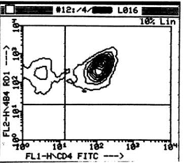
lympho-FIG. 1. Representative contour plot of implant-associated lymphocytes stained with antibodies to CD4 and CD29 (4B4). The vast majority of the cells coexpress CD4 and CD29 (upper right quadrant).
TABLE 3. Phenotype of implant-associated lymphocytes compared with that of paired peripheral blood lymphocytes
Phenotype
% of lymphocytes positivea
Implant Peripheral blood
CD31(T cells) 8964.9 7169.1
CD19 or CD201(B cells)b 0.961.3 1268.3
CD16/561CD32(natural killer cells) 3.663.8 1567.2 aValues are means6standard deviations (n516). For two-tailed probability, P,0.001 (paired t test).
bThe B-lymphocytes in five of the peripheral blood samples were quantitated
with antibodies to CD20 instead of CD19.
TABLE 4. Implant-associated T-cell subsets compared with paired peripheral blood T-cell subsets
Phenotype
% of T cells positivea
2-Tailed probabilityb
Implant Peripheral blood
HLA-DR1 2568.1 7.963.8 ,0.001
CD41CD291 68612 59613 0.043
CD41CD292 0.660.1 3.663.5 0.007
CD41CD45RO1 2.665.2 1967.2 ,0.001
CD41CD45RO2 63613 4168.5 ,0.001
CD81 37610 4269.6 0.115
a
Values are means6standard deviations (n516).
b
Paired t test.
on August 17, 2020 by guest
http://cvi.asm.org/
cytes that are associated with polyurethane-coated and
tex-tured-surface silicone gel-containing breast implants. These
types of implants were specifically chosen for this study
be-cause of the large number of lymphocytes that could be
har-vested from the capsule or from the peri-implant fluid.
Whether or not the inclusion of only textured-surface and
polyurethane-coated implants in our study introduced some
bias cannot be determined from our available data. In all cases,
there was a striking predominance of T cells. Most of the T
cells had the phenotype CD3
1CD4
1CD29
1CD45RO
2.
Fur-thermore, among the T cells there was significant expression of
HLA-DR. Comparison of the implant-associated lymphocytes
with patient-matched peripheral blood lymphocytes, obtained
at the time of surgery, showed that the increase in CD3
1cells,
the increased expression of HLA-DR by the T cells, and the
decrease in expression of CD45RO by the CD4
1cells were all
statistically significant (P
,
0.001 for each comparison). The
increase in the percentage of CD4
1CD29
1T cells among the
implant-associated lymphocytes compared with the level of the
matched peripheral blood lymphocytes was of borderline
sta-tistical significance (P
5
0.043).
The role of textured-surface implants in eliciting an
exuda-tive host response is uncertain. It is known that
textured-sur-face Silastic implants, as well as polyurethane-covered
im-plants, are frequently associated with synovial metaplasia of
the lining of the fibrous capsule (30). Review of histologic
sections from the capsules from each of the patients in this
study confirmed the presence of synovial metaplasia in all
cases. In addition, in most capsules there was a marked
lym-phocytic infiltrate together with foamy macrophages.
The significance of the predominance of CD29
1CD4
1T
cells among the implant-associated lymphocytes is uncertain.
CD4
1T cells can generally be divided into two major
func-tional categories. CD4
1CD29
1T cells proliferate maximally
to soluble antigen and increase antigen-specific antibody
pro-duction (22). In contrast, CD4
1CD45RO
1T cells induce CD8
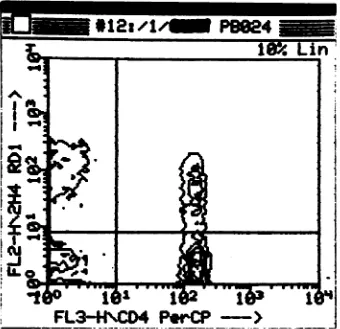
FIG. 2. Representative contour plot of peripheral blood lymphocytes stained with antibodies to CD4 and CD45RO (2H4). A significant fraction of the CD41cells coexpress CD45RO (upper right quadrant). PerCP, PERCP.
TABLE 5. Patient peripheral blood lymphocytes compared with control peripheral lymphocytes
Phenotype
% of lymphocytes positivea
2-Tailed probabilityb
Implant patients (n516)
Controls (n512)
CD3 7169.1 7563.5 0.072
CD31HLA-DR1 5.562.5 3.962.3 0.097
CD41CD291 41610 4569.2 0.301
CD41CD45RO1 1365.5 2169.8 0.022
CD81 3067.6 2567.1 0.081
CD191(or CD201)c 1268.3 1564.1 0.186
CD16/561CD32 1567.2 1165.0 0.069
a
Values are means6standard deviations.
b
Unpaired t test.
c
B lymphocytes in five of the patient peripheral blood samples were quanti-tated with antibodies to CD20 instead of CD19.
on August 17, 2020 by guest
http://cvi.asm.org/
cells to exert suppressor function (23). It is of interest that
CD4
1CD29
1T cells are also the predominant lymphocyte
subset in synovial fluid of patients with rheumatoid arthritis
(24). Furthermore, as we found for implant-associated
lympho-cytes, synovial tissue lymphocytes in patients with rheumatoid
arthritis have increased expression of HLA-DR antigens (8).
The apparent depletion of CD4
1CD45RO
1T cells from the
peripheral blood of patients with silicone gel-containing breast
implants will require confirmation by more extensive
con-trolled studies.
To date, there is very little information that sheds light on
the manner whereby silicone gel-containing breast implants
could potentially cause connective tissue diseases. Some
au-thors have suggested that silicone or silicone-protein
com-plexes may themselves be antigenic (13, 18, 19, 44). Others
have drawn attention to the fact that a significant percentage of
women with silicone breast implants have antinuclear
antibod-ies (5, 28). The results from animal studantibod-ies have been
conflict-ing (4, 25). Clinical studies of patients with silicone gel breast
implants suffer from the fact that patients are often referred
because of symptoms of rheumatic disease. Large
population-based epidemiologic studies are limited by the fact that
‘‘dis-eases’’ potentially caused by silicone gel-containing implants
have not been clearly defined. Most of the patients in this study
reported a symptom complex similar to fibromyalgia. A
pliminary study of 144 patients with breast implants also
re-ported clinical manifestations characteristic of fibromyalgia
(45). Certainly there is no reproducibly specific marker for
patients with silicone gel-associated rheumatic disease. In one
study, which has yet to be substantiated, Vasey and colleagues
reported clinical observations suggesting that rheumatic
dis-ease symptoms in patients with silicone gel implants may be
reversible after implant removal (43). This finding should at
least provide the impetus for continued studies of the
immu-nologic effects of silicone.
There are several inherent limitations in our study regarding
its ability to address the questionable relationship between
silicone breast implants and immunologic diseases. First, our
patient population includes only those patients who are
cur-rently having problems with their implants. Because it is
diffi-cult to form a control group of patients with no local or
sys-temic problems and who wish to undergo implant removal and
capsulectomy, this study does not resolve the question of
whether T-cell activation around polyurethane or
textured-surface breast implants is in fact a pathologic phenomenon.
Although polyurethane-covered implants have been known to
elicit greater inflammatory responses in the capsule than
smooth-shell implants, it is not clear from previous studies
what type of inflammation they elicit (16). Nor is it clear from
previous studies how long the inflammatory responses persist.
Our study has shown that T-cell activation around these
im-plants occurs as early as 1 year after implantation and can
persist for as long as 9 years.
The other unresolved question which this study could not
address is the significance of local T-cell activation in eliciting
local pain and systemic illness. Although all patients studied
had significant pain around their implants and all of the
pa-tients had some concomitant constitutional symptoms, without
a large symptom-free control group, the effect of local T-cell
activation on the systemic immune system remains elusive.
In summary, this study is the first to define the phenotype of
the lymphocytes isolated from the fluid or tissue surrounding
silicone gel-containing breast implants. The lymphocytes are
almost all CD3
1T cells, most of which express CD4 and CD29.
Compared with peripheral blood lymphocytes, the T cells have
significantly increased expression of HLA-DR and significantly
reduced expression of CD45RO. Histologic examination of
capsular tissue, as well as examination of the exudative fluid,
has shown that most of the implant-associated lymphocytes are
present in association with foamy macrophages that are known
to ingest droplets of silicone gel. We have found that these
macrophages strongly and uniformly express HLA-DR (17).
Our results are consistent with the hypothesis that the
silicone-containing macrophages act as antigen-presenting cells to CD4
cells which become activated and subsequently function to
up-regulate an immune response, as indicated by expression of
CD29. Admittedly, this simplistic hypothesis requires extensive
further testing and refinement. In particular, it will be
impor-tant to determine whether the implant-associated CD4
1T
cells belong to the Th1 or Th2 subset. A recent study by
Ojo-Amaize et al. (26) provides additional evidence in support
of the central role of T cells in the immunologic reaction to
silicone breast implants. In any case, if science is to prevail in
the silicone controversy, as Fisher rightly insists (10), then
further analysis of the inflammatory reaction to silicone breast
implants will be necessary.
ACKNOWLEDGMENTS
This research was supported in part by a grant from the Haas Fund, The Mount Sinai Medical Center, Cleveland, Ohio.
We express our gratitude to David Hom of the Department of Epidemiology and Biostatistics at Case Western Reserve University School of Medicine, Cleveland, Ohio, for assistance with the statistical analyses.
REFERENCES
1. Baker, J. L., R. R. LeVier, and D. E. Spielvogel. 1982. Positive identification of silicone in human mammary capsular tissue. Plast. Reconstr. Surg. 69:56– 60.
2. Baldwin, C. M., and E. N. Kaplan. 1983. Silicone-induced human adjuvant disease? Ann. Plast. Surg. 10:270–273.
3. Brandt, B., V. Breiting, L. Christensen, M. Nielsen, and J. L. Thomsen. 1984. Five years experience of breast augmentation using silicone gel prostheses with emphasis on capsule shrinkage. Scand. J. Plast. Reconstr. Surg. 18:311– 316.
4. Brantley, S. K., S. F. Davidson, P. A. St. Arnold, M. B. Johnson, P. J. Talbot,
J. B. Grogan, M. A. Cuchens, H. S. Hsu, and S. K. Das.1990. Assessment of the lymphocyte response to silicone. Plast. Reconstr. Surg. 86:1131–1137. 5. Bridges, A. J., C. Conley, G. Wang, D. E. Burns, and F. B. Vasey. 1993. A
clinical and immunologic evaluation of women with silicone breast implants and symptoms of rheumatic disease. Ann. Intern. Med. 118:929–936. 6. Brozena, S. J., N. A. Fenske, C. W. Cruse, C. G. Espinoza, F. B. Vasey, B. F.
Germain, and L. R. Espinoza.1988. Human adjuvant disease following augmentation mammoplasty. Arch. Dermatol. 124:1383–1386.
7. Cronin, T. D., and F. J. Gerow. 1964. Augmentation mammoplasty: a new ‘‘natural feel’’ prosthesis, p. 41. In Transactions of the Third International Congress of Plastic Surgery. Excerpta Medica, Amsterdam.
8. Cush, J. J., and P. E. Lipsky. 1988. Phenotypic analysis of synovial tissue and peripheral blood lymphocytes isolated from patients with rheumatoid arthri-tis. Arthritis Rheum. 31:1230–1238.
9. Endo, L. P., N. L. Edwards, S. Longley, L. C. Corman, and R. S. Panush. 1987. Silicone and rheumatic diseases. Semin. Arthritis Rheum. 17:112–118. 10. Fisher, J. C. 1992. The silicone controversy: when will science prevail? N.
Engl. J. Med. 326:1696–1698.
11. Gabriel, S. E., W. M. O’Fallon, L. T. Kurland, C. M. Beard, J. E. Woods, and
L. J. Melton.1994. Risk of connective-tissue diseases and other disorders after breast implantation. N. Engl. J. Med. 330:1697–1702.
12. Gayou, R. M. 1979. A histological comparison of contracted and non-con-tracted capsules around silicone breast implants. Plast. Reconstr. Surg. 63: 700–707.
13. Goldblum, R. M., R. P. Pelley, A. A. O’Donell, D. Pyron, and J. P. Heggers. 1992. Antibodies to silicone elastomers and reactions to ventriculoperitoneal shunts. Lancet 340:510–513.
14. Hardt, N. S., L. T. Yu, G. La Torre, and B. Steinbach. 1994. Fourier trans-form infared microspectroscopy used to identify foreign materials related to breast implants. Mod. Pathol. 7:669–676.
15. Hester, T. R., F. Nahai, J. Bostwick, and J. Cukic. 1988. A 5-year experience with polyurethane-covered mammary prostheses for treatment of capsular contracture, primary augmentation, mammoplasty, and breast reconstruc-tion. Clin. Plast. Surg. 15:569–585.
16. Jabaley, M. E., and S. K. Das. 1986. Late breast pain following
on August 17, 2020 by guest
http://cvi.asm.org/
tion with polyurethane-covered implants. Plast. Reconstr. Surg. 78:390–395. 17. Katzin, W. E., and L. J. Feng. 1994. Phenotype of lymphocytes associated with the inflammatory reaction to silicone-gel breast implants. Mod. Pathol.
7:17A. (Abstract.)
18. Kossovsky, N., and C. J. Freiman. 1994. Silicone breast implant pathology: clinical data and immunologic consequences. Arch. Pathol. Lab. Med. 118: 686–693.
19. Kossovsky, N., J. P. Heggers, and M. C. Robson. 1987. Experimental dem-onstration of the immunogenicity of silicone-protein complexes. J. Biomed. Mater. Res. 21:1125–1133.
20. Kumagai, Y., A. Chiyuki, and Y. Shiokawa. 1979. Scleroderma after cosmetic surgery: four cases of human adjuvant disease. Arthritis Rheum. 22:532–537. 21. Marik, P. E., A. L. Kark, and A. Zambakides. 1990. Scleroderma after silicone augmentation mammoplasty: a report of 2 cases. S. Afr. Med. J.
77:212–213.
22. Morimoto, C., N. L. Letvin, A. W. Boyd, M. Hagan, H. M. Brown, M. M.
Kornacki, and S. F. Schlossman.1985. The isolation and characterization of the human helper inducer T cell subset. J. Immunol. 134:3762–3769. 23. Morimoto, C., N. L. Letvin, J. A. Distaso, H. M. Brown, and S. F.
Schloss-man.1986. The cellular basis for the induction of antigen-specific T8 sup-pressor cells. Eur. J. Immunol. 16:198–204.
24. Morimoto, C., P. L. Romain, D. A. Fox, P. Anderson, M. DiMaggio, H.
Levine, and S. F. Schlossman.1988. Abnormalities in CD41T-lymphocyte subsets in inflammatory rheumatic diseases. Am. J. Med. 84:817–825. 25. Naim, J. O., R. J. Lanzafae, and C. J. van Oss. 1993. The adjuvant effect of
silicone-gel on antibody formation in rats. Immunol. Invest. 22:151–161. 26. Ojo-Amaize, E. A., V. Conte, H.-C. Lin, R. F. Brucker, M. S. Agopian, and
J. B. Peter.1994. Silicone-specific blood lymphocyte response in women with silicone breast implants. Clin. Diagn. Lab. Immunol. 1:689–695.
27. Picha, G. J., and J. A. Goldstein. 1990. Analysis of the soft-tissue response to components used in the manufacture of breast implants: rat animal model. Plast. Reconstr. Surg. 87:490–500.
28. Press, R. I., C. L. Peebles, Y. Kumagai, R. L. Ochs, and E. M. Tan. 1992. Antinuclear autoantibodies in women with silicone breast implants. Lancet
340:1304–1307.
29. Raso, D. S. 1994. Breast prostheses, the immune response, and B- and T-lymphocytes. Plast. Reconstr. Surg. 93:649–650.
30. Raso, D. S., L. W. Crymes, and J. S. Metcalf. 1994. Histological assessment of fifty breast capsules from smooth and textured augmentation and recon-struction mammoplasty prostheses with emphasis on the role of synovial metaplasia. Mod. Pathol. 7:310–316.
31. Rowlands, C., and F. H. Y. Green. 1994. Tissue responses to silicone breast implants. Mod. Path. 7:21A. (Abstract.)
32. Rudolph, R., J. Abraham, T. Vecchione, S. Guber, and M. Woodward. 1978. Myofibroblasts and free silicon around breast implants. Plast. Reconstr. Surg. 62:185–196.
33. Sahn, E. E., P. D. Garen, R. M. Silver, and J. C. Maize. 1990. Scleroderma following augmentation mammoplasty. Report of a case and review of the literature. Arch. Dermatol. 126:1198–1202.
34. Sanchez-Guerrero, J., G. A. Colditz, E. W. Karlson, D. J. Hunter, F. E.
Speizer, and M. H. Liang.1995. Silicone breast implants and the risk of connective-tissue disease and symptoms. N. Engl. J. Med. 332:1666–1670. 35. Sanchez-Guerrero, J., P. H. Schur, J. S. Sergent, and M. H. Liang. 1994.
Silicone breast implants and rheumatic disease: clinical, immunologic, and epidemiologic studies. Arthritis Rheum. 37:158–168.
36. Silver, R. M., E. E. Sahn, J. A. Allen, S. Sahn, W. Green, J. C. Maize, and
P. D. Garen.1993. Demonstration of silicon in sites of connective-tissue disease in patients with silicone-gel breast implants. Arch. Dermatol. 129: 63–68.
37. Spiera, H. 1988. Scleroderma after silicone augmentation mammoplasty. JAMA 260:236–238.
38. Thomsen, J. L., L. Christensen, M. Nielsen, B. Brandt, V. B. Breiting, S.
Felby, and E. Nielsen.1990. Histologic changes and silicone concentration in human breast tissue surrounding silicone breast prostheses. Plast. Reconstr. Surg. 85:38–41.
39. Uretsky, B. F., J. J. O’Brien, E. H. Courtiss, and M. D. Beeker. 1979. Augmentation mammaplasty associated with a severe systemic illness. Ann. Plast. Surg. 3:445–447.
40. Van Nunen, S. A., P. A. Gatenby, and A. Basten. 1982. Post-mammoplasty connective tissue disease. Arthritis Rheum. 25:694–697.
41. Varga, J., and S. A. Jimenez. 1990. Augmentation mammoplasty and sclero-derma: is there an association? Arch. Dermatol. 126:1220–1222. 42. Varga, J., R. Schumacher, and S. A. Jimenez. 1989. Systemic sclerosis after
augmentation mammoplasty with silicone implants. Ann. Intern. Med. 111: 377–383.
43. Vasey, F. B., D. Havice, T. Bocanegra, M. J. Seleznick, P. Bridgeford, and
B. F. Germain.1992. Clinical manifestations of fifty consecutive women with silicone breast implants. Arthritis Rheum. 35(Suppl.):S212. (Abstract.) 44. Vojdani, A., N. Brautbar, and A. Campbell. 1994. Immunologic and biologic
markers for silicone. Toxicol. Ind. Health 10:25–42.
45. Ward, C., S. Romero, S. Marlowe, F. B. Vasey, and T. S. Bocanegra. 1994. Clinical manifestations in 144 patients with breast implants (BI). A compar-ison with primary fibromyalgia (PFM). Arthritis Rheum. 36(Suppl.):R28. (Abstract.)
46. Weiner, S. R., and H. E. Paulus. 1986. Chronic arthropathy occurring after augmentation mammaplasty. Plast. Reconstr. Surg. 77:185–187.
47. Weisman, M. H., T. R. Vecchione, D. Albert, L. T. Moore, and M. R. Mueller. 1988. Connective-tissue disease following breast augmentation: a prelimi-nary test of the human adjuvant disease hypothesis. Plast. Reconstr. Surg.
82:626–630.