Diabetes, Metabolic Syndrome and Obesity: Targets and Therapy
Dove
press
O r i g i n a l r e S e a r c h
open access to scientific and medical research
Open Access Full Text Article
Visceral-to-subcutaneous fat ratio as a predictor
of the multiple metabolic risk factors for subjects
with normal waist circumference in Korea
Yun hwan Oh1,2
Ji hyun Moon1,2
hyeon Ju Kim1,3
Mi hee Kong1,3
1Department of Family Medicine,
Jeju national University hospital,
2Department of Medicine, graduate
School of Jeju national University,
3Department of Family Medicine,
School of Medicine, Jeju national University, Jeju, republic of Korea
Purpose: Visceral obesity has been recognized as a predictor of metabolic risk factors. However, few studies have evaluated the metabolic risks in subjects with normal waist circumference (WC). We aimed to examine if the visceral-to-subcutaneous fat ratio (VSR) has diagnostic value to identify multiple metabolic risk factors in subjects with normal WC, compared with visceral fat area (VFA) and subcutaneous fat area (SFA).
Methods: This is a cross-sectional study in which we have compared mean VFA, SFA, and VSR according to each metabolic risk factor. We performed a receiver operating characteristic (ROC) curve analysis for VFA, SFA, and VSR to assess their accuracy in picking out two or more non-adipose factors for metabolic syndrome.
Results: For each metabolic risk factor, mean VSRs were significantly different between groups (risk-absent group vs risk-present group) in men and women, except for men with low high-density lipoprotein. However, mean VFAs and SFAs showed no significant differences between groups. VSR showed superior diagnostic values in predicting at least two non-adipose metabolic risk factors in men and similar diagnostic value in women. Areas under ROC curves for VSR and VFA were 0.705 and 0.649 in men (P=0.028) and 0.798 and 0.785 in women (P=0.321).
Conclusion: For men with a normal WC, VSR appeared to effectively predict the presence of multiple metabolic risk factors. Thus, VSR may serve as an indicator for identifying men who have a normal WC and multiple metabolic risk factors.
Keywords: visceral fat, subcutaneous fat, metabolic syndrome, visceral-to-subcutaneous fat ratio
Introduction
With increasing concerns for obesity because of its reported relationship with risks of developing adverse cardiovascular events and metabolic illnesses, how to define obesity has become an issue to be established. There are known sex differences in body fat distri-bution, fat metabolism, and health risks from obesity and metabolic syndrome.1,2 Based
on accumulated evidence, abdominal obesity has been proven to be more closely associ-ated with cardiovascular and metabolic disorders than general obesity in both sexes.3–7
Subsequently, methods to reflect abdominal obesity have been explored using anthropometric measures, such as waist circumference (WC). In clinical settings, WC is apparently preferred not only because of its great simplicity but also because of its proven accuracy as a reliable predictor for identifying subjects with metabolic aberrance and cardiovascular diseases.8–11
Recently, visceral obesity has been identified as an important factor in the develop-ment of metabolic disorders.12 Several studies have reported that visceral obesity is
correspondence: Mi hee Kong Department of Family Medicine, Jeju national University hospital, 15, aran 13-gil, Jeju-si, Jeju-do, 63241, republic of Korea
Tel +82 64 717 1550 Fax +82 64 717 1581 email kongmihee@jejunu.ac.kr
Journal name: Diabetes, Metabolic Syndrome and Obesity: Targets and Therapy Article Designation: Original Research
Year: 2017 Volume: 10
Running head verso: Oh et al
Running head recto: VSR predicts multiple metabolic risk factors DOI: http://dx.doi.org/10.2147/DMSO.S150914
Diabetes, Metabolic Syndrome and Obesity: Targets and Therapy downloaded from https://www.dovepress.com/ by 118.70.13.36 on 21-Aug-2020
For personal use only.
Dovepress
Oh et al
closely related to metabolic risk factors, including hyperten-sion, impaired glucose tolerance, dyslipidemia, and other cardiovascular risk factors.13,14 In defining obesity and its
validity to predict metabolic risks, focus has changed from central obesity to visceral obesity.
Of several measurements of abdominal visceral fat accu-mulation, visceral fat area (VFA) using computed tomogra-phy (CT) scan of the abdomen was explored extensively for its usefulness in predicting the presence of metabolic risk factors.15–19 On the other hand, considering the differences
of body frame size or shape among people, it seems that the absolute amount of visceral fat may not fully reflect such differences while defining visceral obesity. For this reason, theoretically, visceral-to-subcutaneous fat ratio (VSR) would be a better index to allow evaluating a person’s build. Back in 1987, Fujioka et al20 showed the correlation between
visceral fat accumulation represented by VSR and glucose or lipid metabolism in obese subjects; the study introduced the cut-off point of 0.4 for VSR, above which individuals with obesity are likely to show glucose intolerance and hyperlipidemia. Recently, Kim et al17 demonstrated
diag-nostic accuracy of VFA in predicting metabolic risk factors in Koreans and briefly addressed VSR being inferior to VFA in terms of predictability with its cut-off points. Regarding the association between various metabolic components and visceral obesity, previous studies21–23 have revealed specific
relationships between various factors and visceral obesity. However, the predictability of VSR in identifying individual non-adipose components of metabolic syndrome has never been addressed in the Korean population.
While WC remains a fascinating tool to be used in clinical practice, it has limitations as it cannot distinguish between visceral and subcutaneous fat deposits in the abdo-men. Therefore, if WC is used as the only evaluation index for visceral obesity, potential metabolic risk factors of par-ticipants with normal WC may be overlooked. According to previous research conducted in Iran, about 9.9% of men with a normal WC have metabolic syndrome.24 Another study also
showed that 10% of men and 3.1% of women with metabolic syndrome have a WC within the normal range.25 As a result,
assessing visceral obesity is particularly important in subjects with normal WC because these subjects are sometimes easily considered as having low metabolic risk.
In this context, the aim of this study was to examine whether VFA, subcutaneous fat area (SFA), and VSR in particular have a diagnostic value in identifying individu-als who have a WC within the normal range and multiple metabolic risk factors.
Methods
Study subjects
From the Koreans who spontaneously attended the health promotion center in a local university hospital for their routine health examination from June 1, 2012 to April 30, 2017, we selected those who underwent a CT scan of the abdomen as well as ordinary examinations, for this cross-sectional study. Participants completed a medical history questionnaire, responding to questions about previously diagnosed diseases, prescribed medications, and other topics. Then, they underwent anthropometric evaluation, laboratory tests, and CT scan of the abdomen. Participants with abnormal WC were excluded from the study.
anthropometric measurements
Participants who were fasting underwent anthropometric evaluations: weight, height, and WC. Weight was measured using calibrated electronic scales while subjects wore light clothing and no shoes; height was obtained by a fixed stadio-meter; WC was measured at the level of umbilicus while participants were standing. Body mass index (BMI) was calculated as weight in kilograms divided by the height in meters squared. Blood pressure (BP) was measured using an automatic digital BP monitor FT-700R (Jawon Medical Co. Ltd., Seoul, South Korea) while participants were comfort-ably seated after resting for least 10 min and refraining from smoking and ingesting caffeine.
Definition of metabolic syndrome and
multiple metabolic risk factors
We excluded WC and defined multiple metabolic risk fac-tors as having two or more of the following components of metabolic syndrome according to the modified National Cholesterol Education Program Adult Treatment Panel III:11
fasting blood sugar (FBS) ≥100 mg/dL or taking medication for previously diagnosed diabetes; BP ≥130/85 mmHg or taking medication for previously diagnosed hypertension; serum triglyceride (TG) ≥150 mg/dL; and serum high-density lipoprotein (HDL)-cholesterol <40 mg/dL in men or <50 mg/dL in women. We referred to these factors as hyperglycemia, high BP, high TG, and low HDL-cholesterol, respectively.
Determination of VFa, SFa, and VSr using
cT scan of the abdomen
Abdominal visceral and subcutaneous fat amounts were deter-mined using a single-slice CT scan (SOMATOM Definition;
Diabetes, Metabolic Syndrome and Obesity: Targets and Therapy downloaded from https://www.dovepress.com/ by 118.70.13.36 on 21-Aug-2020
Dovepress VSr predicts multiple metabolic risk factors
Siemens, Munich, Germany), 5 mm thickness, at the L4–5 intervertebral space level with subjects in supine position. SFA and VFA were defined by delineating these areas with a graph pen. Both fat areas were determined by identifying fat surface with attenuation range of −140 to −40 Hounsfield units. VSR was finally calculated using the obtained data.
Statistical analysis
All statistical analyses were conducted with SPSS for Win-dows version 20.0 (IBM, Armonk, NY, USA) and STATA version 13.0 (StataCorp., College Station, TX, USA). The
P-values of all the reported results were two-tailed, and the significance level was set at P<0.05.
First, all the subjects were divided by sex. To compare the basic demographic characteristics, the continuous variables were expressed as means and standard deviations and compared through independent t-test. The categorical variables were expressed as percentages and compared by using the c2 test.
Second, the subjects were divided into two groups according to the presence or absence of each metabolic risk factor (hyperglycemia, high BP, high TG, and low HDL-cholesterol), at least two metabolic risk factors, and metabolic syndrome. Mean VFA, SFA and VSR were calculated. Values were adjusted for age and BMI to control for the effects of the body frame size and aging; differences in adjusted mean VFA, SFA, and VSR between the groups were tested. Addi-tionally, differences in mean VFA, SFA, and VSR between the two groups according to the presence or absence of each metabolic risk factor, at least two metabolic risk factors, and metabolic syndrome were analyzed by independent t-test.
Third, binary logistic regression analysis was used to evaluate the relationship between variables (VFA, VSR, and SFA) and the presence of multiple metabolic risk factors. With a receiver operating characteristic curve, we assessed the predictive accuracy of VFA, SFA, and VSR in identifying individuals with at least two metabolic risk factors. Subse-quently, area under curve (AUC) was used to quantify the accuracy of each test.
ethical approval
The Institutional Review Board of the Jeju National Uni-versity Hospital approved the design of the present study and ensured that individuals were not identifiable by provid-ing linkable anonymous data to the researchers. Waiver of informed consent for this study was approved by the Institu-tional Review Board, and the study was approved (Approval No. 2017-04-014) after it was reviewed for ethical issues.
Results
Baseline characteristics of subjects
The mean age of men and women was 52.1±9.9 and 50.6±9.7 years, respectively. Likewise, there were no significant dif-ferences between sexes in total cholesterol and number of subjects with metabolic syndrome. However, men showed significantly higher values of BMI, WC, systolic BP, diastolic BP, TG, FBS, VFA, and VSR than women. As for HDL-cholesterol and SFA, women showed higher values than men. Compared to women, there were higher numbers of men taking antihypertensive medications or oral hypoglycemic agents and with at least two risk factors other than central besity (Table 1).
analyses of adjusted mean VFas, SFas,
and VSrs according to the presence of
metabolic risk factors
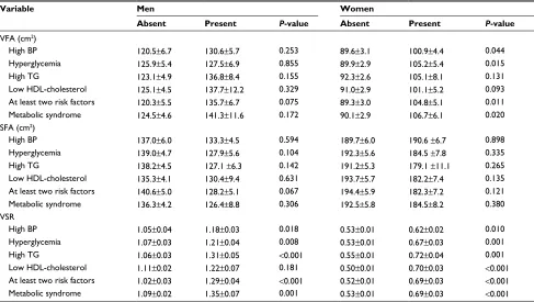
Among men, no significant differences were observed in adjusted mean VFA and SFA according to the presence or absence of each metabolic risk factor, multiple metabolic risks, and metabolic syndrome. On the contrary, groups with metabolic risk factors except low HDL-cholesterol showed significantly higher adjusted mean VSR than groups without risk factors.
Among women, groups with the presence of high BP, hyperglycemia, at least two risk factors, and metabolic syn-drome showed significantly higher adjusted mean VFA than groups without risk factors. There were no significant differ-ences in mean SFA between groups. Regarding VSR, there were significant differences between the groups according to the presence of metabolic risk factors (Table 2).
aUcs for VFa, SFa, and VSr to predict
multiple metabolic risk factors
Based on logistic regression analysis, after adjusting for age and BMI, VSR appeared to be an independent predictor of the presence of multiple metabolic risk factors in both men and women (b=1.42, P<0.001; b=2.51, P<0.001, respec-tively). However, VFA served as a predictor only in women (b=0.01, P=0.034). The AUCs of VSR, VFA, and SFA were 0.705 (95% CI 0.645–0.764), 0.649 (95% CI 0.587–0.712), and 0.636 (95% CI 0.573–0.699) in men and 0.798 (95% CI 0.729–0.867), 0.785 (95% CI 0.716–0.855), and 0.763 (95% CI 0.688–0.838) in women. In men, VSR was superior to VFA (P=0.028). It also seemed that VSR was superior to VFA in women, but it was not statistically significant (P=0.321) (Figure 1).
Diabetes, Metabolic Syndrome and Obesity: Targets and Therapy downloaded from https://www.dovepress.com/ by 118.70.13.36 on 21-Aug-2020
Dovepress
Oh et al
Table 1 Baseline characteristics of study participants according to sex
Variable Normal range Men (n=296) Women (n=239) P-value
age (years) 52.1±9.9 50.6±9.7 0.089
BMi (kg/m2) <25 24.0±2.2 22.4±2.1 <0.001
Wc (cm) Men <90, women <85 83.1±4.9 76.60±5.2 <0.001
SBP (mmhg) <130 128.2±11.7 118.0±13.5 <0.001
DBP (mmhg) <85 81.9±9.0 74.2±9.5 <0.001
Triglyceride (mg/dl) <150 132.0±108.3 89.8±60.1 <0.001
hDl-cholesterol (mg/dl) Men ≥40, women ≥50 52.9±13.0 60.3±14.3 <0.001
FBS (mg/dl) <100 103.8±29.1 93.3±14.4 <0.001
Taking antihypertensive, n (%) 50 (16.8) 19 (7.9) 0.002
Taking Oha, n (%) 25 (8.4) 7 (2.9) 0.007
high BP, n (%) 183 (61.8) 73 (30.5) <0.001
hyperglycemia, n (%) 116 (39.1) 49 (20.5) <0.001
high Tg, n (%) 80 (27.0) 21 (8.7) <0.001
low hDl-cholesterol, n (%) 36 (12.1) 51 (21.3) 0.004
at least two risk factors other than central obesity, n (%) 128 (43.2) 54 (22.5) <0.001
Metabolic syndrome, n (%) 41 (13.8) 37 (15.4) 0.595
VFa (cm2) 133.3±74.5 84.9±43.2 <0.001
SFa (cm2) 121.0±60.5 157.2±52.6 <0.001
VSr 1.34±3.63 0.54±0.25 <0.001
Notes: Values are presented as mean ± standard deviation or number (%). P-values were calculated using independent t-test for the continuous variables and chi-square test for categorical variables. high BP: blood pressure ≥130/85 mmhg or taking medication for previously diagnosed hypertension; hyperglycemia: fasting blood sugar 100 mg/dl or taking medication for previously diagnosed diabetes; high Tg: serum triglycerides ≥150 mg/dl; low hDl-cholesterol: serum high-density lipoprotein cholesterol <40 mg/ dl in men and <50 mg/dl in women.
Abbreviations: BMi, body mass index; Wc, waist circumference; SBP, systolic blood pressure; DBP, diastolic blood pressure; hDl, high-density lipoprotein; FBS, fasting blood sugar; Oha, oral hypoglycemic agent; BP, blood pressure; Tg, triglyceride; VFa, visceral fat area; SFa, subcutaneous fat area; VSr, visceral-to-subcutaneous fat ratio.
Table 2 comparison of adjusted mean VFa, SFa, and VSr by metabolic risk factors
Variable Men Women
Absent Present P-value Absent Present P-value
VFa (cm2)
high BP 120.5±6.7 130.6±5.7 0.253 89.6±3.1 100.9±4.4 0.044
hyperglycemia 125.9±5.4 127.5±6.9 0.855 89.9±2.9 105.2±5.4 0.015
high Tg 123.1±4.9 136.8±8.4 0.155 92.3±2.6 105.1±8.1 0.131
low hDl-cholesterol 125.1±4.5 137.7±12.2 0.329 91.0±2.9 101.1±5.2 0.093
at least two risk factors 120.3±5.5 135.7±6.7 0.075 89.3±3.0 104.8±5.1 0.011
Metabolic syndrome 124.5±4.6 141.3±11.6 0.172 90.1±2.9 106.7±6.1 0.020
SFa (cm2)
high BP 137.0±6.0 133.3±4.5 0.594 189.7±6.0 190.6 ±6.7 0.898
hyperglycemia 139.0±4.7 127.9±5.6 0.104 192.3±5.6 184.5 ±7.8 0.335
high Tg 138.2±4.5 127.1 ±6.3 0.142 191.2±5.3 179.1 ±11.1 0.265
low hDl-cholesterol 135.3±4.1 130.4±9.4 0.631 193.7±5.7 182.2±7.4 0.135
at least two risk factors 140.6±5.0 128.2±5.1 0.067 194.4±5.9 182.3±7.2 0.121
Metabolic syndrome 136.3±4.2 126.4±8.8 0.306 192.5±5.8 184.5±8.2 0.380
VSr
high BP 1.05±0.04 1.18±0.03 0.018 0.53±0.01 0.62±0.02 0.010
hyperglycemia 1.07±0.03 1.21±0.04 0.008 0.53±0.01 0.67±0.03 0.001
high Tg 1.06±0.03 1.31±0.05 <0.001 0.55±0.01 0.72±0.04 0.001
low hDl-cholesterol 1.11±0.02 1.22±0.07 0.181 0.50±0.01 0.70±0.03 <0.001
at least two risk factors 1.02±0.03 1.29±0.04 <0.001 0.52±0.01 0.69±0.03 <0.001
Metabolic syndrome 1.09±0.02 1.35±0.07 0.001 0.53±0.01 0.69±0.03 <0.001
Notes: Values are presented as mean ± standard error and are adjusted for age and BMi. P-value was calculated using independent t-test.
Abbreviations: VFa, visceral fat area; SFa, subcutaneous fat area; VSr, visceral-to-subcutaneous fat ratio; BP, blood pressure; Tg, triglyceride; hDl, high-density lipoprotein; BMi, body mass index.
Diabetes, Metabolic Syndrome and Obesity: Targets and Therapy downloaded from https://www.dovepress.com/ by 118.70.13.36 on 21-Aug-2020
Dovepress VSr predicts multiple metabolic risk factors
Figure 1 rOc curves for VSr, VFa, and SFa to identify at least two components of metabolic syndrome other than waist circumference in normal waist-circumference (A) men and (B) women.
Abbreviations: rOc, receiver operating characteristic; VSr, visceral-to-subcutaneous fat ratio; VFa, visceral fat area; SFa, subcutaneous fat area; aUc, area under curve.
0.0
0.0 0.2 0.4 0.6
1 – specificity
0.8 1.0
VSR VFA SFA
VSR 0.70 (0.64–0.76) 0.65 (0.58–0.71) 0.63 (0.57–0.70) VFA
SFA
Reference line
AUC (95% CI)
VSR VFA SFA
VSR 0.79 (0.72–0.86)
0.78 (0.71–0.85) 0.76 (0.68–0.87) VFA
SFA
Reference line
AUC (95% CI)
0.0 0.2 0.4 0.6
1 – specificity
0.8 1.0
0.2 0.4
Sensitivit
y 0.6
0.8 1.0 A
B
0.0 0.2 0.4
Sensitivit
y 0.6
0.8 1.0
Diabetes, Metabolic Syndrome and Obesity: Targets and Therapy downloaded from https://www.dovepress.com/ by 118.70.13.36 on 21-Aug-2020
Dovepress
Oh et al
Discussion
Our study showed differences in adjusted mean VFA, SFA, and VSR according to the status of each metabolic risk factor; our study also compared VFA, SFA, and VSR of subjects with normal WC in association with the presence of metabolic risk factors. Additionally, we evaluated the diagnostic value of VSR to predict multiple metabolic risk factors and proved that it was superior to VFA in this regard.
WC or BMI, commonly used as an indicator for predict-ing metabolic risk, is a powerful and convenient tool to use, but it varies widely according to the individual’s body frame size and provides little or no information regarding the rela-tive distribution of body fat, especially visceral adiposity. Moreover, some studies have reported that subcutaneous fat tissue might have protective effects against cardiovascular disease.26,27 Obviously, there were some limitations in using
WC as a representative index to predict cardiovascular and metabolic risks. Especially in subjects with normal WC and metabolic risk factors, using WC as an index for diagnosing metabolic syndrome can be disadvantageous.
For these reasons, VFA might be an alternative to accu-rately measure the visceral adipose tissue of the body, but it is also affected by the individual’s body frame size, such as their height, weight, WC, and BMI.28 As a result, it is hard to
apply VFA in individuals with diverse body types. Moreover, it cannot fully represent the individual’s propensity to store fat viscerally or subcutaneously because it only suggests absolute amounts of single fat deposits and does not reflect relative distribution of fat in the body. In this respect, our hypothesis was that VSR could represent an individual’s propensity to store fat and be less affected by body type than VFA.
In this respect, our study showed that VSR has diagnostic value as a unique indicator to predict multiple metabolic risk factors regardless of the subject’s age and BMI. After adjust-ing for age and BMI, mean VSRs were significantly different between groups according to each metabolic risk factor, except for low HDL-cholesterol in men. However, mean VFAs and SFAs showed no significant differences between groups. When we compared mean VFAs, some differences between groups were found in women. Additionally, the diag-nostic value of VSR was higher than VFA in men, but similar in women. These findings implicated that the propensity for visceral fat deposits (as opposed to subcutaneous) is associ-ated with metabolic risk factors regardless of age and BMI. Diagnostic value of VSR to predict multiple metabolic risk factors was higher in women than in men. The mecha-nism behind this sex-specific result is unclear, but it is
probably due to the decreased bioavailability of estrogen in women.22 Further study is required to elucidate causality of
this finding.
Our study has some strengths. First, we used standard-ized and reproducible methods to assess fat deposit amounts through CT scan. Second, all our study subjects had a normal WC, believed in the past to indicate fewer or no metabolic risks. As a result of this study’s design, we confirmed the need for a new index to replace WC, as well as the clinical use-fulness of direct measurement of visceral and subcutaneous adipose tissue. So far, to our knowledge, no other studies have shown VSR to be a valuable indicator in predicting metabolic risk components in both sexes regardless of age and BMI.
There are some limitations to our study. First, we per-formed the study with a relatively small sample size in a single center, which prevents the generalization of results to the entire Korean population. Second, as a cross-sectional study, our data showed only cross-sectional associations between variables and metabolic risk factors. Finally, our study subjects were mostly middle-aged men and women, so we might not be able to generalize our findings to other age groups.
The design of future investigations needs to be on a larger scale, prospective, and include other clinical parameters that better reflect visceral obesity. Ultimately, researchers need to address the direct relationships between health indices and cardiovascular diseases. Additionally, the abdominal cavity should be subdivided into several compartments, and researchers should analyze possible associations of subdivided areas with metabolic disorders or cardiovascular disease.
Disclosure
The authors report no conflicts of interest in this work.
References
1. Power ML, Schulkin J. Sex differences in fat storage, fat metabolism, and the health risks from obesity: possible evolutionary origins. Br J Nutr. 2008;99(5):931–940.
2. Pradhan AD. Sex differences in the metabolic syndrome: implications for cardiovascular health in women. Clin Chem. 2014;60(1):44–52. 3. Lee CM, Huxley RR, Wildman RP, Woodward M. Indices of abdominal
obesity are better discriminators of cardiovascular risk factors than BMI: a meta-analysis. J Clin Epidemiol. 2008;61(7):646–653.
4. Zamboni M, Armellini F, Sheiban I, et al. Relation of body fat distri-bution in men and degree of coronary narrowings in coronary artery disease. Am J Cardiol. 1992;70(13):1135–1138.
5. Björntorp P. Abdominal obesity and the development of noninsulin-dependent diabetes mellitus. Diabetes Metab Rev. 1988;4(6):615–622. 6. Folsom AR, Kaye SA, Sellers TA, et al. Body fat distribution and 5-year
risk of death in older women. JAMA. 1993;269(4):483–487.
Diabetes, Metabolic Syndrome and Obesity: Targets and Therapy downloaded from https://www.dovepress.com/ by 118.70.13.36 on 21-Aug-2020
Dovepress
Diabetes, Metabolic Syndrome and Obesity: Targets and Therapy
Publish your work in this journal
Submit your manuscript here: https://www.dovepress.com/diabetes-metabolic-syndrome-and-obesity-targets-and-therapy-journal
Diabetes, Metabolic Syndrome and Obesity: Targets and Therapy is an international, peer-reviewed open-access journal committed to the rapid publication of the latest laboratory and clinical findings in the fields of diabetes, metabolic syndrome and obesity research. Original research, review, case reports, hypothesis formation, expert
opinion and commentaries are all considered for publication. The manuscript management system is completely online and includes a very quick and fair peer-review system, which is all easy to use. Visit http://www.dovepress.com/testimonials.php to read real quotes from published authors.
Dove
press
VSr predicts multiple metabolic risk factors
7. Folsom AR, Prineas RJ, Kaye SA, et al. Incidence of hypertension and stroke in relation to body fat distribution and other risk factors in older women. Stroke. 1990;21(5):701–706.
8. Katzmarzyk PT, Janssen I, Ross R, et al. The importance of waist circumference in the definition of metabolic syndrome: prospective analyses of mortality in men. Diabetes Care. 2006;29(2):404–409. 9. Han T, van Leer EM, Seidell JC, Lean M. Waist circumference action
levels in the identification of cardiovascular risk factors: prevalence study in a random sample. BMJ. 1995;311(7017):1401–1405. 10. Pouliot MC, Després JP, Lemieux S, et al. Waist circumference and
abdominal sagittal diameter: best simple anthropometric indexes of abdominal visceral adipose tissue accumulation and related cardiovas-cular risk in men and women. Am J Cardiol. 1994;73(7):460–468. 11. Expert Panel on Detection, Evaluation, and Treatment of High Blood
Cholesterol in Adults. Executive summary of the Third Report of the National Cholesterol Education Program (NCEP) expert panel on detection, evaluation, and treatment of high blood cholesterol in adults (Adult Treatment Panel III). JAMA. 2001;285(19):2486–2497. 12. Fox CS, Massaro JM, Hoffmann U, et al. Abdominal visceral and
sub-cutaneous adipose tissue compartments: association with metabolic risk factors in the Framingham Heart Study. Circulation. 2007;116(1):39–48. 13. Hayashi T, Boyko EJ, Leonetti DL, et al. Visceral adiposity and the risk of impaired glucose tolerance: a prospective study among Japanese Americans. Diabetes Care. 2003;26(3):650–655.
14. Rosito GA, Massaro JM, Hoffmann U, et al. Pericardial fat, visceral abdominal fat, cardiovascular disease risk factors, and vascular calci-fication in a community-based sample: the Framingham Heart Study.
Circulation. 2008;117(5):605–613.
15. Kim JA, Choi CJ, Yum KS. Cut-off values of visceral fat area and waist circumference: diagnostic criteria for abdominal obesity in a Korean population. JKoreanMed Sci. 2006;21(6):1048–1053.
16. Seo JA, Kim BG, Cho H, et al. The cutoff values of visceral fat area and waist circumference for identifying subjects at risk for metabolic syndrome in elderly Korean: Ansan Geriatric (AGE) cohort study. BMC Public Health. 2009;9(1):443.
17. Kim HI, Kim JT, Yu SH, et al. Gender differences in diagnostic values of visceral fat area and waist circumference for predicting metabolic syndrome in Koreans. J Korean Med Sci. 2011;26(7):906–913.
18. Hayashi T, Boyko EJ, McNeely MJ, Leonetti DL, Kahn SE, Fujimoto WY. Minimum waist and visceral fat values for identifying Japa-nese Americans at risk for the metabolic syndrome. Diabetes Care. 2007;30(1):120–127.
19. Oka R, Kobayashi J, Yagi K, et al. Reassessment of the cutoff values of waist circumference and visceral fat area for identifying Japanese subjects at risk for the metabolic syndrome. Diabetes Res Clin Pract. 2008;79(3):474–481.
20. Fujioka S, Matsuzawa Y, Tokunaga K, Tarui S. Contribution of intra-abdominal fat accumulation to the impairment of glucose and lipid metabolism in human obesity. Metabolism. 1987;36(1):54–59. 21. Hayashi T, Boyko EJ, McNeely MJ, Leonetti DL, Kahn SE, Fujimoto
WY. Visceral adiposity, not abdominal subcutaneous fat area, is associ-ated with an increase in future insulin resistance in Japanese Americans.
Diabetes. 2008;57(5):1269–1275.
22. Taskinen MR, Adiels M, Westerbacka J, et al. Dual metabolic defects are required to produce hypertriglyceridemia in obese subjects. Arterioscler Thromb Vasc Biol. 2011;31(9):2144–2150.
23. Sironi AM, Gastaldelli A, Mari A, et al. Visceral fat in hypertension: influence on insulin resistance and beta-cell function. Hypertension. 2004;44(2):127–133.
24. Hadaegh F, Zabetian A, Harati H, Azizi F. Metabolic syndrome in normal-weight Iranian adults. Ann Saudi Med. 2007;27(1):18–24. 25. Motala AA, Esterhuizen T, Pirie FJ, Omar MA. The prevalence of
metabolic syndrome and determination of the optimal waist circumfer-ence cutoff points in a rural South African community. Diabetes Care. 2011;34(4):1032–1037.
26. Tran TT, Yamamoto Y, Gesta S, Kahn CR. Beneficial effects of sub-cutaneous fat transplantation on metabolism. Cell Metab. 2008;7(5): 410–420.
27. Porter SA, Massaro JM, Hoffmann U, Vasan RS, O’donnel CJ, Fox CS. Abdominal subcutaneous adipose tissue: a protective fat depot? Diabetes Care. 2009;32(6):1068–1075.
28. Camhi SM, Bray GA, Bouchard C, et al. The relationship of waist circumference and BMI to visceral, subcutaneous, and total body fat: sex and race differences. Obesity (Silver Spring). 2011;19(2): 402–408.
Diabetes, Metabolic Syndrome and Obesity: Targets and Therapy downloaded from https://www.dovepress.com/ by 118.70.13.36 on 21-Aug-2020