O R I G I N A L R E S E A R C H
Effects of losartan and atorvastatin on the
development of early posttraumatic joint stiffness
in a rat model
This article was published in the following Dove Press journal: Drug Design, Development and Therapy
Andreas Baranowski 1
Ludwig Schlemmer1
Katharina Förster1
Ekaterina Slotina1
Tim Mickan1
Sebastian Truffel1
Anja Klein1
Stefan G Mattyasovszky1
Alexander Hofmann1,2
Ulrike Ritz1
Pol M Rommens1
1Department of Orthopaedics and Traumatology, Biomatics Group, University Medical Centre of the Johannes Gutenberg University, Mainz, Germany;2Department of Traumatology and Orthopaedics 1, Westpfalz-Medical Centre Kaiserslautern, Kaiserslautern, Germany
Background:After a trauma, exuberant tissue healing withfibrosis of the joint capsule can
lead to posttraumatic joint stiffness (PTJS). Losartan and atorvastatin have both shown their antifibrotic effects in different organ systems.
Objective:The purpose of this study was the evaluation of the influence of losartan and
atorvastatin on the early development of joint contracture. In addition to joint angles, the change in myofibroblast numbers and the distribution of bone sialoprotein (BSP) were assessed.
Study design and methods:In this randomized and blinded experimental study with 24
rats, losartan and atorvastatin were compared to a placebo. After an initial joint injury, rat knees were immobilized with a Kirschner wire. Rats received either losartan, atorvastatin or a placebo orally daily. After 14 days, joint angle measurements and histological assessments were performed.
Results: Losartan increased the length of the inferior joint capsule. Joint angle and other
capsule length measurements did not reveal significant differences between both drugs and the placebo. At cellular level both losartan and atorvastatin reduced the total number of myofibroblasts (losartan: 191±77, atorvastatin: 98±58, placebo: 319±113 per countingfield,
p<0.01) and the percentage area of myofibroblasts (losartan: 2.8±1.8% [p<0.05], atorvastatin: 2.5±1.7% [p<0.01], vs control [6.4±4%], respectively). BSP was detectable in equivalent amounts in the joint capsules of all groups with only a trend toward a reduction of the BSP-stained area by atorvastatin.
Conclusion: Both atorvastatin and losartan reduced the number of myofibroblasts in the
posterior knee joint capsule of rat knees 2 weeks after trauma and losartan increased the length of the inferior joint capsule. However, these changes at the cellular level did not translate an increase in range of motion of the rats´ knee joints during early contracture development.
Keywords: posttraumatic joint stiffness, knee joint contracture, rat model, myofibroblast,
antifibrotic drugs, bone sialoprotein
Introduction
In case of joint dislocation or joint-related fractures, tissue integrity is compro-mised and mechanisms for the rapid restoration of mechanical stability are
initiated.1–3 Exuberant tissue healing can lead to posttraumatic joint stiffness
(PTJS) and a reduced quality of life of those affected. PTJS is a fibrotic joint
disorder that can occur in most joints after injury or trauma. It is characterized by excessive extracellular matrix (ECM) production and synovial adhesions that
Correspondence: Andreas Baranowski Department of Orthopaedics and Traumatology, University Medical Center Mainz, Langenbeckstraße 1, Mainz 55131, Germany
Tel +49 613 117 7292 Fax +49 613 117 4043
Email [email protected]
Drug Design, Development and Therapy
Dove
press
open access to scientific and medical research
Open Access Full Text Article
Drug Design, Development and Therapy downloaded from https://www.dovepress.com/ by 118.70.13.36 on 21-Aug-2020
result in restricted joint motion. Synonyms for this dis-order are joint contracture, stiff knee, stiff elbow or
arthrofibrosis.4,5 In our study, PTJS of the knee was
defined as a limited passive extension of the knee joint
after trauma, which was not caused by incongruency of the joint. Prolonged physiotherapeutic treatment or operative removal of the joint capsule (with certain asso-ciated risks) are often capable of improving the range of motion in PTJS. Still, there is no medical prophylaxis or treatment for this crippling condition. It has been shown that joint contracture is associated with an increased
number of myoblastic differentiated fibroblasts, the
so-called myofibroblasts.3,6 Myofibroblasts are fi broblast-like cells that form intracellular stressfiber microfilament bundles. These bundles express contractile proteins, such
as alpha-smooth muscle actin (α-SMA), and allow the
cells to have a high contractile activity.1,7 In addition,
myofibroblasts develop integrins and large focal
adhe-sions to link the cytoskeleton to the ECM.8In this way,
cell forces can be transmitted to the ECM and, vice versa, extracellular mechanical stress can be sensed by the
myofibroblast. The mechanical load of the ECM is
par-tially generated by the myofibroblasts and leads to a
positive feedback, which, together with stimulation by TGF-ß1, results in their persistent activity. Persistent
myofibroblasts were found in posttraumatic joint
contrac-tures or pathological scar tissue. Since they are known to
synthesize large amounts of ECM, fibrogenic mediators
and protease inhibitors, the myofibroblast is thought to be
a paramount effector cell in many fibrotic diseases.9
Besides the increase of ECM production and the
forma-tion of synovial adhesions, heterotopic ossifications
(HOs) are often formed in PTJS.10,11Bosse et al proved
the presence of bone sialoprotein (BSP) in HOs and in the activity centers of early osteogenetic areas with
accumu-lation in fibroblasts and preosteoblasts.12 Hegyi et al
could confirm the presence of BSP in stromal cells and
endothelial cells of fibroproliferative lesions in patients
withfibrodysplasia ossificans progressiva.13Within these
fibroproliferative lesions, which are known to progress to
form HO, many stromal cells expressed α-SMA as well.
In a rat model of ectopic bone formation in a muscle pouch of the thigh, we could show that BSP is capable of inducing HO (unpublished data). BSP can usually be
found in subchondral bone, but its serum and joint fluid
levels are increased after knee injury.14,15 Its role and distribution in the development of PTJS remain unclear. Our hypothesis is that the early expression of BSP in the
posttraumatic joint capsule could lead to the later forma-tion of HO and that the reducforma-tion of early BSP expression might attenuate their development.
Apart from their occurrence in posttraumatic joint con-tractures, myofibroblasts are also a characteristic forfi bro-sis of organs such as liver, heart, lung or kidney.
Significant progress has been made in recent years in the
study of the regulatory mechanisms of myofibroblast
acti-vation. From thesefindings, new approaches to treatfi
bro-tic diseases of various organ systems have been developed.
To date, antibodies against TGF-ß1 or the α2 chain of
collagen I have been tested in rabbit models of posttrau-matic joint contracture, which successfully led to the
reduction of postoperative joint contracture.16,17
However, the low half-life of the antibodies necessitated continuous intra articular administration by pumping. Similarly, in a rabbit model, mast cell inhibition via
decreased release of profibrotic growth factors (basic
fibroblast growth factor, platelet-derived growth factor,
nerve growth factor and TGF-ß1) resulted in decreased
postoperative joint contracture.18 In a rat model of
post-operative contracture, it was shown that intra articular administration of special interfering RNA molecules suc-cessfully disrupted a profibrotic signaling cascade, leading to decreased contracture.19
Numerous in vitro studies have shown that inhibition of profibrotic factors or their signaling cascades can inhibit the proliferation and differentiation of myofibroblasts.9,20–22In recent in vitro studies, the drugs losartan, an angiotensin receptor antagonist, and atorvastatin, an inhibitor of
HMG-CoA reductase, proved to be able to inhibit myofibroblast
differentiation.23,24 Klein et al showed that atorvastatin concentration-dependently lowered the mRNA levels of profibrotic cytokines (TGF-β1 and connective tissue growth factor [CTGF]).24In a rat model of liverfibrosis, Trebicka et al demonstrated that early atorvastatin administration not
only retards activation of myofibroblasts and reduces
sub-sequent collagen deposition, but also reduces the prolifera-tion rate of myofibroblasts in fibrosis.25 The anti fibrotic effect of atorvastatin could also be demonstrated forfibrotic
remodeling of the heart or for fibrotic remodeling of the
lung.26,27 TGF-β1 is more extensively expressed in tissue injury, but is also stimulated by the oligopeptide hormone angiotensin II through the angiotensin type 1 (AT-1) recep-tor in fibroblasts.28,29 AT-1 receptor antagonists, such as losartan, can abolish this stimulation and have been effec-tive in reducing fibrosis on various organ systems.30–34 It was shown that the blockade of AT-1 receptors by losartan
Drug Design, Development and Therapy downloaded from https://www.dovepress.com/ by 118.70.13.36 on 21-Aug-2020
reduced the fibrosis in skeletal muscle.35–37 However, the
applicability of these results to fibrosis in PTJS remains
unclear. In a former study, we successfully established a
reproducible model of PTJS in rats.38 First, we used a
hyperextension of−45° of the knee joint in order to disrupt the posterior joint capsule. Second, a 2-mm-thick and 4-mm-deep hole was drilled into the non cartilaginous part of the lateral femoral condyle to create a standardized intraar-ticular bony lesion and a hemarthros. Finally, the knee joint
wasfixed in a kneeflexion angle of 145° for 4 weeks. The
reliability of this model and the easy handling of rats render it particularly suitable for the evaluation of medical treat-ment options. To clarify the anti fibrotic effects of losartan and atorvastatin on the joint capsule, we undertook an in vivo study in our aforementioned rat model. Our objective was to determine if the daily oral administration of atorvas-tatin or losartan was associated with a reduced number of myofibroblast and a reduction in joint contracture
develop-ment after 2 weeks. The primary efficacy endpoint was the
reduction of joint contracture. Secondary endpoints were differences in cell numbers and changes in the morphology of the posterior joint capsule. Our hypothesis was that atorvastatin and losartan reduce posttraumatic joint contrac-ture via reduction of myofibroblast numbers in the posterior joint capsule in rats. This might lead to a prophylactic treatment of posttraumatic and postoperative joint stiffness in orthopedic and trauma surgery.
Methods
Study design
24 male Sprague Dawley rats (Janvier Labs, Saint-Berthevin Cedex, France) with a mean weight of 391±15 g and an age of 10 weeks were used for this in vivo study. All animals were kept individually in Makrolon type IV cages (Zoonlab, Castrop-Rauxel, Germany) at room tem-perature in a 12-hr light/dark cycle. Experimental setup was planned with the Experimental Design Assistant (https://
eda.nc3rs.org.uk/). The rats were randomized with
RandList (V 1.2, http://randomisation.eu/) into three equal groups (n=8), two drug groups and a placebo control group. The number of animals was based on sample size
calcula-tions based on our previous study.38Animal caretakers and
operating surgeons were blinded and not informed whether a drug or a placebo was administered. Medications were grounded in mortars and mixed with white chocolate spread, which was offered to the animals. The animals gave the treat priority over the regular chow and consumed
it immediately and completely. All animals underwent the same surgical procedure and differed in the administered drug only (losartan [30 mg/kg/day orally], atorvastatin [15 mg/kg/day orally] and control [placebo]), which was given once daily for 14 days from operation day till euthanasia. The doses of the drugs were selected based on previous
small animal models.39–44This study was approved by the
local ethics committee (ID 23 177–07/G 13–1-043).
Injury model and surgical procedure
Standardized capsular and bony knee injuries were surgi-cally created to imitate injury patterns seen in human joint injuries that can lead to PTJS, as described previously.38In brief, after measurement of the initial range of motion of the joint, the left knee of the anesthetized rats was hyperex-tended to −45° in order to disrupt the posterior joint cap-sule. After surgical prepping, the animals were operated on the left limb under sterile conditions. A 2-mm-thick and 4-mm-deep hole was drilled into the non cartilaginous por-tion of the lateral femoral condyle (without lateral collateral ligament damage). After that, the knee joint was retained ina 145° flexed position with a K-wire (Kirschner wire),
which was driven through tibia and femur and bent around
the bones (Figure 1). Accidental fractures were ruled out
Figure 1K-wire fixation and immobilization of a knee joint in a 145° flexed
position. The black spot reflects the hole in the lateral femoral condyle.
Drug Design, Development and Therapy downloaded from https://www.dovepress.com/ by 118.70.13.36 on 21-Aug-2020
andfixation angle was checked viafluoroscopy. During the 2-week immobilization period, the animals had free cage activity and access to water ad libitum. Drinking water was supplemented with tramadol (1 mg/mL) 3 days before and 5 days after surgery. Access to food was free after admin-istering the daily dose of the respective drug or placebo. Animals were evaluated at least every second day to iden-tify symptoms of pain or distress and to ensure proper wound healing of the constrained legs. Two weeks after the initial operation, the K-wires were removed and all animals underwent joint angle measurements immediately.
All animals were sacrificed after joint angle measurements
using carbon dioxide (CO2) inhalation. Knee joints were
preserved for histological and immunohistochemical
examinations.
Joint angle measurements
The joint contracture angle was defined as the deviation
between the physiological extension of the knee joint of each rat before the operation and the remaining degree of extension after 2 weeks of joint immobilization. All angle measurements were performed under general anesthesia after K-wire removal. The rats were placed on a custom-built mechanical testing system and 35 N mm extension torque was applied to the lower leg. Lateral radiographs were taken (MX-20 cabinet X-ray system, Faxitron, DOM 2009) and analyzed with ImageJ (Version 1.50e). Additional details regarding the positioning of the animals and measurement
of the angles were previously reported.38 After that first
measurement of the extension of the knee joint, all soft tissues were circumferentially cut 10 mm proximal and distal to the joint line. The gain in joint extension through that incision was defined as myogenic contracture. The difference between the extension after the soft tissue cut and the initial physiological extension of the knee joint was interpreted as arthrogenic or capsular contracture.
Histological assessment
Tissue preparation
For histological analysis and morphometrical evaluation,
the knee joints were removed andfixed in 4.5%
formalde-hyde solution (Carl-Roth, Karlsruhe, Germany) and stored at room temperature after completion of joint angle
mea-surements. The joints were subsequently decalcified in
17.7% EDTA (Applichem, Darmstadt, Germany) solution buffered with TRIS (Applichem) at room temperature for
6 weeks. Knee joints were embedded in paraffin and cut
into 5 µm sagittal sections with a microtome. For
orientation, everyfifth section was stained with HE accord-ing to a standard protocol. Slides were chosen from the para-median mid-region of the joint. A slice was considered appropriate when all the relevant structures were present (ie, femoral condyle, tibial plateau, triangular cross section of the posterior part of the meniscus with adjacent posterior joint capsule, untorn synovial folds) and if there were no artifacts from tissue preparation. For the morphometrical evaluation of joint parameters, n=3 slides in the control and n=4 slides in the losartan and atorvastatin group, respectively, were evaluated independently by two blinded investigators. Cell count and immunohistochemical evalua-tion were performed by a third blinded investigator with the same number of slices. The samples evaluated all came from different animals. Appropriate slices were processed as follows for immunohistological analysis: hydrated slices were incubated with protein kinase K (Dako, Hamburg, Germany) for 10 mins followed by two 5-min washing steps in PBS. Endogenous peroxidases were blocked with 3% hydrogen peroxide (Merck, Darmstadt, Germany), fol-lowed by several washing steps with PBS, an incubation in
10% horse serum for 30 mins and incubation with thefirst
antibody overnight at 4°C (α-SMA [Arigo Biolaboratories,
Hsinchu City, Taiwan] and BSP [Immundiagnostik,
Bensheim, Germany]). Controls were incubated in PBS with 1% BSA. On the next day, the samples were incubated
with a biotinylated link (Dako), followed by streptavidin–
horseradish peroxidase (HRP, Dako) with several washing steps in between. Incubation and conversion with 3,3′
-dia-minobenzidine (DAB) substrate–chromogen (Dako) were
concurrently stopped with distilled water after the appear-ance of a brown-colored precipitate in the positive control. Finally, the samples were counterstained with hematoxylin.
Morphometrical evaluation of joint parameters
Sagittal sections of the knee joints were photographed for
histological observation and analyzed with ImageJ
(Version 1.50e). Three parameters of the capsule were measured in detail according to a previously reported method: the postero superior synovial length, the poster-oinferior synovial length and the capsular thickness behind the posterior border of the meniscus (Figure 2).45,46Knee joint contracture appears to be evoked by a shortened posterior synovial length.47–51
Cell count and immunohistochemical evaluation
Cell nuclei were visualized by counterstaining with
hema-toxylin; α-SMA- and BSP-positive cells were visualized
Drug Design, Development and Therapy downloaded from https://www.dovepress.com/ by 118.70.13.36 on 21-Aug-2020
by immunohistological staining after treatment with the
respective antibodies. In brief, an area of 97,035.9 µm2
(countingfield with side lengths of 361.4 µm x 268.5μm)
of the posterior joint capsule, which was located directly behind the meniscus, was chosen for the cell count. This area is referred to as the staining area. For cell count, the images were converted into binary images via a threshold application and counted with ImageJ (Version 1.50e). We have used a cell diameter threshold for cell counting in order not to count small artifacts. The area of the cells was
computed and set in relation to the total area.
Myofibroblasts, pericytic cells and smooth muscle cells
of blood vessels express α-SMA.7 α-SMA-positive cells
outside of blood vessels were defined as myofibroblasts,
whereas all luminal structures with immunostaining of α
-SMA in their surrounding cells were interpreted as blood vessels.52Since BSP does not only occur in cells, but also in the ECM, a clear differentiation of the cells is not
possible as with α-SMA. Therefore, for BSP, we only
determined the staining intensity and computed the BSP-positive area in relation to the total area. Assessment of staining intensity was performed with ImageJ (Version 1.50e) and measured in folds of intensity in comparison to the blank control.
Statistical analysis
Statistical analysis was conducted with SPSS V 23.0 (SPSS Inc., Chicago, IL, USA). We conducted a one-way ANOVA
with Tukey post-hoc analysis. A Welch-ANOVA with
Games–Howell post-hoc analysis was carried out when the
assumption of variance homogeneity was violated. Extreme outliers in the data (values above the 95th or below the 5th percentile) were replaced by the 95th or 5th percentile, respectively (90% winsorization). Quantitative results in the text are presented as means ± SD. Quantitative results in the
figures are depicted as a box and whiskers chart. The box
represents the IQR and the ends of the whisker represent the respective minimum or maximum value (or, in case of out-liers, the ends of the whiskers represent one and a half times the IQR). The median is represented by a horizontal bar in the box. Outliers (>1.5 SDs from the mean) are represented by circles.p-values <0.05 were considered as statistically sig-nificant. The correctness of the applied statistical methods has been verified by the Institute for Medical Biostatistics, Epidemiology and Informatics, University Medical Center Mainz, Germany.
Results
Perioperative weight development and
complications
All 24 rats that were initially randomized to losartan, atorvastatin and placebo group underwent surgery without any complications. Before the operation, the average weight was 391±15 g. Seven rats lost weight during the
first week, but all of them recovered during the second
week. The mean weight at the time of joint angle measure-ment was 436±21 g. We did not see any infections, frac-tures or K-wire dislocations.
Joint angle measurement
Development of joint contracture
Rats have a physiological deficit of joint extension and do not reach an extension of 0°. At the beginning of the study and before any operation, the rats´ physiological extension
deficit was 32.4° ±5.8° in the losartan group, 32.3° ±8.8°
in the control and 31.3° ±8.7° in the atorvastatin group
without significant differences. Joint angle measurements
were repeated after 2 weeks, followed by immediate euthanization. After K-wire removal, 35 N mm of sion torque was applied to the ankle and knee joint
exten-sion was measured. In this first step, the periarticular
muscles were left intact and thus the combined contracture
of muscles and joint capsule/ligaments was quantified. We
observed a significant increase in joint extension deficit in all groups at week 2 (losartan: 96.1° ±6.7°, control: 93.1° F
T
M
1000 µm *
***
**
Figure 2Morphometrical parameters in a HE-stained section in the sagittal plane: *postero superior synovial length between the meniscus and the femur-synovial
junction; **posteroinferior synovial length between the meniscus and the tibia–
synovial junction; ***capsule thickness between the posterior border of the menis-cus and the muscle layer.
Abbreviations:F, femoral condyle; T, tibial head; M, meniscus.
Drug Design, Development and Therapy downloaded from https://www.dovepress.com/ by 118.70.13.36 on 21-Aug-2020
±7.8°, atorvastatin: 99.7° ±12°, p<0.001). However, there was no difference detectable between the groups at week 2 (Figure 3).
Myogenic contracture
After the first measurement of the joint contracture, the
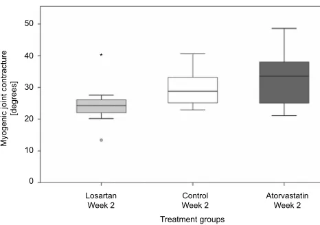
skin was removed and a periarticular myotomy 10 mm proximal and distal to the joint line was performed. This muscular release led to an improved extension of the knee joint, which corresponded to the extent of joint contracture caused by periarticular muscles. There was no observable
significant difference in myogenic contracture between the
treatment groups and the control (losartan: 24.8° ±7.6°
[p=0.43 to control], control: 29.7° ±5.9°, atorvastatin:
32.9° ±9.4° [p=0.70 to control]). Furthermore, the effect
of losartan on myogenic contracture was not different from that of atorvastatin (p=0.12, Figure 4).
Arthrogenic contracture
A persisting extension deficit after myotomy is caused by joint capsule contracture, ligament shortening and synovial adhe-sions, summarized as arthrogenic contracture. About half of the total joint contracture can be attributed to arthrogenic contracture. With 31.2° ±14.0° in the control, the arthrogenic contracture was not significantly reduced by administration of both losartan (37.2° ±9.5°,p=0.38 to control) and atorvastatin
(31.0° ±14.7°, p=0.99 to control). As with the results for
myogenic contracture, arthrogenic contracture did not vary between both treatment groups (p=0.36,Figure 5).
Histological assessment
Joint capsule length
In absence of structural damage to joint surfaces, arthrogenic contracture is mostly ascribed to a shortened joint capsule and synovial adhesions. Joint tissue sections were harvested after 2 weeks and dyed with H&E. Digitized and calibrated pictures were used to determine the capsule parameters.
We observed a significantly longer inferior capsule in
the losartan group (2.8 mm ±1.3 mm) in comparison to the control (1.1±0.3 mm,p<0.05) and to atorvastatin (0.9±0.3 mm, p<0.05). Atorvastatin did not increase the length of the inferior capsule in comparison to the control (p=0.74).
For the superior capsule, we only found a non significant
trend toward a longer capsule under medical treatment with both losartan and atorvastatin (losartan: 2.0 mm ±0.6 mm, control: 1.7 mm ±1.4 mm, atorvastatin: 1.9 mm ±1.0 mm,p>0.85,Figure 6).
Joint capsule thickness
When we compared the two treatment groups, we
observed a significant reduction in capsule thickness
120
100
80
60
40
Extension deficit
[degrees]
20
0
Losartan Week 0
Control Week 0
Control Week 2 Losartan
Week 2 Atorvastatin
Week 0
Treatment groups
Atorvastatin Week 2
Figure 3Comparison of the extension deficit of knee joints before treatment and
after 2 weeks. Joint contracture (the difference between extension deficit after 2
weeks and week 0) developed in all groups without significant differences between
the groups.
Control Week 2 Losartan
Week 2
Atorvastatin Week 2
Treatment groups 10
20
Myogenic joint contracture
[degrees]
30
40 *
50
0
Figure 4 Myogenic joint contracture did not differ significantly between the treatment groups and the control. Outliers are represented by circles and extreme outliers by an asterisk.
10 20 30
Arthrogenic joint contracture
[degrees]
40 60
50
0
Control Week 2 Losartan
Week 2
Atorvastatin Week 2
Treatment groups
Figure 5Arthrogenic contracture after administration of losartan, atorvastatin or placebo (control). Outliers are represented by circles.
Drug Design, Development and Therapy downloaded from https://www.dovepress.com/ by 118.70.13.36 on 21-Aug-2020
under atorvastatin treatment (0.7 mm ±0.2 mm, p<0.05 vs losartan with 1.1 mm ±0.4 mm). The posterior joint capsule behind the meniscus tended to be thicker when the rats were treated with losartan in comparison to
control (0.8 mm ±0.3 mm, p=0.20), while atorvastatin
tended to reduce the capsules thickness (p=0.50 to
con-trol). However, the differences between treatment
groups and the control were not significant at week 2
(Figure 7).
Immunohistochemistry
Immunohistochemistry of α-SMA
Myofibroblasts, pericytic cells and smooth muscle cells of
blood vessels in the joint capsule expressα-SMA.7,53After staining with the respective antibodies, we examined the
histological sections. α-SMA-positive cells were found in
abundance at the transition from the posterior capsule to the meniscus (Figure 8).
α-SMA-positive cells covered 6.4% ±4% of the
cap-sule area in the control. Both losartan and atorvastatin
reduced the area covered by α-SMA-stained cells (2.8%
±1.8% [p<0.05] and 2.5% ±1.7% [p<0.01], respectively). This constellation is also resembled by the absolute and
relative number of myofibroblasts. We counted 319±113
myofibroblasts, accounting for 79% of all 406±157 cells
per countingfield in the control. In rats receiving losartan,
the myofibroblast numbers decreased to 191±77 (61% of
all cells). Atorvastatin lowered the myofibroblast numbers
even more to 98±58 (24% of all cells,Table 1).
Immunhistochemistry of BSP
BSP is usually situated in the ECMof bone and plays a role during remodeling of bone and during the formation of ectopic ossifications.54,55BSP was detectable in small amounts in all groups. It was mostly found close to the
synovial membrane and in the superficial cartilage areas,
as can be seen in the overview image (Figure 9). The
assessment of staining intensity showed no major differ-ences between the treatment groups and the control (losartan: 1.3±0.4, control: 1.2±0.8 and atorvastatin: 1.0±0.4 [measured in folds of intensity in comparison to blank control]). Atorvastatin treatment reduced the BSP-stained area by trend (9.7% ±9.9%) in comparison to the control (19.8% ±15.6%) and to losartan (17.8% ±10.0%).
Discussion
Joint stiffness is a disabling condition and occurs mostly after trauma or operation.4,56Excessive active exercises or
stretching might even aggravate the fibrosis and
subse-quent motion restriction.57–59Although the surgical treat-ment of joint stiffness can improve joint motion, there is an urgent need for a non invasive prophylactic orfirst-line treatment.
In recent years, several new animal models of immobi-lity-induced contractures and PTJS were established (Table 2). Many research groups are interested in under-standing the underlying processes of contracture develop-ment and how drug application might slow it down or even stop it. After injury of the joint, several different cell types (eg, mast cells, macrophages, neutrophils, platelets,
leuko-cytes andfibroblasts) migrate into the damaged tissue and
induce the production of cytokines and growth factors.60,61
This leads to excessive collagen production and finally to
fibrosis of the joint capsule.62,63 Immobilization, local
hypoxia and intraarticular hematoma promote the
Treatment groups
1 2
Joint capsule length
[mm]
3 4
Losartan superior capsule
Week 2 Control superior capsule
Week 2
Control inferior capsule
Week 2 Losartan inferior capsule
Week 2 Atorvastatin superior capsule
Week 2
Atorvastatin inferior capsule
Week 2
Figure 6Depiction of the length of the superior and inferior parts of the posterior
knee joint capsule after 2 weeks (*p<0.05).
Control
Losartan Atorvastatin Treatment groups
0 1 1
Posterior joint capsule thickness
[mm]
2
2 *
Figure 7Comparison of the thickness of the posterior knee joint capsule after 2
weeks (*p<0.05).
Drug Design, Development and Therapy downloaded from https://www.dovepress.com/ by 118.70.13.36 on 21-Aug-2020
development of contracture.52,64,65 Myofibroblasts play a
decisive role in contracture development and fibrotic
capsule remodeling by the expression of collagen and the contractile proteinα-SMA.22,66
A
FM
500 µm 50 µm
T
B
Figure 8(A) Overview of the histological sagittal section of the posterior knee joint with femur (F), tibia (T), meniscus (M),αSMA-positive vessels (green arrows), superior
and inferior capsular regions (black arrows) and synovial lining (white arrows). (B) Detailed view of the central region behind the meniscus, in which the 361.4×268.5 µm
countingfield was placed.αSMA-positive cells appear brown and either belong to vessels (green arrows) or are defined as myofibroblasts (red arrows).
Table 1Total amount and ratio of myofibroblasts
Number of myofibroblasts Total cell number Ratio of myofibroblasts/ total cell number
p-value
Losartan 191±77 312±133 61% p<0.01 vs control
p<0.05 vs atorvastatin
Control 319±113 406±157 79% p<0.01 vs losartan
p<0.001 vs atorvastatin
Atorvastatin 98±58 410±108 24% p<0.001 vs control
p<0.05 vs losartan
A
F
M
500 µm 50 µm
T
B
Figure 9(A) BSP-positive staining is situated mostly in and close to the synovial membrane (white arrows) and in the superficial cartilage of the tibia (T) and the meniscus
(M). Superior and inferior capsular regions are pointed out by black arrows. In the enlarged view of the capsule behind the meniscus, (B) BSP-stained areas can be found
close to the meniscal border and around vessels (green arrows). Abbreviation:F, femur.
Drug Design, Development and Therapy downloaded from https://www.dovepress.com/ by 118.70.13.36 on 21-Aug-2020
T able 2 Compr ehensiv e list of in viv o models of joint stiffness with medical tr eatment in chr onological or der Author/y ear Animal, joint Induction of trauma Drug Application Reduction of contractur e Decr eased m y o fi br o-blast numbers Clark et al, 112 1971 Rat, knee No T riamcinolone Intraarti cular injection Y es Not measur ed Namba et al, 95 1992 Rabbit, ankle Y es T riamcinolone Singular intraarticular injection Y es Not measur ed Kikuchi et al, 104 1993 Rabbit, knee No High-molecular -w eight sodium h ya-lur onate (SL-1010) Intraarti cular injection Y es Not measur ed Fukui et al, 16 2000 Rabbit, knee Y es TGF-β 1 neutralizing antibody Continuous intraarticular injec-tion via implanted pumps Y es Not measur ed Fukui et al, 96 2001 Rabbit, knee Y es Pr oteoglycan decorin Continuous intraarticular injec-tion via implanted pumps Y es Not measur ed Brunelli et al, 97 2005 Rabbit, knee Y es Cr oss link ed h yalur onan (Hyaloglide) Singular intraarticular injection Not measur ed, but reduction of capsular adhesion obser ved. Not measur ed Monument et al, 18 2010 Rabbit, knee Y es K etotifen fumarate Subcutaneous injection twice daily Y es Y es K ocoaglu et al, 98 2011 Rat, knee Y es Mitom ycin C W eekly intraar ticular injection Not measur ed, but reduction of arthr o fi br osis obser ved. Not measur ed Emami et al, 99 2012 Rabbit, knee Y es Be vacizumab One or tw o intraarticu lar injections Y es, but only after tw o injections Not measur ed (but fi br o-blast numbers decr eased) Li et al, 108 2013 Rabbit, knee Y es Celeco xib Orally , twice daily Y es Not measur ed (but fi br o-blast numbers decr eased) Li et al, 2013 19 Rat, knee Y es Lentivirus (L V)-mediated ERK2 siRNA T w o intraarticu lar injections Y es Y es E fi rd et al, 109 2014 Rat, knee Y es a) Montelukast, b) Forsk olin or c) T riamcinolone a) orally , daily b and c) tw o intraarticular injections Y es Not measur ed Kanaza wa et al, 100 2015 Rat, knee No Hyalur onic acid with high molecular w eight Singular intraarticular injection Not measur ed, but mor e elasti-city obser ved Not measur ed Sotoba yashi et al, 64 2016 Rat, knee No Ribbon-type deco y oligodeoxynu-cleotides for HIF-1 Singular intraarticular injection Y es Not measur ed Oza wa et al, 110 2016 Rat, knee No Celeco xib Orally , daily No Not measur ed Barlow et al, 111 2016 Rabbit, knee Y es Rosiglitazone Subcutaneous injection on the fi rst 2 da ys, afterwar d orally daily No No Steple wski et al, 17 2017 Rabbit, knee Y es Non modi fi ed anti-α 2Ct antibody (A C A) and PEGylated A C A (P-A C A) Continuous intraarticular injec-tion via implanted pumps Y es Not measur ed Gao et al, 101 2017 Rat, knee Y es Botulinum to xin A Singular intraarticular injection Y es Not measur ed (but fi br o-blast numbers decr eased) Kaneguchi et al, 113 2018 Rat, knee No Dexamethasone Daily by subcutaneous injection Partially Y es Ita ya et al, 102 2018 Rat, knee No Pulsed ultrasound 20 mins daily Y es Not measur ed W ong et al, 103 2018 Rat, knee No Collagenase Singular intraarticular injection Y es Not measur ed
Drug Design, Development and Therapy downloaded from https://www.dovepress.com/ by 118.70.13.36 on 21-Aug-2020
Most of the existing studies focus on anti-inflammatory drugs in order to protect the periarticular connective tis-sues from posttraumatic or immobility-induced regional
inflammation (Table 2). In this blinded and randomized
study, we sought to find out, if and how orally
adminis-tered losartan and atorvastatin affect the early phase (first 2 weeks) of the development of joint contracture. Both drugs
have shown to successfully inhibit the TGF-β1-triggered
inflammatory cascade and subsequentfibrosis in different
organ systems, but their effect on the development of joint contracture was unclear.24–27,30–32 TGF-β1 is involved in the formation of fibrosis in many tissues.67,68 It leads to
the differentiation of myofibroblasts and increases their
synthesis of proteins of the ECM.69In addition, it induces
the production of CTGF, which also has a strong fibrotic
effect.70By inhibiting matrix metalloproteinases (MMPs),
TGF-β1 inhibits the degradation of the newly synthesized
ECM.71Losartan is an AT1 receptor blocker and prevents
direct activation of TGF-β as well as the angiotensin
II-induced phosphorylation of Smad2 and Smad3 via the ERK/p38/MAPK pathway. Normally, the phosphorylated Smad2 and Smad3 complex with Smad4 and translocate into the nucleus of the cell. This leads to a transcription of TGF-β, procollagen I and III and fibronectin.67,72,73 Atorvastatin is a 3-hydroxy-3-methylglutaryl coenzyme A (HMG-CoA) reductase inhibitor and is able to inhibit
inflammatory responses and collagen fiber production,
possibly by inhibiting the expression of TGF- β1.74
Atorvastatin also inhibits the RhoA/ROCK (Rho-asso-ciated coiled-coil-containing protein kinase) pathway, which plays an important role in the signal transduction offibrotic and sclerotic diseases.75–77
In the current study, we laid a focus on assessing joint
angles, synovial configuration and cell numbers by
visua-lization of myofibroblasts and BSP-positive cells. In a
study by Remst et al, a fibrosis of the synovium was
found in over 50% of patients with osteoarthritis (OA), and other studies have found an association between OA andfibrosis as well.78–80There is evidence that high
num-bers of fibroblasts in knee synovium can prompt infl
am-mation due to overexpression of TGF-β.81,82 BSP is
usually located in the ECM of bone and involved in the
primary bone formation during embryonic development.83
It could be shown that BSP stimulated osteoblast differ-entiation, bone growth and vascularization in murine cal-varial defect models.84,85Increased levels of BSP are not only reported in the synovialfluid of inflamed knee joints after injury, in rheumatoid arthritis or in early OA, but also
in HOs.12,86,87 Since HO often occurs in PTJS, it appears
that BSP is increasingly expressed during inflammation
after trauma and promotes the formation of HO. Yet, the presence and distribution pattern of BSP in the posttrau-matic joint is still elusive. In the current study, we created a substantial PTJS after 2 weeks in all groups. BSP was not significantly expressed in the posterior capsule; neither the intensity of antibody staining nor the area coverage of positively stained cells was significantly different between the groups. In our experiments, we could not observe any development of HO 2 weeks after the trauma. It could be that BSP accumulates in the capsule and leads to HO only in a later stage of PTJS and that the time window of 2 weeks in the current study is too narrow to notice a corresponding effect.
Since myofibroblast numbers are increased during the
development of a joint contracture and reach a peak level about 2 weeks after trauma in human tissues and animal models, we have chose a period of 2 weeks for our experiment.6,88In our study, the number of myofibroblasts in the posterior joint capsule fell by 40% in rats receiving treatment with losartan and by nearly 70% after atorvasta-tin administration in comparison to the control. Likewise,
the area percentage of myofibroblasts was significantly
reduced by about 55% (losartan) and 60% (atorvastatin),
respectively. These findings are in accordance with the
observations of Abdel et al, who found the number of
myofibroblasts at its peak in the contracted limb 2 weeks
after the initial operation in their rabbit model of PTJS.6In
immobilization-induced contractures without induced
trauma, the limitation to knee range of motion after 2
weeks of immobilization is mostly muscular.47,89 This
contrasts with observations in our posttraumatic contrac-ture, where the posterior capsule accounted for about half of the total joint contracture (Figures 3and4). Morrey et al hypothesize that it may be the combination of soft tissue trauma, instability and bony trauma that leads to a more
clinically relevant contracture in humans.90 Nesterenko et
al found a more severe and permanent contracture in their rabbit model of PTJS after ligamentous and capsular dis-ruption. The combination of capsular contracture, adhe-sions between bony surfaces and the capsule and intraarticular adhesions could be the reason for the
pre-dominance of the arthrogenic component of joint
contracture.91 However, there are controversial findings
from Lake et al, who observed similar histological char-acteristics for all three groups (ie, sham, minor joint injury and major joint injury) in their rat model of PTJS of the
Drug Design, Development and Therapy downloaded from https://www.dovepress.com/ by 118.70.13.36 on 21-Aug-2020
elbow.92 There are not many in vivo studies on medical treatment of joint stiffness that included the cell count of myofibroblasts (Table 2, right column). In the studies that
include myofibroblast or fibroblast count, a concordance
between decreasing cell numbers and decreasing contrac-ture could be observed. All these studies consider a longer period than we do in our study, usually between 4 and 8
weeks, in some cases longer. It appears that myofibroblast
numbers might not be responsible for the early onset of
PTJS, since the drug-induced changes in myofibroblast
numbers (Table1) and partially in capsule length
(Figure 6) did not translate directly into a significant improvement in range of motion. Accordingly, neither synovial length increased nor synovial thickness decreased after 2 weeks of daily drug application. Scarring at the posterior aspect of the joint after the initial trauma with indirect capsular injury might be responsible for that early arthrogenic joint contracture. It is possible that inhibition
of myofibroblasts influences contracture development at a
later stage, when the physiological wound healing
response is fading and elevated numbers of myofibroblasts
result in exuberantfibrotic remodeling.93,94
Several working groups have performed experiments on inhibition of joint contracture in rabbits and rats (Table 2). The reservation must be made, however, that the results are hardly comparable due to differences in the research methodology. Around half of the above-men-tioned studies concentrate on immobilization-induced con-tractures and did not induce a trauma to create a PTJS. Although some experimental results suggest that immobi-lization alone might be adequate to create a stable joint contracture, these models do not fully reproduce the
mechanism of PTJS in humans.46,52,89 Since there is no
comprehensive standard model for the creation of joint stiffness, the various studies differ in several aspects, such as the duration of immobilization, the method of joint contracture measurements, the applied extension tor-que during the measurements, the distinction between myogenic and arthrogenic factors, the administration of medication and the animal model, to name but a few.
In the existing studies on medical inhibition of the
development of joint contracture (Table 2), an
improve-ment of the range of motion could be achieved mostly after intraarticular application of the respective active agents.16,17,64,95–104 Although these results constitute an opportunity for the development of a medical treatment of patients with PTJS, the intraarticular application is unfavorable for a number of reasons: i) it is a painful
and invasive procedure for the patient, ii) it bears the risk of septic arthritis, iii) it might damage the articular cartilage by chondrotoxic effects or iv) weaken the capsule
and ligaments of the knee.105–107 Based on the rabbit
models of PTJS of Hildebrand et al and Nesterenko et al, our group established a rat model of PTJS of the knee joint that included a rupture of the posterior joint capsule, a hemarthrosis, an intraarticular bone damage and a tempor-aryfixation of the joint.38,65,91In order to avoid the poten-tial risks of intraarticular injections, which might lessen their applicability and acceptance in the treatment of patients, we administered the drugs orally.
There are only two published animal experiments that
focus on oral drug administration in PTJS.108,109 In the
study of Efird et al, montelukast significantly improved
joint motion of rats´ knee joints 2 weeks after scraping the soft tissues on the medial and lateral condyles and the cartilage of the trochlea. This massive structural damage to the joint might have led to extensive scarring and
inflammation, which might explain the effect on joint
contracture of the leukotriene receptor antagonist monte-lukast. In comparison, we sought to prevent structural damage to the cartilage joint surfaces by setting an extra-cartilaginous bony lesion to be able to examine the cap-sular contracture only and rule out impairment on joint motion based on an incongruence of the joint partners.
Myofibroblast numbers and synovial length were not
mea-sured in the study of Efird et al. In the study of Li et al,
celecoxib improved joint motion significantly, but myofi
-broblast numbers and synovial length were not measured. In the following two studies by Ozawa et al and Barlow et al. the drugs were also administered orally. However, they
differ from the two studies mentioned above by Efird et al
and Li et al, since Ozawa et al used a non traumatic rat model, whereas Barlow et al initially performed an intra articular injection before switching to oral administration. Ozawa et al measured the synovial length in their non traumatic rat model of knee joint contracture under oral celecoxib treatment after 3 weeks and did neither observe an inhibition of the shortening of the joint capsule nor a reduction of the joint contracture.110Barlow et al
adminis-tered rosiglitazone subcutaneously during thefirst 2 days
and then switched to oral drug administration in their rabbit model of PTJS, but did not observe an effect on joint contracture development.111
There are some limitations to our study that need to be mentioned. First, the manual application of the extension torque using a spring scale might add a human error factor.
Drug Design, Development and Therapy downloaded from https://www.dovepress.com/ by 118.70.13.36 on 21-Aug-2020
Second, the medication was not administered in different concentrations and serum and joint concentrations of the drugs were not measured. Third, the initial trauma is not really standardized, because hyperextension of the knee joint and drilling of the femoral defect are individual proce-dures, which might as well lead to bias. Fourth, the immobi-lization model (ie, using an internal K-wire for jointfixation)
does not exactly reflect the human situation and a cast or
external fixation might be more suitable. Fifth, the small
number of animals in each group might add to the observed variability in the data and mask significant differences. Sixth,
immunohistochemistry was limited to myofibroblasts only.
Finally, repetitive measurement of the joints is needed to distinguish between muscular and capsular components of the contracture, but might weaken the involved joint struc-tures and lead to larger angles.
Conclusion
In our placebo-controlled, randomized and blinded experi-mental study with 24 rats, we examined the effects of the anti fibrotic drugs losartan and atorvastatin on the development of early PTJS. Both losartan and atorvastatin reduced the area
covered by α-SMA-stained cells (myofibroblasts) and the
number of myofibroblasts in the joint capsule 2 weeks after trauma. Losartan increased the length of the inferior joint capsule, but not of the superior capsule and had no effect on capsule thickness. Atorvastatin had no significant influence on capsule length and thickness in comparison to the control.
Despite some effects on myofibroblast count and joint
cap-sule, both drugs did not decrease myogenic and arthrogenic joint contracture. BSP was not significantly expressed in the posterior capsule; neither the intensity of antibody staining nor the area coverage of positively stained cells was signifi -cantly affected by drug administration. In our future studies, we plan to analyze if this early reduction of myofibroblast numbers affects the long-term contracture development after
4 and more weeks and how atorvastatin and losartan infl
u-ence gene expression patterns in the posterior knee joint capsule.
Ethics approval
This study was approved by the local ethics committee
“Landesuntersuchungsamt Rheinland-Pfalz”(ID 23 177-07/
G 13-1-043). We have followed the German“Legal
ordi-nance on the Protection of Animals Used for Experimental
Purposes or for Other Scientific Purposes (Experimental
Animal Welfare Regulation - TierSchVersV)”(https://www.
gesetze-im-internet.de/tierschversv/BJNR312600013.html).
Data availability
The datasets used and/or analyzed during the current study are available from the corresponding author on reasonable request.
Abbreviations list
α-SMA, alpha smooth muscle actin; ACA, anti-α2Ct
anti-body; AT-1, angiotensin type 1; BSP, bone sialoprotein;
CO2, carbon dioxide; CTGF, connective tissue growth
factor; DAB, 3,3′-diaminobenzidine; ECM, extracellular
matrix; HIF-1, hypoxia-inducible factor 1; HMG-CoA, 3-hydroxy-3-methyl-glutaryl-coenzyme A; HO, heterotopic
ossification; HRP, horseradish peroxidase; K-wire,
Kirschner wire; MMP, matrix metalloproteinases; mRNA,
messenger ribonucleic acid; P-ACA, PEGylated anti-α2Ct
antibody; PTJS, posttraumatic joint stiffness; ROCK, Rho-associated coiled-coil-containing protein kinase; TRIS, tris (hydroxymethyl)aminomethane.
Acknowledgments
The authors extend special thanks to Angelika Ackermann for technical support and Anke Grünow for graphic illus-trations. We thank Dr Irene Schmidtmann from the Institute for Medical Biostatistics, Epidemiology and
Informatics (IMBEI) for statistical consulting. Our
research was funded in 2015 from the Johannes
Gutenberg-University Mainz within the
“Inneruniversitäre Forschungsförderung Stufe I”program
and in 2016 from the German Society of Shoulder and Elbow Surgery (DVSE). The work was performed at the
“Molekulares Forschungszentrum operativer Fächer
(MFO)”of the University Medical Center Mainz.
Author contributions
All authors made substantial contributions to conception and design, acquisition of data, or analysis and interpreta-tion of data; took part in drafting the article or revising it
critically for important intellectual content; gave final
approval of the version to be published; and agree to be accountable for all aspects of the work.
Disclosure
Dr Andreas Baranowski reports grants from the Johannes Gutenberg-University Mainz and the German Society of Shoulder and Elbow Surgery (DVSE), during the conduct of the study. Dr Anja Klein reports grants from the German Society of Shoulder and Elbow Surgery (DVSE)
Drug Design, Development and Therapy downloaded from https://www.dovepress.com/ by 118.70.13.36 on 21-Aug-2020
and Inneruniversitäre Forschungsförderung Stufe I, during the conduct of the study. The authors report no other conflicts of interest in this work.
References
1. Hinz B. The myofibroblast: paradigm for a mechanically active cell.J Biomech.2010;43(1):146–155. doi:10.1016/j.jbiomech.2009.09.020 2. Desmouliere A, Chaponnier C, Gabbiani G. Tissue repair,
contrac-tion, and the myofibroblast.Wound Repair Regen.2005;13(1):7– 12. doi:10.1111/j.1067-1927.2005.130102.x
3. Hildebrand KA, Zhang M, van Snellenberg W, King GJ, Hart DA, Myofibroblast numbers are elevated in human elbow capsules after trauma.Clin Orthop Relat Res.2004;(419):189–197. doi:10.1097/ 00003086-200402000-00031
4. Mellema JJ, Lindenhovius AL, Jupiter JB. The posttraumatic stiff elbow: an update.Curr Rev Musculoskelet Med. 2016;9(2):190– 198. doi:10.1007/s12178-016-9336-9
5. Usher KM, Zhu S, Mavropalias G, Carrino JA, Zhao J, Xu J. Pathological mechanisms and therapeutic outlooks for arthrofi bro-sis.Bone Res.2019;7:9.
6. Abdel MP, Morrey ME, Barlow JD, et al. Myofibroblast cells are preferentially expressed early in a rabbit model of joint contracture.
J Orthop Res.2012;30(5):713–719. doi:10.1002/jor.21588 7. Darby I, Skalli O, Gabbiani G. Alpha-smooth muscle actin is
transiently expressed by myofibroblasts during experimental wound healing.Lab Invest.1990;63(1):21–29.
8. Hinz B, Dugina V, Ballestrem C, Wehrle-Haller B, Chaponnier C. Alpha-smooth muscle actin is crucial for focal adhesion maturation in myofibroblasts. Mol Biol Cell. 2003;14(6):2508–2519. doi:10.1091/mbc.e02-11-0729
9. Kis K, Liu X, Hagood JS. Myofibroblast differentiation and survi-val in fibrotic disease. Expert Rev Mol Med. 2011;13:e27. doi:10.1017/S1462399411001967
10. Charalambous CP, Morrey BF. Posttraumatic elbow stiffness.J Bone Joint Surg Am.2012;94(15):1428–1437. doi:10.2106/JBJS.K.00711 11. Freeman TA, Parvizi J, Dela Valle CJ, Steinbeck MJ. Mast cells
and hypoxia drive tissue metaplasia and heterotopic ossification in idiopathic arthrofibrosis after total knee arthroplasty.Fibrogenesis Tissue Repair.2010;3:17. doi:10.1186/1755-1536-3-17
12. Bosse A, Wuisman P, Jones DB, Schwarz K. Noncollagenous proteins in heterotopic ossification. Immunohistochemical analysis in 15 paraplegies.Acta Orthop Scand.1993;64(6):634–638. 13. Hegyi L, Gannon FH, Glaser DL, Shore EM, Kaplan FS, Shanahan
CM. Stromal cells offibrodysplasia ossificans progressiva lesions express smooth muscle lineage markers and the osteogenic transcrip-tion factor Runx2/Cbfa-1: clues to a vascular origin of heterotopic ossification?J Pathol.2003;201(1):141–148. doi:10.1002/path.1413 14. Lohmander LS, Saxne T, Heinegard D. Increased concentrations of
bone sialoprotein in jointfluid after knee injury.Ann Rheum Dis.
1996;55(9):622–626. doi:10.1136/ard.55.9.622
15. Larsson E, Mussener A, Heinegard D, Klareskog L, Saxne T. Increased serum levels of cartilage oligomeric matrix protein and bone sialoprotein in rats with collagen arthritis.Br J Rheumatol.
1997;36(12):1258–1261.
16. Fukui N, Tashiro T, Hiraoka H, Oda H, Nakamura K. Adhesion formation can be reduced by the suppression of transforming growth factor-beta1 activity. J Orthop Res. 2000;18(2):212–219. doi:10.1002/jor.1100180208
17. Steplewski A, Fertala J, Beredjiklian PK, et al. Blocking collagen
fibril formation in injured knees reducesflexion contracture in a rabbit model.J Orthop Res.2017;35(5):1038–1046. doi:10.1002/ jor.23369
18. Monument MJ, Hart DA, Befus AD, Salo PT, Zhang M, Hildebrand KA. The mast cell stabilizer ketotifen fumarate lessens contracture severity and myofibroblast hyperplasia: a study of a rabbit model of posttraumatic joint contractures.J Bone Joint Surg Am.2010;92(6):1468–1477. doi:10.2106/JBJS.I.00684
19. Li F, Liu S, Fan C. Lentivirus-mediated ERK2 siRNA reduces joint capsulefibrosis in a rat model of post-traumatic joint contracture.Int J Mol Sci.2013;14(10):20833–20844. doi:10.3390/ijms141020833 20. Mattyasovszky SG, Hofmann A, Brochhausen C, et al. The effect
of the pro-inflammatory cytokine tumor necrosis factor-alpha on human joint capsule myofibroblasts.Arthritis Res Ther.2010;12(1): R4. doi:10.1186/ar2902
21. Kulkarni AA, Thatcher TH, Olsen KC, Maggirwar SB, Phipps RP, Sime PJ. PPAR-gamma ligands repress TGFbeta-induced myofi -broblast differentiation by targeting the PI3K/Akt pathway: impli-cations for therapy of fibrosis. PLoS One. 2011;6(1):e15909. doi:10.1371/journal.pone.0015909
22. Hinz B, Phan SH, Thannickal VJ, et al. Recent developments in myofibroblast biology: paradigms for connective tissue remodeling.
Am J Pathol. 2012;180(4):1340–1355. doi:10.1016/j.ajpath.20 12.02.004
23. Leask A. Potential therapeutic targets for cardiacfibrosis: tGFbeta, angiotensin, endothelin, CCN2, and PDGF, partners infibroblast activation. Circ Res. 2010;106(11):1675–1680. doi:10.1161/ CIRCRESAHA.110.217737
24. Klein S, Klösel J, Schierwagen R, et al. Atorvastatin inhibits proliferation and apoptosis, but induces senescence in hepatic myofibroblasts and thereby attenuates hepatic fibrosis in rats.
Lab Invest. 2012;92(10):1440–1450. doi:10.1038/labinvest.20 12.106
25. Trebicka J, Hennenberg M, Odenthal M, et al. Atorvastatin attenu-ates hepaticfibrosis in rats after bile duct ligation via decreased turnover of hepatic stellate cells.J Hepatol.2010;53(4):702–712. doi:10.1016/j.jhep.2010.04.025
26. Geng J, Zhao Z, Kang W, Wang W, Zhang Y, Zhiming GE. Atorvastatin reverses cardiac remodeling possibly through regula-tion of protein kinase D/myocyte enhancer factor 2D activaregula-tion in spontaneously hypertensive rats.Pharmacol Res. 2010;61(1):40– 47. doi:10.1016/j.phrs.2009.08.004
27. Jiang BH, Tardif JC, Sauvageau S, et al. Beneficial effects of atorvastatin on lung structural remodeling and function in ischemic heart failure. J Card Fail. 2010;16(8):679–688. doi:10.1016/j. cardfail.2010.03.003
28. Kane CJ, Hebda PA, Mansbridge JN, Hanawalt PC. Direct evidence for spatial and temporal regulation of transforming growth factor beta 1 expression during cutaneous wound healing.
J Cell Physiol. 1991;148(1):157–173. doi:10.1002/jcp.104 1480119
29. Campbell SE, Katwa LC. Angiotensin II stimulated expression of transforming growth factor-beta1 in cardiacfibroblasts and myofi -broblasts.J Mol Cell Cardiol.1997;29(7):1947–1958. doi:10.1006/ jmcc.1997.0435
30. Shibasaki Y, Nishiue T, Masaki H, et al. Impact of the angiotensin II receptor antagonist, losartan, on myocardialfibrosis in patients with end-stage renal disease: assessment by ultrasonic integrated backscatter and biochemical markers. Hypertens Res. 2005;28 (10):787–795. doi:10.1291/hypres.28.787
31. De Mello WC, Specht P. Chronic blockade of angiotensin II AT1-receptors increased cell-to-cell communication, reduced fibrosis and improved impulse propagation in the failing heart. J Renin Angiotensin Aldosterone Syst. 2006;7(4):201–205. doi:10.3317/ jraas.2006.038
32. Yoshiji H, Kuriyama S, Yoshii J, et al. Angiotensin-II type 1 receptor interaction is a major regulator for liverfibrosis develop-ment in rats.Hepatology.2001;34(4 Pt 1):745–750. doi:10.1053/ jhep.2001.28231
Drug Design, Development and Therapy downloaded from https://www.dovepress.com/ by 118.70.13.36 on 21-Aug-2020
33. Ishidoya S, Morrissey J, McCracken R, Reyes A, Klahr S. Angiotensin II receptor antagonist ameliorates renal tubulointersti-tial fibrosis caused by unilateral ureteral obstruction. Kidney Int.
1995;47(5):1285–1294.
34. Molteni A, Moulder JE, Cohen EF, et al. Control of radiation-induced pneumopathy and lungfibrosis by angiotensin-converting enzyme inhibitors and an angiotensin II type 1 receptor blocker.Int J Radiat Biol.2000;76(4):523–532.
35. Bedair HS, Karthikeyan T, Quintero A, Li Y, Huard J. Angiotensin II receptor blockade administered after injury improves muscle regen-eration and decreasesfibrosis in normal skeletal muscle.Am J Sports Med.2008;36(8):1548–1554. doi:10.1177/0363546508315470 36. Cabello-Verrugio C, Morales MG, Cabrera D, Vio CP, Brandan E.
Angiotensin II receptor type 1 blockade decreases CTGF/CCN2-mediated damage andfibrosis in normal and dystrophic skeletal muscles.J Cell Mol Med.2012;16(4):752–764. doi:10.1111/j.1582-4934.2011.01354.x
37. Burks TN, Andres-Mateos E, Marx R, et al. Losartan restores skeletal muscle remodeling and protects against disuse atrophy in sarcopenia.
Sci Transl Med.2011;3(82):82ra37. doi:10.1126/scitranslmed.3002227 38. Baranowski A, Schlemmer L, Forster K, et al. A novel rat model of stable posttraumatic joint stiffness of the knee.J Orthop Surg Res.
2018;13(1):185. doi:10.1186/s13018-018-1026-4
39. Trebicka J, Hennenberg M, Laleman W, et al. Atorvastatin lowers portal pressure in cirrhotic rats by inhibition of RhoA/Rho-kinase and activation of endothelial nitric oxide synthase. Hepatology.
2007;46(1):242–253. doi:10.1002/hep.21673
40. Black AE, Hayes RN, Roth BD, Woo P, Woolf TF. Metabolism and excretion of atorvastatin in rats and dogs. Drug Metab Dispos.
1999;27(8):916–923.
41. Wassmann S, Laufs U, Baumer AT, et al. HMG-CoA reductase inhibitors improve endothelial dysfunction in normocholesterole-mic hypertension via reduced production of reactive oxygen spe-cies.Hypertension.2001;37(6):1450–1457.
42. Heller J, Shiozawa T, Trebicka J, et al. Acute haemodynamic effects of losartan in anaesthetized cirrhotic rats. Eur J Clin Invest.2003;33(11):1006–1012.
43. Kaneko K, Susic D, Nunez E, Frohlich ED. Losartan reduces cardiac mass and improves coronary flow reserve in the sponta-neously hypertensive rat.J Hypertens.1996;14(5):645–653. 44. Gohlke P, Linz W, Scholkens BA, Wiemer G, Unger T. Cardiac and
vascular effects of long-term losartan treatment in stroke-prone spon-taneously hypertensive rats.Hypertension.1996;28(3):397–402. 45. Ando A, Hagiwara Y, Onoda Y, et al. Distribution of type A and B
synoviocytes in the adhesive and shortened synovial membrane during immobilization of the knee joint in rats. Tohoku J Exp Med.2010;221(2):161–168.
46. Ando A, Suda H, Hagiwara Y, Onoda Y, Chimoto E, Itoi E. Remobilization does not restore immobilization-induced adhesion of capsule and restricted joint motion in rat knee joints.Tohoku J Exp Med.2012;227(1):13–22.
47. Trudel G, Uhthoff HK. Contractures secondary to immobility: is the restriction articular or muscular? An experimental longitudinal study in the rat knee.Arch Phys Med Rehabil.2000;81(1):6–13. 48. Hagiwara Y, Ando A, Onoda Y, et al. Expression patterns of
collagen types I and III in the capsule of a rat knee contracture model.J Orthop Res.2010;28(3):315–321. doi:10.1002/jor.20997 49. Hagiwara Y, Saijo Y, Chimoto E, et al. Increased elasticity of
capsule after immobilization in a rat knee experimental model assessed by scanning acoustic microscopy. Ups J Med Sci.
2006;111(3):303–313. doi:10.3109/2000-1967-054
50. Moriyama H, Yoshimura O, Kawamata S, Takemoto H, Saka Y, Tobimatsu Y. Alteration of knee joint connective tissues during contracture formation in spastic rats after an experimentally induced spinal cord injury. Connect Tissue Res.2007;48(4):180– 187. doi:10.1080/03008200701413512
51. Lee S, Sakurai T, Ohsako M, Saura R, Hatta H, Atomi Y. Tissue stiffness induced by prolonged immobilization of the rat knee joint and relevance of AGEs (pentosidine).Connect Tissue Res.2010;51 (6):467–477. doi:10.3109/03008201003686941
52. Onoda Y, Hagiwara Y, Ando A, et al. Joint haemorrhage partly accelerated immobilization-induced synovial adhesions and capsu-lar shortening in rats. Knee Surg Sports Traumatol Arthrosc.
2014;22(11):2874–2883. doi:10.1007/s00167-013-2659-9 53. Azuma K, Ichimura K, Mita T, et al. Presence of alpha-smooth
muscle actin-positive endothelial cells in the luminal surface of adult aorta.Biochem Biophys Res Commun.2009;380(3):620–626. doi:10.1016/j.bbrc.2009.01.135
54. Bouleftour W, Juignet L, Bouet G, et al. The role of the SIBLING, Bone Sialoprotein in skeletal biology - Contribution of mouse experimental genetics. Matrix Biol. 2016;52–54:60–77. doi:10.1016/j.matbio.2015.12.011
55. Kaden JJ, Bickelhaupt S, Grobholz R, et al. Expression of bone sialoprotein and bone morphogenetic protein-2 in calcific aortic stenosis.J Heart Valve Dis.2004;13(4):560–566.
56. Frontera WR, Silver JK, Rizzo TD.Essentials of Physical Medicine and Rehabilitation: Musculoskeletal Disorders, Pain, and Rehabilitation. Oxford: Elsevier Health Sciences;2018.
57. Moseley AM, Herbert RD, Nightingale EJ, et al. Passive stretching does not enhance outcomes in patients with plantarflexion contrac-ture after cast immobilization for ankle fraccontrac-ture: a randomized controlled trial. Arch Phys Med Rehabil. 2005;86(6):1118–1126. doi:10.1016/j.apmr.2004.11.017
58. Kaneguchi A, Ozawa J, Kawamata S, Yamaoka K. Development of arthrogenic joint contracture as a result of pathological changes in remobilized rat knees. J Orthop Res. 2017;35(7):1414–1423. doi:10.1002/jor.23419
59. Kaneguchi A, Ozawa J, Minamimoto K, Yamaoka K. Active exercise on immobilization-induced contractured rat knees devel-ops arthrogenic joint contracture with pathological changes. J Appl Physiol (1985). 2018;124(2):291–301. doi:10.1152/ japplphysiol.00438.2017
60. Morrey ME, Abdel MP, Riester SM, et al. Molecular landscape of arthrofibrosis: microarray and bioinformatic analysis of the tem-poral expression of 380 genes during contracture genesis. Gene.
2017;610:15–23. doi:10.1016/j.gene.2017.01.025
61. Kumar V, Abbas AK, Fausto N, Robbins SL, Cotran RS.Robbins and Cotran Pathologic Basis of Disease. 7th ed. Philadelphia: Elsevier Saunders;2005:xv, 1525 p.
62. Monument MJ, Hart DA, Salo PT, Befus AD, Hildebrand KA. Posttraumatic elbow contractures: targeting neuroinflammatory
fibrogenic mechanisms. J Orthop Sci. 2013;18(6):869–877. doi:10.1007/s00776-013-0447-5
63. Hildebrand KA, Zhang M, Germscheid NM, Wang C, Hart DA. Cellular, matrix, and growth factor components of the joint capsule are modified early in the process of posttraumatic contracture formation in a rabbit model. Acta Orthop. 2008;79(1):116–125. doi:10.1080/17453670710014860
64. Sotobayashi D, Kawahata H, Anada N, Ogihara T, Morishita R, Aoki M. Therapeutic effect of intra-articular injection of ribbon-type decoy oligonucleotides for hypoxia inducible fac-tor-1 on joint contracture in an immobilized knee animal model. J Gene Med. 2016;18(8):180–192. doi:10.1002/jgm.2 891
65. Hildebrand KA, Sutherland C, Zhang M. Rabbit knee model of post-traumatic joint contractures: the long-term natural history of motion loss and myofibroblasts. J Orthop Res.2004;22(2):313– 320. doi:10.1016/j.orthres.2003.08.012
66. Kendall RT, Feghali-Bostwick CA. Fibroblasts infibrosis: novel roles and mediators. Front Pharmacol. 2014;5:123. doi:10.3389/ fphar.2014.00123
Drug Design, Development and Therapy downloaded from https://www.dovepress.com/ by 118.70.13.36 on 21-Aug-2020