A N e w Family of Genes Coding for an Antigen
Recognized by Autologous Cytolytic T Lymphocytes
on a Human Melanoma
By Benoit Van den Eynde, Olivier Peeters, Olivier De Backer,
B6atrice Gaugler, Sophie Lucas, and Thierry Boon
From the Ludwig Institute for Cancer Research, Brussels Branch; and Cellular Genetics Unit, Universitd Catholique de Louvain, B1200 Brussels, Belgium
Summary
Human melanoma MZ2-MEL expresses several distinct antigens that are recognized by autologous cytolytic T lymphocytes (CTL). Some of these antigens are encoded by genes MAGE-1, MAGE-3,
and BAGE, which are expressed in a large fraction of tumors of various histological types but are silent in normal adult tissues with the exception of testis. We report here the identification of the gene coding for MZ2-F, another antigen recognized by autologous CTL on MZ2-MEL cells. This gene, which was named GAGE-l, is not related to any presently known gene. It belongs to a family of genes that are expressed in a variety of tumors but not in normal tissues, except for the testis. Antigenic peptide YILPRPRRY, which is encoded by GAGE-l, is recognized by anti-MZ2-F CTL on class I molecule HLA-Cw6. The two genes of the GAGE family that code for this peptide, namely GAGE-1 and GAGE-2, are expressed in a significant proportion of melanomas (24%), sarcomas (25%), non-small cell lung cancers (19%), head and neck tumors (19%), and bladder tumors (12%). About 50% of melanoma patients carry on their tumor at least one of the presently defined antigens encoded by the MAGE, BAGE, and GAGE genes.
H
uman melanoma cells bear antigens that are recognized by autologous CD8 + CTL, which can be derived ei- ther from blood lymphocytes or from tumor-infiltrating lym- phocytes (1). Two distinct strategies have been used to char- acterize these antigens. The first is a genetic approach based on transfection of genomic or cDNA libraries to identify the gene encoding the antigen (2-7). The antigenic peptide can then be identified on the basis of the protein sequence encoded by the gene (6, 8-12). The second approach involves acid elu- tion of peptides from immunoprecipitated HLA molecules followed by separation of the peptides, evaluation of their ability to sensitize target cells to CTL, and sequencingof
the active peptide (13, 14).
A first class of antigens that are recognized on melanoma by autologous CTL is encoded by genes that are expressed in various tumors but that are completely silent in normal adult tissues, except the testis. These genes include genes
MAGE-1, MAGE-3, and BAGE (2, 6, 9, 11, 15). The mouse gene P/A, which codes for a tumor rejection antigen of mouse tumor P815, follows the same pattern of expression (16). The tumor specificity of these antigens may make them targets of choice for cancer immunotherapy based on specific immu- nization.
A second class of antigens represents differentiation an- tigens encoded by genes that are expressed in melanoma and
in normal mdanocytes. Antigens derived from tyrosinase were the first examples of this class, which also comprises antigens encoded by Melan-A ~ART-1, gplOOmet17, and gp75 TRPI (3, 4, 7, 10, 12, 13, 17, 18).
We report here the identification of a new member of the first class of tumor antigens.
Materials and Methods
Cell Lines. Melanoma cell line MZ2-MEL was derived from an abdominal metastasis of patient MZ2 and a number of subdones were obtained. Subdone MZ2-MEL.3.0 was obtained by limiting dilution. Subline MZ2-MEL.3.1 was obtained by extending the cul- ture of subclone MZ2-MEL.3.0 for more than 150 generations. Subline MZ2-MEL.43 was derived by limiting dilution from MZ2- MEL.3.0 cells that had survived to a mutagen treatment (19, 20). Clonal subline M7_2-MEL.4, which does not express antigen MZ2-F, was selected in vitro from subline MZ2-MEL.3.1 with autologous anti-MZ2-F CTL clone 76/6 (20). MZ2-E-negative donal subline MZ2-MEL.2.2 was sdected from subline MZ2-MEL3.1 with an autologous anti-MZ2-E CTL clone (20). MZ2-MEL2.2 was fur- ther treated in vitro with anti-MZ2-F CTL done 76/6 and clonal subline MZ2-MEL.2.2.5, which does not express antigen MZ2-F was obtained. Melanoma ceU lines were grown as previously de- scribed (20, 21). Autologous CTL clone 76/6 was derived from PBL of patient MZ2 by in vitro stimulation with irradiated MZ2- 689 J. Exp. Med. 9 The Rockefeller University Press 9 0022-1007/95/09/0689/10 $2.00
Volume 182 September 1995 689-698
on April 1, 2017
MEL.3.1 cells (20). Lymphoblastoid cell lines MZ2-EBV and LB33- EBV were derived from the PBL of patients MZ2 and LB33 by standard techniques. IGR3-MEL, a melanoma line derived from an HLA-Cw6 patient (22), was kindly provided by Dr. D. Rimoldi (Ludwig Institute for Cancer Research, Lausanne, Switzerland). HeLa-S3 cells were obtained from the American Type Culture Col- lection. They were cotransfected by the calcium phosphate precipi- tate method with the GAGE.1 cDNA cloned in plasmid PeDNA3 (InVitrogen, San Diego, CA), which contains the neomycin resis- tance gene, and with the HLA-Cw*0601 cDNA cloned in plasmid pcDNAI/Amp (InVitrogen). A clonal subline was isolated from a G418-resistant transfected population.
Lysis Test. Lytic activity of CTL was tested in chromium re- lease assays as previously described (23). We used indifferently as positive control target MZ2-MEL.43 (see Fig. 1) or MZ2-MEL.3.1 (see Fig. 6). Negative control target was MZ2-MEL.4.
Construction of the cDNA Library. Poly-A + RNA was extracted from MZ2-MEL.43 cells using mRNA extraction kit FasTrack (In- Vitrogen). mRNA was converted to cDNA with the Superscript Choice System (GIBCO BILL, Gaitbersburg, MD) using an oligo- dT primer containing a NotI site at its 5' end. cDNA were then ligated to BstXI adaptors and digested with NotI. After size frac- tionation, the cDNA were unidirectionally cloned into the BstXI and NotI sites of plasmid pcDNAI/Amp. Recombinant plasmids were electroporated into Escherichia coli DH5-c~ and selected with ampicillin (50/~g/ml). The library was divided into 1,500 pools of'~100 cDNA clones. Each pool of bacteria was amplified to satu- ration, and plasmid DNA was extracted by a simplified alkaline lysis method without phenol extraction (24).
Transfection of COS-7 Cells. Transfection experiments were per- formed by the DEAE-dextran-chloroquine method (3, 4, 25). Briefly, 1.5 x 10⁴ COS-7 cells were transfected with '~100 ng of plasmid DNA of a pool of the cDNA library and either 30 ng of plasmid pcD-SRot (26) containing the HLA-AI gene (27), 50 ng of plasmid pcDNAI/Amp containing the autologous HLA-B37
cDNA, or 75 ng of plasmid pcDNAI/Amp containing the autolo- gnus HLA.Cw*0601 cDNA. The HLA-AI gene, derived from an- other patient, was kindly provided by Dr. Girdlestone (Medical Research Council Centre, Hills Road, Cambridge, UK). The HLA- B37 and the HLA-Cw*0601 cDNA derive from patient MZ2 and were isolated from the cDNA library described above by hybrid- ization with specific oligonucleotides. Transfected COS-7 cells were tested in a CTL stimulation assay after 24-48 h. The data shown in Figs. 2 and 7 were obtained by transfecting 100 ng of the HLA- Cw*0601 construct with 100 ng of the GAGE cDNA, and by test- ing the transfected COS-7 cells after 24 h.
CTL Stimulation Assay. Transfectants were tested for their ability to stimulate the production of TNF by the CTL as described (21). Briefly, 3,000 CTL were added to microwells containing transfected cells in 100/~1 of Iscove's modified Dulbecco's medium (GIBCO BRL) containing 10% human serum and 30 U/ml rhIb2. After 18 h, the supernatant was collected and its TNF content was de- termined by testing its cytotoxic effect on WEHI 164 clone 13 cells (28) in a 3-[4,5-dimethylthiozol-2-yl]-2,5-diphenyl-tetrazolium bromide (MTT) colorimetric assay (21, 29). Positive control stimu- lator cells were indifferently MZ2-MEL.3.1 (see Fig. 2) or MZ2- MEL.43 (see Fig. 7). Negative control stimulator cells were MZ2- MEL.2.2.5. The inhibition with mAb W6/32 (30) was performed by addition of a 1/20 dilution of W6/32 ascites to the CTL stimu- lation assay.
DNA Sequence Analysis. DNA sequencing was performed by the dideoxy-chain termination method (T7 Sequencing Kit; Phar- macia LKB, Uppsala Sweden and ATaqTMCycle-Sequencing Kit; U.S. Biochemical Corp., Cleveland, OH) using specific oligonu-
cleotides as primers. The computer search for sequence homology was done with program blast @ncbi.nlm.nih.gov. Sequence align- ments were performed with Geneworks | software (Intelligenetics, Mountain View, CA).
Northern Blot Analysis. Total RNA was isolated by the gnanidineqsothiocyanate procedure as described (31). Northern blots were prepared as described (16) and were hybridized with 32p. labeled cDNA 2D6. The membranes were washed twice for 5 min at room temperature in 2 x SSC and twice for 15 rain at 60~ in 2 x SSC supplemented with 1% SDS, and were autoradiographed overnight. Control hybridization was performed on the same mem- brane with a mouse tS-actin probe.
PCR Assay for the Expression of GAGE Genes. Total RNA was extracted by the guanidine-isothiocyanate procedure as described (31). Reverse transcription was performed on 2/~g of total RNA in a reaction volume of 20/~1 with 4/~1 of 5 x reverse transcriptase buffer (GIBCO BILL), 1/zl each of 10 mM deoxynucleotides, 2 /zl of a 20-/zM solution of oligo(dT)ls primer, 20 U of RNasin (Promega Biotech, Madison, WI), 2/zl of 0.1 M dithiotreitol, and 200 U of Moloney murine leukemia virus reverse transcriptase (GIBCO BRL). The reaction was incubated at 42~ for 60 min. 1/20 of the cDNA product was then supplemented with 5/zl of 10 x thermostable DNA polymerase buffer (Finnzymes OY, Espoo, Finland), 1/~1 each of 10 mM dNTP, 2 #1 each of 20-/~M primer solutions, 1 U of DynaZyme TM (Finnzymes OY), and water to a final volume of 50/zl. The PCR primers for the amplification of all GAGE genes were either 5'-GCGGCCCGAGCAGTTCA-3' (VDE43, sense, nucleotide 126--142) or 5'-AGACGCTACGTAGAG- CCT-Y (VDE18, sense, nucleotide 88-105), as indicated, and anti- sense primer 5'-CCATCAGGACCATCTTCA-Y (VDE24, anti- sense, nucleotide 309-326). For the specific amplification of GAGE-I
and GAGE-2, sense primer 5'-GACCAAGACGCTACGTAG-Y (VDE44, sense, nudeotide 83-100) was used with antisense primer VDE24. A first denaturation step was done for 5 min at 94~ 30 cycles were then performed as follows: 1 min at 94~ 2 min at 56~ (55~ when primer VDE18 was used), and 3 min at 72~ A final extension step of 15 min at 72~ was done. 10-/zl aliquots of the PCR products were size separated on agarose gels. RNA integrity was checked by reverse transcription and amplification of the B-actin mRNA.
Production of Truncated GAGE-1 cDNA. Progressive 3' deletions were produced with the Erase-a-Base System (Promega) as described (11). A minigene containing the first 118 nucleotides of GAGE-1
was constructed by PCK using specific primers and was cloned in plasmid pcDNA3.
Antigenic Peptides and CTL Assay. Peptides were synthesized on solid phase using F-moc for transient NH2-terminal protection, as described by Atherton et al., and they were characterized by mass spectrometry (32). All peptides were >90% pure as indicated by analytical HPLC. Lyophilized peptides were dissolved at 20 mg/ml in DMSO, diluted at 2 mg/ml in 10 mM acetic acid, and stored at -80~ Peptides were tested in CTL stimulation assays with COS-7 cells transfected with HLA-Cw*0601 and incubated with the peptides. They were also tested in chromium release assays, where 1,000 SlCr-labeled target cells were incubated for 30 min at 37~ in 96-well microplates with various concentrations of peptide be- fore addition of CTL 76/6 at a lymphocyte/target ratio of 10:1. The assay was terminated after a 4-h incubation at 37~
l~esu|t$
A panel of stable autologous C T L clones was obtained against the human melanoma cell line MZ2-MEL by stim-
on April 1, 2017
ulating PBL from patient MZ2 with irradiated autologous tumor cells (19). Tumor cell variants selected in vitro for resistance to some of these CTL clones remained sensitive to others, indicating that several different antigens were present on the MZ2-MEL cells (20). One of these antigens, named MZ2-F, was recognized by CD8 + CTL done 76/6, which lysed autologous MZ2-MEL cells, but neither autologous EBV-B cells nor NK target K562 cells (Fig. 1). Upon con- tact with MZ2 melanoma cells, this CTL clone released TNF, and this was inhibited by an mAb directed against HLA class I molecules (Fig. 2).
Identification of a eDNA Encoding Antigen MZ2-E To clone the gene encoding antigen MZ2-F, we used KNA from MZ2- MEL to prepare a eDNA library with plasmid pcDNAI/Amp. This plasmid contains the origin of replication of SV40 resulting in considerable episomal multiplication in COS-7 cells and therefore high expression of doned genes (25). To allow presentation of the antigen to CTL, COS cells must express the appropriate HLA molecule, and this can be achieved by cotransfection of the relevant gene (3, 4). A prerequisite for this method is therefore the knowledge of the restricting MHC molecule. For antigen MZ2-F, only three of the six HLA class I specificities of patient MZ2, namely A1, B37, and Cw*0601, were possible candidates. This came from the finding that subline MZ2-MEL.3.1, which had lost the HLA- A29, B'4403, and Cw'1601 genes, was still lysed by CTL 76/6 (6, 11, 20).
The library was divided into pools of 100 recombinant clones, and DNA from each pool was cotransfected into microcultures of COS-7 cells with either the HLA-A1 gene, the HLA-B37 eDNA, or the HLA-Cw*060I eDNA cloned in pcDNAI/Amp. 48 h later, we screened the transfectants for the expression of antigen MZ2-F by adding CTL 76/6 to the microcukures and by measuring TNF production after 18 h. One out of the 500 cDNA pools that were tested proved positive when cotransfected with HLA-Cw*0601. By sub- doning the bacteria of this pool and repeating the transfec- tion and screening procedures outlined above with individual plasmid DNA, we obtained several clones that transferred the expression of the antigen. The result obtained with rep-
~ 4 0 ,
9 ~ 30 -
o 2 0 .
TARGETS
MZ2-MEL MZ2-MEL.F" MZ2-EBV K562 IGR3-MEL
lO.F
/
0.i I1' lib I,~ I i I l b l t i I 1' Ilo I).'1 I i II'ol .i I ~ I1"ol
0.3 3 30 0.3 3 30 03 3 30 0.3 3 30 03 3 30 Effector to target ratio
Figure 1. Specific lysis of autologous melanoma cell line MZ2-MEL
by CTL 76/6. Variant MZ2-MEL.F- was selected in vitro for resistance
to CTL 76/6. Control targets include the autologous EBV-transformed
lymphoblastoid line MZ2-EBV and the NK-sensitive line K562. IGR3-
MEL is an allogeneic melanoma line expressing GAGE-l~2 and HLA-
Cw6. Chromium release was measured after 4 h. Results are pooled from two representative experiments.
resentative cDNA clone 2D6 is shown in Fig. 2. When this done was cotransfected into HeLa cells with the HLA- Cw*0601 cDNA, it produced stable transfectants that were also recognized by CTL 76/6 (Fig. 2).
This eDNA was 650 bp long and appeared to be full length, since it hybridized with a similarly sized band on a Northern blot prepared with KNA from MZ2-MEL (Fig. 3). Its se- quence did not show significant homology with any gene reported in data banks, and this new gene was named GAGE.
It contained an open reading frame coding for a protein of 138 amino adds.
The expression of gene GAGE was analyzed by Northern blot with RNA from various tissues. No GAGE mRNA was detected in the normal tissues that were tested, but it was found in a number of mdanoma lines derived from different patients (Fig. 3). It appeared therefore that expres- sion of antigen MZ2-F results from the activation in mda- noma ceils of a gene that is silent in normal tissues.
The GAGE Gene Family. By hybridizing 5,000 dones of the MZ2-MEL eDNA library with the GAGE eDNA, we obtained 20 other eDNA dones carrying five new ho- mologous sequences that were 80-98% identical to the GAGE eDNA. Accordingly, we renamed GAGE-I the gene corre- sponding to our first eDNA and named the homologous se- quences GAGE-2-6. An alignment of the six eDNA sequences is shown in Fig. 4. They differ mainly by single nucleotide substitutions scattered throughout the sequence. This lea-
Stimulator cells
MZ2-MEL
MZ2-MELF -
MZ2-MEL
with mAb W6/32 COS
(3OS + HLA-Cw6 Ii COS + cDNA2D6 C06 + HLA-Cw6
+ c D N A 2 D 6
HeLa
HeLa + HLA-Cw6 + cDNA 2D6
9 - - | - - - i - - - i - - - i - - - i - - . i
20 40 80 80 100 120 T N F r e l e a s e d b y C T L 7 6 / 6 ( p g / m l )
Figure 2. Stimulation of CTL 76/6 by cells transfected with the HLA-
Cw.0601 eDNA and with eDNA 2196. COS-7 cells were transiently trans- fected with the indicated eDNA cloned in pcDNAI/Amp. HeLa cells were stably transfected with the indicated eDNA. The production of TNF by CTL 76/6 was measured after 18 h of coculture with the transfi.-cted cells by testing toxicity of the supernatants for TNF-seasitive cells WEH1164.13. Control stimulator cells included autologous MZ2-MEL and variant MZ2- MEL.F-, which was selected in vitro for resistance to CTL 76/6. The
production of TNF by CTL 76/6 was inhibited in the presence of mAb
W6/32, which binds to all HLA class I molecules.
691 Van den Eynde
on April 1, 2017
Figure 3. Northern blot analysis of the expression of gene GAGE. Each lane contains 10 #g of total RNA of the calls indicated on top. MZ2- MEL.3.0 is a done derived from melanoma MZ2-MEL. MZ2-CTL.82/30 is a CTL done derived from patient MZ2. LB34-MEL, MI13443-MEL, MI10221-MEL, LB33-MEL, and SK33-MEL are mdanoma lines derived from other patients. Hybridizations were performed successively with
GAGE cDNA 2D6 and with a ~-actin probe. A series of other mdanoma
lines were tested similarly and several of them expressed the GAGE mes- sage (not shown).
ture makes it unlikely that the different CAGE cDNA result from alternative splidng of distinct exons of a single gene, but some of them could correspond to different alleles of the same gene, so that the actual number of GAGE genes may not exceed 3.
As shown in Fig. 4, a sequence of 143 nudeotides that is located near the termination codon of the GAGE-1 coding sequence is absent in the other GAGE cDNA. Because of this insertion, the GAGE-1 putative protein is 20-22 amino acids longer than the five other predicted proteins (Fig. 5). The first 35 bases of this stretch (nudeotide 376-410) show significant homologies with Alu repeats, and could therefore result from the lack of splidng of an intron (33). However, the sequence of a genomic GAGE done suggests that the GAGE-1 stretch rather corresponds to an additional exon that is homologous to a small region of an intron of the other GAGE genes. A similar situation has been observed with the genes MACE-1 and MACE-2 (34).
Identification of
the Antigenic Peptide Encoded by GeneGAGE. To identify the GAGE-I;-encoded peptide presented by HLA-Cw*0601 to anti-MZ2-F CTL, we generated pro- gressive deletions from the 3' end of the GAGE-1 cDNA by digestion with exonuclease III. Plasmids containing the truncated cDNA were then cotransfected into COS-7 cells with the HLA-Cw'0601 cDNA, and the transfected cells were tested for recognition by the CTL in a TNF produc- tion assay. The smallest truncated cDNA that was positive contained the first 168 nucleotides of GAGE-1. Since the open reading frame started in position 49, this result localized the peptide in the first 40 amino adds of the protein. A minigene
GAGE I . . . C T ~ C G T C C ~ A C T C ~ T T ~ C C T C T A C T G A G A T T C A 3 6 GAG~ 4 . . . CGCC~CC.~AG C ' ~ ' O ~ C A G ~ C C T G ~ G ~ T T C C ~ C G ~ C C C - ~ A C T C T T T T T C C ~ C Z A C ~ G A T ~ r C A ; 0
V D ~ 4 4 V D E 1 8 9 9
~ o ~ ~ T C ~ G ~ T A T G A C T ~ S ~ r T C G A C C - - - T ~ T C ~ S C C T A G A C C ~ O A C G C TAC~TAGAOCCTCCTC, A ~ A ~ T ~ C C T 123
SAGE 2 ~ C T S T ~ T G ~ A ~ _ _ A ~ T T S S C G A ~ G A TCSACC T A T C G G C C T A C ~ C C ~ G A C S C T A C G T ~ A ~ C T C C T S ~ T G A ~ C ~ C C T ~ S ~ g - 3 TTCACACASAT~.._.~TCTCASTA~C-GAAAA T C G A C C T A T T A T T C ~ C C T A ~ A C C ~ C ~ T A T S T A C A 3 C C T C C ~ G T S A T T O ~ C T ~S0 GAGE 4 T C ~ T G T G ~ T A T G A G ? ~ C ~ 3 C S A ~ S A TC~ACCTATTATT~CCTAC, A C C ~ C ~ C G C T A T S T A C A G C C T C C T ~ A A A T ~ T T ~ G C C T ~60 GAGE-5 T C T S T O T ~ T A T O _ _ _ _ A G q ' ~ C ~ C ~ A ~ T C G A C C ? A ~ A T T ~ C C T A G A C C A A ~ S C G C T A T G T A C A G C C T C C ~ G T C - A T T G C - S C C T 152
SAng-6 T C ' ~ 3 T G T G A A A T A T G A G ~ C C ~ C ~ A A G A T C G A C C T A ~ A T T C . C . C C T A ~ A C r T A T G T A C A G C C T C C T G ~ G ' ~ A T T G G O C C T Z59
V D s 4 3
SAG~ I A ~ ' C , C ~ C C r ~ C C A G C ~ A C A ~ T G ~ G ~ C C A C ~ A A C ~ r 2 ~ 3 OAO~ 2 A T G C ~ C C ~ C A S T T C A O T S A ~ G T G G ~ C C A G C ~ C A C C T ~ G ~ C ~ C C A O C A ~ C T C ~ C G T C A G G A ~ C C T S C A G C T ~ C T 2~8
GAGg-3 A T G C O G C C C G A G C A G T T C A ~ T G A T G ~ G T G G A A C C A G C ~ C A C C T O ~ G ~ G S C ~ C C A G C ~ C T C ~ C G T C A ~ A T C C T G C A G C ~ C T 2~0 GAG~ 4 ATSCC~3CCCGAGCAOTTCA~T~TGAAGTS G A A r G C ~ C T r 2~0
GAG~ S ATSCC-GCCCGAGCAOTTCAGTGATG~STG G A A r G C A ~ C T C ~ C G T C A G G A T r 2~2
GAGE 6 A T O C ~ C C G A C - C A G T T C A G T G A T ~ G T G C ~ C C A G C ~ C A C C ~ C , A A ~ C C A G C ~ C T C ~ C G T C A C ~ A T r 1 6 2 249
OA~E i C A C ~ A ~ a A C ~ A T ~ A C ~ S A G C A ~ C A C ~ C ~ C G ~ C ~ E ~ A T A ~ C A ~ C ~ C ~ S G T C A C ~ A C ~ G ~ C ~ C ~ T ~0~ GAGE 2 C A C ~ G A C . G A T G A G G G A G C A T C T C C A G G T C ~ C C G ~ G C C T G A A G C T C A T A ~ C A G G A A C A G ~ T C A C C C A C A G A C T G G G T G T )~B ~ G E - 3 CAGGAGGGAGA~ATGAGC-GACCATCTGCA C G T C ~ G G G C C G A A G C C T ~ G C T G A T A G C C A G ~ C A ~ T C A C C C A C A G A C ~ T G T 36O GAGE 4 C A G G A ~ A ~ A T G A C . G G A C C A T C T ~ A GGTCAAGGGCCG~GCCTG~GCTGATAfiC CAGG~CAGCGTCACCCACAGACTC-GGTGT 340 GAGE 5 C A G G A ~ G G A T G A C . G G A ~ A T C T ~ A C ~ 3 T C ~ G ~ C C G ~ G C C T C ~ G C T G A T A G C CAOGAACAGGGTCACCCACAGACTGGGTOT B32
GAGE-6 CAC-GACC~GAC~TGAC, C-GA~ATCTGCA G G T C ~ C C ~ G C C T G A A G C T ~ T A G C CAC.GAACAC.C, G T C A C C C A C A G A C T ~ O ~ T 33S V O E Z 4
GAGg 1 ~ G T ~ T G ~ O A ~ T C ~ T S G ~ C A ~ A G A T r A C O C C T G ~ G ~ G A G A T ~ A ~ T ~ C A C T A T 3~3
GAGE 2 ~ G T G T G ~ G A ~ T C C T G A T G ~ C A ~ A G A T ~ C C C G C C ~ T C C A G A ~ A ~ A C G C C T ~ G ~ G ... 411
SAGE 3 G A ~ T G T G ~ G A T C ~ T r 1 6 2 A T ~ C C C ~ C C ~ C C A ~ A C ~ A ~ ACSCCTC~AGAAS ... ~ 3
GAGE-4 ~ G T G T G ~ G A ~ T C C ~ A T ~ C A ~ G A T ~ C C C G C C ~ T C C A ~ A ~ A C G C r ... 413
~ G E - 5 G A ~ T ~ T G ~ A ~ T C C ~ A ~ C A ~ A T ~ C C C G C C ~ T C C A G A ~ A ~ T G ~ A C ~ C C ~ G ~ G . . . ~05
~ O E 6 G A G T G T G ~ G A T ~ T C C ~ C A ~ G G ~ C C C ~ C ~ T C C A G A ~ A ~ T G ~ A C ~ C T G ~ G ~ G ... 412
a A G ~ - I G ~ G C e C A G A C ~ A ~ r C ~ C ~ C . ~ C ' I ~ ' r a A ~ C ~ T G C T T C T T ~ e C T ~ T C C C C A C ~ O ~ C C ~ G ~ G T C ~ C r e ~ . ~ e A ~ C , " . ~ T ~ e ~ G A G E - 2 . . . 4 i t ~ A ~ ~ . .. .. .. .. .. .. .. . .. .. .. .. .. .. .. .. .. .. . .. .. .. .. 4 1 3 CAGE 5 .. . .. .. . .. .. . .. .. . .. .. . .. .. . .. .. . .. .. . .. .. . .. 4 0 5 GA~E ~ ... 4 ~
SAOE I O G C ~ A G A C C G ~ A ~ ' ~ T C C T A ~ C A T C T ~ T C ~ C A ~ C . ~ T C A C ~ G ~ AG~V, G A C A T G C T ~ T G T T G C ~ C , GCTOCT ~ GAGE 2 . . . G ~ G C ~ T C A C A G T G T T ~ A G ~ O A C A C G T ~ T G A T O C A ~ C T 465 GAGE- 3 ... G T G ~ G C ~ T C A C A G T G T T ~ A G N A C G T N ~ T G A T G C A ~ N C T 487 G A G g . 4 . . . G T G ~ T C A C A G T S T T ~ A G ~ C A C G T T G ~ T ~ T G C A ~ C T 4 6 7 GAGE 5 . . . G T G ~ G C ~ T C A C A G T G T T ~ A G ~ G f i C A C G T T G ~ A T G C A ~ C T ~ T 4 5 9 GAGE 6 . . . G T G ~ G C ~ T C A C A G T G T T ~ A G ~ G A C A C G T T G ~ A T G C A ~ C T ~ T *66 ~AGE 1 C C T ~ T O ~ , b ~ T T C T T C A T T G ~ G T T C T C C C ~ T ~ G C T T T A C A G C C T ~ C ~ C , ~ X ~ GAGE 2 C C T A T G ~ T T T G T T C A T T ~ T T C T C C C ~ T ~ G C T T T A C A G C C ~ C ~ C ~ GAGE-3 C C T A T G T T ~ T ~ G T T C A T T ~ T T C T C C C ~ T ~ G C ~ A C ~ C C ~ C ~ G ~ GAGE 4 C C T A T G T T ~ T T C A T T ~ T T C T C C C ~ T ~ G C ~ A C A ~ C ~ C ~ GAGE 5 C C T A T G T T ~ T T C A T T ~ T T C T C C C ~ T ~ A C A ~ C ~ C T G C ~ o ~ GAGE 6 C C T A T G T T ~ C A ~ T T C T C C C ~ T ~ T ~ A C A ~ C T T C T G C ~ 54~ 5 3 8 560 540 5 3 2 5 3 9 Figure 4. Alignment of the nudeotide sequences of the six GAGE cDNA. Bold lines indicate the regions that are conserved in the six se- quences. The initiation and termination codons are underlined. Primers VDE44, VDE18, VDE43, and VDE24 used for the analysis of GAGE ex- pression by PCR are indicated by arrows. The nudeotide sequences of the six GAGE cDNA are available from EMBL/Genbank/DDBJ under accession numbers U19142, U19143, U19144, U19145, U19146, and U19147. encoding only the first 23 amino acids of GAGE-1 was syn- thesized by PCK, cloned into plasmid pcDNA3, and tested similarly. It conferred expression of the antigen. Two over- lapping synthetic peptides containing residues 1-15 and 8-23 were synthesized and tested for their ability to render COS-7 cells, which had been transfected with the HLA-Cw*0601 cDNA, capable of stimulating the release of TNF by CTL 76/6. The second peptide was effective. Several peptides of nine residues induded in this peptide were tested, and antigenic peptide G A G E - 1 M S - W R G R S T - y R p R p R R Y V E p p E M I G P ~ P E Q F S D K ' V ~ P A T P E E G E P A T Q R Q D p A A A Q E G E D E G A S A G Q G p K p E A 73 ~ G E - 2 M S ~ G R S T - y R p R p ~ p p ~ I G ~ P E Q F S D ~ P A T P E ~ E P A ~ R Q D p ~ Q ~ E D ~ A S A ~ G p K p ~ '73 G A G E 3 M N L S R G K S T Y Y W P R p R R Y V Q p P E V I G P ~ P E Q F S D E V E p A T P E E G E P A T Q R Q D p A A A Q E G E D E G A S A G Q G P K p E A 7 5 G A G E 4 M S - % g n R G R S T ' f ~ P R p R R Y V Q P p ~ I G p ~ I ~ p E Q F S D E ~ / K P A T p E E G E p A T Q R Q D p A A . K Q ~ E D D ~ A S A G Q G P K p K A 7 4 G A G E - 5 M S - W R G R S T Y Y W p R p R R Y V Q p pK'VI G p ~ P E Q F S D E V ~ P A T p E E G E P A T Q RQDpAA.AQ B G E D E G A S A G Q G p K p K A 74 G A G E - 6 M S - W R G R S T Y Y W p R P R R Y V Q p P E V I G p M R P E Q F S D E V E P A T p E E G E P A T Q R Q D p A A A Q E G K D E G A S A G Q G P K p E A 7 4 G A G E - 1 D S Q E Q G H p Q T G C E C E D G P D G Q ~ P p N p E E V ~ T P E E ~ S H Y V A Q T G I L W L L ~ C F L N L S P ILK P 1 3 8 G A G E - 2 H S Q E Q G H p Q T G C E C E D G p D G Q E~IDp p N F E E D ' K T P E E G E K Q S Q C . . . 116
~ G E 3 D S Q E Q O H P ~ E C ~ G ~ Q ~ P P N P ~ T P E ~ E K Q $ ~ . . . i~8 G A G E - 4 D S Q E Q G H P Q T C C E C F / ) G P D G Q ~ q D p p N p EK'VKT p E E G E K Q S Q C . . . 117
G A G E 5 D S Q E Q G H P Q T G C E C E D G p D G Q E M D p P N p E E V K T P E E G E K Q S Q C . . . i19
G A G E - 6 D S Q E Q ~ p Q T G C E C E D G p D G Q E V D p p N p E E V K T p E E G E K Q S Q C . . . i17
Figure 5. Alignment of the protein sequences encoded by the six GAGE
cDNA. Bold lines indicate the regions that are conserved in the six se- quences. The antigenic peptide derived from the GAGE-1 and GAGE-2 proteins is underlined.
on April 1, 2017
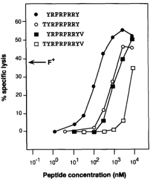
two overlapping nonamers, namely TYRPRPRRY and YRPRPRRYV, were positive. We then synthesized the oc- tameric peptide common to the two nonamers and the deca- meric peptide containing both. The four peptides were com- pared for their ability to sensitize HLA-Cw*0601 + EBV lymphoblastoid cells to lysis by CTL 76/6 (Fig. 6). Octamer YRPRPRRY was found to be the optimal peptide. Half max- imal lysis was obtained at a peptide concentration of ,o100 nM. The two nonamers were one order of magnitude less efficient and the decamer was even less efficient. Another anti-MZ2-F CTL clone derived from a different blood sample of patient MZ2 also lysed HLA-Cw*0601 + lymphoblastoid cells pulsed with peptide YKPRPRKY (data not shown). The gene GAGE-2 codes for the same antigenic peptide
as GAGE-l, but the homologous peptides encoded by GAGE-
3-6 have tryptophan instead of arginine in position 2 (Fig. 5). When transfected into COS-7 cells with the HLA-
Cw*060I cDNA, only GAGE-1 and GAGE-2 cDNA were
able to confer recognition by CTL 76/6, showing that the arginine in position 2 is an essential element of the MZ2-F antigenic peptide (Fig. 7). 9 of the 21 G A G E cDNA were either GAGE-1 or GAGE-2 sequences.
Expression of G A G E Genes. The expression of the G A G E
genes was tested in a panel of normal tissues by reverse tran- scription PCR (RT-PCK) 1 using primers common to the six GAGE sequences. We found no expression in any normal adult tissue except testis (Table 1). With another set of primers that amplified only GAGE-I and GAGE.2, we found that a significant proportion of tumors of various histological types express at least one of these genes (Table 1). The highest proportions of positive tumors were found among sarcomas (25%), melanomas (24%), non-small cell lung carcinomas (19%), head and neck tumors (19%), and testicular semi- nomas (five out of six), but the genes are also expressed in bladder tumors and breast tumors. No expression was found in colorectal carcinomas and renal cell carcinomas. Melanoma line IGR3-MEL, which expresses GAGE-I~2 and HLA-Cw6, triggered TNF release by CTL 76/6 (data not shown) and was lysed by it (Fig. 1).
Discussion
Two members of the G A G E gene family, GAGE-1 and
GAGE-2, code for a tumor-specific antigenic peptide presented
to CTL by HLA-Cw*0601. Previous results obtained with mouse tumors revealed two mechanisms that can produce such antigens. One is the occurrence of a point mutation (35-37); the other is the activation in the tumor of a gene that is silent in normal cells (16). For GAGE, all our evi- dence supports the second mechanism. G A G E genes are ex- pressed in a number of tumors, but not in normal tissues, except the testis. Furthermore, the fact that two distinct
' Abbreviation used i, this paper: RT-PCR, reverse transcription PCR.
9 Y R P R P R R Y
60 O T Y R P R P R R Y
9 Y R P R P R R Y V
50 [] T Y R P R P R R Y V
40 ( F +
, ~ ~
I I I I I I
10 "1 100 101 102 103 104
Peptlde conoentratlon (nM)
Figure 6. Lysis by CTL 76/6 of HLA-Cw*0601 + cells pulsed with
GAGE-encoded peptides. Chromium-hbded HLA-Cw*0601 + EBV-
transformed lymphoblastoid cells (LB33-EBV) were pulsed 30 min with the indicated peptides at various concentrations before addition of CTL
76/6 at an E/T ratio of 10. Chromium release was measured after 4 h.
The arrow indicates the level of lysis of MZ2.MEL cells (F +) at the same E/T ratio. Similar results were obtained when autologous lymphoblastoid cells MZ2-EBV were used as peptide-pulsed target cells.
members of the GAGE gene family code for this antigen rules out the possibility that the antigen appeared by mutation. Because of its specific expression in tumors, the GAGE an- tigen may constitute a useful target for specific cancer im- munotherapy. The expression of G A G E genes in testis, how- ever, raises the issue of undesirable auto-immune effects. The
Stimulator ceils MZ2-MEL
MZ2-MELF" I COS + HLA-Cw6
COS + HLA-Cw6 + GAGE-1
COS + HLA-Cw6 + GAGE-2
COS + HLA-Cw6 + GAGE-3
COS + HLA-Cw6 + GAGE-4 COS + HLA-Cw6 + GAGE-8
COS + HLA-Gw6 + GAGE-6
TNF released by CTL 76/6 (pg/ml)
Figure 7. Transfection of the GAGE.J-6 cDNA into COS cells. The
GAGE eDNA were transiently cotransfected into COS-7 cells with the
HLA.Cw*0601 cDNA. CTL 76/6 was added after 24 h and the produc-
tion of TNF was measured 18 h hter. Control stimulator cells included autologous MZ2-MEL and variant MZ2-MEL.F-, which was selected in vitro for resistance to CTL 76/6.
693 Van den Eynde
on April 1, 2017
Table 1. Expression of G A G E Genes by Normal and Tumoral Tissues
Normal tissues
Histological type
Expression of
GAGE.I-6*
Tumors
Histological type
Number of tumors expressing
GAGE-I-6* GAGE.l~2 s
Adult tissues Adrenal gland Benign naevus Bone marrow Brain Breast Cerebellum Colon Heart Kidney Liver Lung Melanocytes Muscle Ovary Prostate Skin Splenocytes Stomach Testis Thymocytes Urinary bladder Uterus
Fetal tissues I Fibroblasts Brain Liver Spleen Thymus
Testis +
- Tumor samples
- Melanomas primary lesions
- metastases
- Sarcomas
- Lung carcinomas (NSCLCII
- Bladder tumors superficial
- infiltrating
- Head and neck tumors
- Mammary carcinomas
- Testicular seminomas
- Prostatic carcinomas
- Colorectal carcinomas
- Leukemias and Lymphomas
- Renal carcinomas
- Tumor cell lines
+
- Melanomas
- Sarcomas
- Lung carcinomas NSCLC II
SCLC** Mesotheliomas
Head and neck tumors Mammary carcinomas Bladder tumors Colorectal carcinomas Leukemias
Lymphomas Renal carcinomas
5/39 5/39 (13%)
47/130 36/129 (28%)
7/24 6/24 (25%)
18/77 15/77 (19%)
1/36 1/36
10/39 8/39 (21%)
14/59 11/58 (19%)
18/162 14/162 (9%)
6/6 5/6
2/20 2/20
0/43 1/71 0/45
45/74 40/74 (54%)
1/4 1/4
1/2 1/2
7/24 7/24 (29%)
5/19 5/19 (26%)
0/2
1/4 0/4
0/3
5/13 5/13
3/6 1/6
0/6 0/6
* Expression of genes GAGE-I-6 was tested by RT-PCR on total RNA with sense primer VDE43 and antisense primer VDE24 (Fig. 4). These
primers are common to the six GAGE sequences. They are located in different exons and amplify a 201-base product that is not observed when
genomic DNA is tested.
* Expression of genes GAGE-l-6 was tested by RT-PCR on total RNA with sense primer VDE18 and antisense primer VDE24 (Fig. 4). These
primers amplify the six GAGE sequences. They are located in different exons and give a 239-bp product that is not observed when genomic DNA is tested.
s Expression of GAGE-l~2 was tested by RT-PCR on total RNA with sense primer VDE44 and antisense primer VDE24 (Fig. 4). These primers
amplify GAGE-I and GAGE-2, but not GAGE-3-6. They are located in different exons and give a 244-bp product that is not observed when genomic
DNA is tested.
II NSCLC, non-small cell lung carcinoma. I Fetal tissues derived from fetuses older than 20 w.
"" SCLC, small cell lung carcinoma.
on April 1, 2017
testis is an immunoprivileged site, however, and germ-line cells do not express M H C class I molecules and therefore should not express antigens recognized by T cells (38, 39). In male mice, it was possible to generate CTL responses against a tumor antigen encoded by gene PIA, which is also expressed in testis but not in other normal tissues. We observed nei- ther inflammation of testicular tissues nor reduction of fer- tility (Uyttenhove, C., B. Leth6, T. Boon, and B. Van den Eynde, manuscript in preparation). In our view, it is there- fore likely, but not certain, that immunization against the GAGE antigen will not produce autoimmune effects.
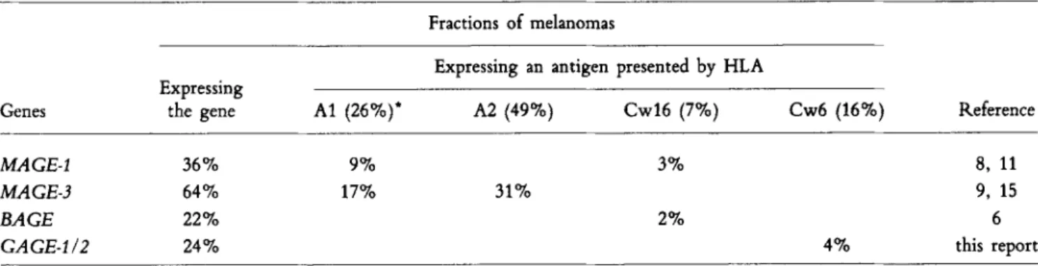
Tumors expressing GAGE-l~2 and HLA-Cw6 can be iden- tified by typing patients for HLA and by testing the expres- sion of GAGE-l~2 by RT-PCK amplification on KNA from a small tumor sample. Because HLA-Cw*0601 is present in 16% of Caucasian individuals and 24% of melanomas ex- press GAGE-I or GAGE-2, ,,04% of all melanomas should express this antigen. This brings to "~51% the fraction of Caucasian melanoma patients eligible for immunotherapy directed against defined tumor antigens encoded by genes
MAGE-1, MAGE-3, BAGE, or GAGE (Table 2). The histo- logical distribution of GAGE-positive tumors is rather similar to that of MAGE-1- or MAGE-3-positive tumors. In view of the very high incidence of non-small cell lung cancer, it is noteworthy that 49% of these cancers express at least one of the MAGE-I, MAGE-3, BAGE, GAGE-l, or GAGE-2
genes (40). Accordingly, patients with lung cancer represent the largest cohort of patients that could benefit from spe- cific immunotherapy directed against antigens encoded by these genes.
Melanoma lines studied in vitro were found to simultane- ously express several antigens recognized by CTL (20, 41, 42). Many tumors expressing GAGE-l~2 also express MAGE- 1, MAGE-3, or BAGE (data not shown). Some of the pa- tients bearing such tumors could therefore be immunized simultaneously against several antigens encoded by these genes. This might ensure a more effective tumor rejection response.
It should also reduce the emergence of antigen loss variants arising by loss of antigen expression, since it is unlikely that tumor cells could simultaneously delete or mutate several of these genes or lose their expression. Nevertheless, the simul- taneous loss of several antigens could still occur after the loss of M H C class I molecules. Fortunately, MHC-negative vari- ants appear to be highly sensitive to N K cells, which may eliminate these variants (43, 44). In support of this notion, Levitsky et al. observed the very frequent emergence of MHC- negative variants in immunized mice that had been depleted of NK1 + cells before challenge with tumor cells. These vari- ants were less frequently observed in mice that had not been N K depleted (45).
To the best of our knowledge, the GAGE peptide is the first antigenic peptide presented by HLA-Cw6 that has been identified. A consensus motif for binding to HLA-Cw6 was proposed by Falk et al. on the basis of peptide elution studies (46). A dominant residue of this motif was L at position 9. Strong residues were P in position 4, I or L in position 5, and V, I, or L in position 6. Our peptide does not fit this motif. A first difference is the presence of tyrosine instead of leucine at the C O O H terminus of the GAGE peptide. Al- though tyrosine was also detected at position 9 in the pool of eluted peptides, it was not considered a dominant or strong residue. Another difference is the fact that the GAGE pep- tide that is most effective in vitro is an octamer rather than a nonamer, but this does not prove that the octamer is the natural peptide. The cells used by Falk et al. (46) for the pep- tide elution expressed the HLA-Cw*0602 allele, which was first reported to be distinct from Cw*0601 (47). However, after correction of sequence uncertainties, the two Cw6 se- quences proved identical (48). In any case, allelic diversity could not explain the divergence of the GAGE peptide from the proposed motif because the cells used by Falk et al. (46) can present the GAGE peptide to CTL 76/6 (data not shown). Among the class I binding antigenic peptides that have so far been identified in humans, most are presented by HLA-A
Table 2. Proportion of Melanoraas Expressing Antigens Encoded by the MAGE, BAGE, or GAGE genes
Fractions of melanomas
Expressing an antigen presented by HLA Expressing
Genes the gene A1 (26%)* A2 (49%) Cw16 (7%) Cw6 (16%) Reference
MAGE-1 36% 9% 3% 8, 11
MAGE-3 64% 17% 31% 9, 15
BAGE 22% 2% 6
GAGE-l~2 24% 4% this report
Corrected* total of melanomas expressing at least one antigen: 51%
" The frequency of each HLA specificity in Caucasians is indicated in parentheses.
After correction for melanomas expressing both HLA-A1 and HLA-A2, and for melanomas expressing both MAGE-1 and MAGE-3.
695 Van den Eynde
on April 1, 2017
or HLA-B molecules, and only very few by HLA-C (49-52). Since surface expression of HLA-C molecules was reported to be lower than that of HLA-A and HLA-B, this led to the suggestion that HLA-C molecules do not contribute significandy to antigen presentation (53). However, in MZ2- MEL melanoma cells, we have observed that among five an- tigens recognized by autologous CTL, three are presented by HLA-C molecules, and both HLA-C alleles are involved: HLA-Cw*1601 presents a peptide derived from MAGE-1 and another derived from BAGE, whereas HLA-Cw*0601 presents
a GAGE-encoded antigen (6, 11).
Like MAGE and RAGE, the GAGE genes form a family of very closely related genes. The M A G E family is made up of 12 genes, none of which are expressed in normal adult tissues besides the testis (6, 34). In addition to these genes
located in the q27-qter region of the X chromosome, several additional related genes are located in the p21.3 region of the same chromosome (54, 55). Hydrophobic cluster anal- ),sis of the proteins encoded by the different MAGE genes showed a remarkable conservation of the main hydrophobic regions, suggesting conservation of function of these pro- teins. Higher variation was observed in the promoter region of the MAGE genes, and this led to the suggestion that dupli- cation of a MAGE gene into a large gene family placed the same function under different transcriptional controls, pos- sibly to allow it to occur at a number of very specific times and locations (34). The apparent absence of expression in adult somatic tissues and in fetuses older than 20 wk raises the possibility that MAGE, BAGE, and GAGE gene products play a role during early stages of embryonic development.
The excellent technical assistance of Miss Anne Authom is gratefully acknowledged. We thank Dr. F. Brasseur and Mrs. M. Swinarska for preparation of KNA. We also appreciated helpful discussion with Drs. P. van der Bruggen, P. Coulie, and E. de Plaen. We also thank Saida Khaoulali for her invaluable help in the preparation of the manuscript.
This work was partially supported by the Fonds J. Maisin (Belgium), by the Caisse Grnrrale d'Epargne et de Retraite-Assurances (Belgium), and by the Association Contre le Cancer (Belgium). B. Gaugler was supported by an European Economic Community Grant, and O. Peeters was supported by a fellow- ship from the Institut pour l'Encouragement de la Recherche Scientifique dans l'Industrie et l'Agriculture (Belgium). S. Lucas is supported by the Fonds National de la Recherche Scientifique (Belgium). Address correspondence to B. Van den Eynde, Ludwig Institute for Cancer Research, Brussels Branch, 74 Avenue Hippocrate, UCL 74.59, B1200 Brussels, Belgium.
Received for publication 8 March 1995 and in revised form 13
April
1995.References
1. Boon, T., J.-C. Cerottini, B. Van den Eynde, P. van der Bruggen, and A. Van Pel. 1994. Tumor antigens recognized by T lymphocytes. Annu. Rev. lmmunol. 12:337-365. 2. van der Bruggen, P., C. Traversari, P. Chomez, C. Lurquin,
E. De Plaen, B. Van den Eynde, A. Knuth, and T. Boon. 1991. A gene encoding an antigen recognized by cytolytic T lym- phocytes on a human melanoma. Science (Wash. DC). 254: 1643-1647.
3. Brichard, V., A. Van Pel, T. Wrlfel, C. Wrlfel, E. De Plaen, B. Lethe, P. Coulie, and T. Boon. 1993. The tyrosinase gene codes for an antigen recognized by autologous cytolytic T lym- phocytes on HLA-A2 melanomas, f Extx Med. 178:489-495. 4. Coulie, P.G., V. Brichard, A. Van Pel, T. Wrlfel, J. Schneider, C. Traversari, S. Mattei, E. De Plaen, C. Lurquin, J.-P. Szikora, et al. 1994. A new gene coding for a differentiation antigen recognized by autologous cytolytic T lymphocytes on HLA- A2 melanomas. J. Exl~ Med. 180:35-42.
5. Kawakami, Y., S. Eliyahu, C.H. Delgado, P.F. Robbins, L. tLivoltini, S.L. Topalian, T. Miki, and S.A. Rosenberg. 1994. Cloning of the gene coding for a shared human melanoma antigen recognized by autologous T cells infiltrating into tumor.
Proc. Natl. Acad. Sci. USA. 91:3515-3519.
6. Bo~l, P., C. Wildmann, M.-L. Senti, R. Brasseur, J.-C. Renauld, P. Coulie, T. Boon, and P. van der Bruggen. 1995. BAGE, a new gene encoding an antigen recognized on human melanomas by cytolytic T tymphocytes. Immunity. 2:167-175. 7. Wang, R.-F., P.F. Robbins, Y. Kawakami, X.-Q Kang, and S.A. Rosenberg. 1995. Identification of a gene encoding a mela- noma tumor antigen recognized by HLA-A31-restricted tumor- infiltrating lymphocytes. J. Extx Med. 181:799-804. 8. Traversari, C., P. van der Bruggen, I.F. Luescher, C. Lurquin,
P. Chomez, A. Van Pel, E. De Plaen, A. Amar-Costesec, and T. Boon. 1992. A nonapeptide encoded by human gene MAGE-1 is recognized on HLA-A1 by cytolytic T lymphocytes directed against tumor antigen MZ2-E. J. Exlx Med. 176:1453-1457. 9. Gaugler, B., B. Van den Eynde, P. van der Bruggen, P. Romero, J.J. Gaforio, E. De Plaen, B. Lethe, F. Brasseur, and T. Boon. 1994. Human gene MAGE-3 codes for an antigen recognized on a melanoma by autologous cytolytic T lymphocytes.J. Exl~ Med. 179:921-930.
10. Kawakami, Y., S. Eliyahu, K. Sakaguchi, P.F. Robbins, L. Rivoltini, J.R. Yannelli, E. Appella, and S.A. Rosenberg. 1994. Identification of the immunodominant peptides of the MART-1 human melanoma antigen recognized by the majority of HLA-
on April 1, 2017
A2-restricted tumor infiltrating lymphocytes. J. Extx Med.
180:347-352.
11. van der Bruggen, P., J.-P. Szikora, P. Bo~l, C. Wildmann, M. Somville, M. Scnsi, and T. Boon. 1994. Autologous cytolytic T lymphocytes recognize a MAGE-1 nonapeptide on mela- nomas expressing HLA-Cw*1601. Eur. J. Immunol. 24:2134- 2140.
12. W61fel, T., A. Van Pal, V. Brichard, J. Schneider, B. Seliger, K.-H. Meyer zum Bfischenfelde, and T. Boon. 1994. Two tyro- sine nonapeptides recognized on HLA-A2 melanomas by au- tologous cytolytic T lymphocytes. Eur.J. Immunol. 24:759-764. 13. Cox, A.L., J. Skipper, Y. Chen, R.A. Henderson, T.L. Darrow, J. Shabanowitz, V.H. Engelhard, D.F. Hunt, and C.L. Slinghuff, Jr. 1994. Identification of a peptide recognized by five melanoma-specific human cytotoxic T cell lines. Science
(Wash. DC). 264:716-719.
14. CasteUi, C., W.J. Storkus, M.J. Maeurer, D.M. Martin, E.C. Huang, B.N. Pramanik, T.L. Nagabhushan, G. Parmiani, and M.T. Lotze. 1995. Mass spectrometric identification of a natu- rally processed melanoma peptide recoghized by CD8 * cyto- toxic T lymphocytes. J. Exlx Med. 181:363-368.
15. van der Bruggen, P., J. Bastin, T. Gajewski, P.G. Coulie, P. Bo~l, C. De Smet, C. Traversari, A. Townsend, and T. Boon. 1994. A peptide encoded by human gene MAGE-3 and presented by HLA-A2 induces cytolytic T lymphocytes that recognize tumor cells expressing MAGE-3. Eur. J. Immunol.
24:3038-3043.
16. Van den Eynde, B., B. Lethe, A. Van Pel, E. De Plaen, and T. Boon. 1991. The gene coding for a major tumor rejection antigen of tumor P815 is identical to the normal gene of syn- geneic DBA/2 mice. J. Ex F Med. 173:1373-1384.
17. Bakker, A.B.H., M.W.J. Schreurs, A.J. de Boer, Y. Kawakami, S.A. Rosenberg, G.J. Adema, and C.G. Figdor. 1994. Melano- cyte lineage-specific antigen gpl00 is recognized by melanoma- derived tumor-infiltrating lymphocytes. J. Ex F Med. 179: 1005-1009.
18. Robbins, P.F., M. EI-Gamil, Y. Kawakami, and S.A. Rosen- berg. 1994. Recognition of tyrosinase by tumor-infiltrating lym- phocytes from a patient responding to immunotherapy. Cancer
Res. 54:3124-3126.
19. H6rin, M., C. Lemoine, P. Weynants, F. Vessi~re, A. Van Pel, A. Knuth, R. Devos, and T. Boon. 1987. Production of stable cytolytic T-cell clones directed against autologous human meh- noma. Int. J. Cancer. 39:390-396.
20. Van den Eynde, B., P. Hainaut, M. H6rin, A. Knuth, C. Le- moine, P. Weynants, P. van der Bruggen, R. Fauchet, and T. Boon. 1989. Presence on a human melanoma of multiple an- tigens recognized by autologous CTL. Int. J. Cancer. 44: 634-640.
21. Traversari, C., P. van der Bruggen, B. Van den Eynde, P. Hainaut, C. Lemoine, N. Ohta, L. Old, and T. Boon. 1992. Transf~ction and expression of a gene coding for a human mela- noma antigen recognized by autologous cytolytic T lympho- cytes. Immunogenetics. 35:145-152.
22. Carrel, S., R. Toil, N. Gross, N. Tanigaki, A.L. Carmagnola, and R.S. Accolla. 1981. Subsets of human Ia-like molecules defined by monoclonal antibodies. Mol. Immunol. 18:403-411. 23. Boon, T., J. Van Snick, A. Van Pel, C. Uyttenhove, and M. Marchand. 1980. Immunogenic variants obtained by mutagen- esis of mouse mastocytoma P815. II. T lyphocyte-mediated cytolysis. J. EXl~ Med. 152:1184-1193.
24. Ausubel, F.M., R. Brent, R.E. Kingston, D.D. Moore, J.G. Scidman, J.A. Smith, and K. Struhl, editors. 1993. Minipreps
of phsmid DNA. In Current Protocols in Molecular Biology. 2 Vols. John Wiley & Sons, Inc., New York. 1.6.2.-1.6.3. 25. Seed, B., and A. Aruffo. 1987. Molecular cloning of the CD2 antigen, the T-cell erythrocyte receptor, by a rapid immunoselec- tion procedure. Proc Natl. Acad. Sci. USA. 84:3365-3369. 26. Takebe, Y., M. Sciki, J.-I. Fujisawa, P. Hoy, K. Yokota, K.-I.
Arai, M. Yoshida, and N. Arai. 1988. SRc~ promoter: an efficient and versatile mammalian cDNA expression system composed of the simian virus 40 early promoter and the K-U5 segment of human T-cell leukemia virus type I long terminal repeat. Mol. Cell. Biol. 8:466-472.
27. Girdlestone, J. 1990. Nucleotide sequence ofan HLA-A1 gene.
Nucleic Acids Res. 18:6701.
28. Espevik, T., andJ. Nissen-Meyer. 1986. A highly sensitive cell line, WEHI 164 clone 13, for measuring cytotoxic factor/tumor necrosis factor from human monocytes. J. Immunol. Methods.
95:99-105.
29. Hansen, M.B., S.E. Nielsen, and K. Berg. 1989. Re-examination and further development of a precise and rapid dye method for measuring cell growth/ceU kill. J. Immunol. Methods.
119:203-210.
30. Parham, P., C.J. Barnstable, and W.F. Bodmer. 1979. Use of a monoclonal antibody (W6/32) in structural studies of HLA- A,B,C antigens. J. Immunol. 123:342-349.
31. Davis, L.G., M.D. Dibner, and J.F. Battey. 1986. Guanidine isothiocyanate preparation of KNA. In Basic Methods in Mo- lecular Biology. Elsevier Science Publishing Co., Inc., New York. pp. 130-135.
32. Atherton, E., C.J. Logan, and R.C. Sheppard. 1981. Peptide synthesis. Part 2. Procedures for solid phase synthesis using N~-fluroenylmethysoxycarbamylamino-acid on polymide sup- ports. Synthesis of substance P and of acyl carrier protein 65- 74 decapeptide. J. Chem. Soc. Lond. Perkin Trans. 1:538. 33. Karl)a, Y., K. Kato, Y. Ha)ashizaki, S. Himeno, S. Tarui, and
K. Matsubara. 1986. Expression of human gastrin gene in normal and gastrinoma tissues. Gene (Amst.). 50:345-352. 34. De Plaen, E., K. Arden, C. Traversari, J.J. Gaforio, J.-P. Szikora,
C. De Smet, F. Brasseur, P. van der Bruggen, B. Leth6, C. Lurquin et al. 1994. Structure, chromosomal localization and expression of twelve genes of the MAGE family. Immunogenetics.
40:360-369.
35. De Plaen, E., C. Lurquin, A. Van Pel, B. Mariam6, J.-P. Szikora, T. W61fel, C. Sibille, P. Chomez, and T. Boon. 1988. Immuno- genic (tum-) variants of mouse tumor P815: cloning of the gene of tum- antigen P91A and identification of the tum- mutation. Proa Natl. Acad. Sci. USA. 85:2274-2278. 36. Boon, T. 1992. Toward a genetic analysis of tumor rejection
antigens. Adv. Cancer Res. 58:177-210.
37. Mandelboim, O., G. Berke, M. Fridkin, M. Feldman, M. Eisen- stein, and L. Eisenbach. 1994. CTL induction by a tumour- associated antigen octapeptide derived from a murine lung car- cinoma. Nature (Lond.). 369:67-71.
38. Barker, C.F., and K.E. Billingham. 1977. Immunologically privileged sites. Adv. Immunol. 25:1-54.
39. Haas, G.G., Jr., O.J. D'Cruz, and L.E. De Bault. 1988. Distri- bution of human leukocyte antigen-ABC and -D/DR antigens in the unfixed human testis. Am.J. Reprod. Immunol. Microbiol.
18:47-51.
40. Weynants, P., B. Leth6, F. Brasseur, M. Marchand, and T. Boon. 1994. Expression of MAGE genes by non-small-cell lung car- cinomas. Int. J. Cancer. 56:826-829.
41. W61fel, T., E. Klehmann, C. M/iller, K.-H. Schfitt, K.-H. Meyer zum Bfischenfelde, and A. Knuth. 1989. Lysis of human 697 Van den Eynde
on April 1, 2017
melanoma cells by autologous cytolytic T cell clones. Iden- tification of human histocompatibility leukocyte antigen A2 as a restriction element for three different antigens.J. Exl~ Med.
170:797-810.
42. Lehmann, F., M. Marchand, P. Hainaut, P. Pouillart, X. Sastre, H. Ikeda, T. Boon, and P.G. Coulie. 1995. Differences in the antigens recognized by cytolytic T cells on two successive metastases of a melanoma patient are consistent with immune selection. Eur. J. Immunol. 25:340-347.
43. Ljunggren, H.G., N.J. Stam, C. Ohlen, J.J. Neefjes, P. H6glund, M.T. Heemels, J. Bastin, T.N.M. Schumacher, A. Townsend, K. K~rre, and H.L. Ploegh. 1990. Empty MHC class I molecules come out in the cold. Nature (Lond.). 346: 476-480.
44. Glas, g., K. Sturmh6fel, G.J. H~mmerling, K. K~rre, and H.-G. Ljunggren. 1992. Restoration of a tumorigenic pheno- type by 32-microglobulin transfection to EL-4 mutant cells.
J. Exp. Med. 175:843-846.
45. Levitsky, H.I., A. Lazenby, R.J. Hayashi, and D.M. Pardoll. 1994. In vivo priming of two distinct antitumor effector popu- lations: the role of MHC class I expression. J. Exp. Med.
179:1215-1224.
46. Falk, K., O. R6tzschke, B. Grahovac, D. Schendel, S. Steva- novic, V. Gnau, G. Jung, J.L. Strominger, and H.-G. Ram- mensee. 1993. Allele-specific peptide ligand motifs of HLA-C molecules. Proc. Natl. Acad. Sci. USA. 90:12005-12009. 47. Zemmour, J., and P. Parham. 1993. HLA class I nucleotide
sequences, 1992. Immunogenetics. 37:239-250.
48. Vilches, C. R. de Pablo, M.J. Herrero, M.E. Moreno, and M. Kreisler. 1993. Molecular cloning and polymerase chain reac- tion-sequence-specific oligonucleotide detection of the allele encoding the novel allospecificity HLA-Cw6.2 (Cw'1502) in
Spanish gypsies. Hum. Immunol. 37:259-263.
49. Littaua, R.A., M.B.A. Oldstone, A. Takeda, C. Debouck, J.T. Wong, C.U. Tuazon, B. Moss, F. Kievits, and F.A. Ennis. 1991. An HLA-C-restricted CD8 § cytotoxic T-lymphocyte clone recognizes a highly conserved epitope on human immuno- deficiency virus type 1 gag. J. Virol. 65:4051-4056. 50. Schendel, D.J., C. Reinhardt, P.J. Nelson, B. Maget, L. Pullen,
G.W. Bornkamm, and A.E. Steinle. 1992. Cytotoxic T lym- phocytes show HLA-C-restricted recognition of EBV-bearing cells and allorecognition of HLA class I molecules presenting self-peptides. J. lmmunol. 149:2406-2414.
51. Johnson, R.P., A. Trocha, T.M. Buchanan, and B.D. Walker. 1993. Recognition of a highly conserved region of human im- munodeficiency virus type I gp120 by an HLA-Cw4-restricted cytotoxic T-lymphocyte clone. J. Virol. 67:438-445. 52. Engelhard, V.H. 1994. Structure of peptides associated with
MHC class I molecules. Cuw. Opin. Immunol. 6:13-23. 53. Neefjes, J.J., and H.L. Ploegh. 1988. Allele and locus-specific
differences in cell surface expression and the association of HLA class I heavy chain with 32-microglobulin: differential effects of inhibition of glycosylation on class I subunit association.
Eur. j. Immunol. 18:801-810.
54. Oaks, M.K., J.P. Hanson, Jr., and D.P. O'Malley. 1994. Mo- lecular cytogenetic mapping of the human melanoma antigen
(MAGE) gene family to chromosome region Xq27-qter: im-
plications for MAGE immunotherapy. Cancer Res. 54:1627- 1629.
55. Muscatelli, F., A.P. Walker, E. De Plaen, A.N. Stafford, and A.P. Monaco. 1995. Isolation and characterisation of a new MAGE gene family in the Xp21.3 region. Proc. Natl. Acad. Sci.
USA. 92:4987-4991.
on April 1, 2017