Dr Aradhana Sahoo et al JMSCR Volume 07 Issue 05 May 2019 Page 207
Original Research Article
Serum Magnesium Status among Type 2 Diabetes Mellitus and Its Chronic
Complications
Authors
Dr Aradhana Sahoo
1, Dr Butungeshwar Pradhan
2*
1
Senor Resident, Department of Medicine, VIMSAR, Burla, Sambalpur, Odisha, India
2
Associate Professor, Department of Medicine, VIMSAR, Burla, Sambalpur, Odisha, India *Corresponding Author
Dr Butungeshwar Pradhan
Department of Medicine, VIMSAR, Burla, Sambalpur, Odisha, India Mobile No.: 9437243697, Email: butungeshwar@gmail.com
Abstract
Background: Magnesium is a necessary cofactor of several enzymes involved in glucose metabolism. Insulin resistance or deficiency may exacerbate renal magnesium wasting and hyperglycemia per se induces higher osmotic urinary excretion of magnesium. High prevalence of hypomagnesaemia in T2DM and its chronic complications have been reported in different studies. Hence this study was undertaken to know the incidence of hypomagnesaemia in T2DM patients and its chronic complications.
Material and Methods: In consecutive 100 T2DM patients and 100 control healthy people, serum magnesium was estimated by Calmagite dye Caloric method at one time contact. Opthalmoscopy and 24 hours total urinary protein excretion was measured in T2DM patients. Data were collected and compared by Chi-square test and comparison of mean values were performed by unpaired student ‘t’ test. All statistical data were analysed by SPSS version 16 soft ware for windows.
Results: There was no significant difference in incidence of hypomagnesaemia with age and sex (p>0.05). Hypomagnesaemia was present in 66 (66%) T2DM patients and 8(8%) in healthy people. (p<0.05). Serum magnesium levels were inversely related to FBS, 2hr PPBS and HbA1c levels (p<0.05). Total cholesterol, TG and LDL were inversely related to magnesium level and HDL was positively related (p<0.05). T2DM patients with hypomagnesaemia had higher incidence of proteinuria in 66(99.9%) and diabetic retinopathy 53(80.3%). (p <0.05).
Conclusion: Hypomagnesaemia was common in T2DM patients and its chronic complications.
Keywords: Hypomagnesaemia, T2DM, Glycemic status, Retinopathy, Albuminuria, Dyslipidimia.
Introduction
Magnesium (Mg) is the fourth most abundant mineral present in human body and the second intracellular cation in living cells after potassium. Most Mg is intracellular (99%) and only 1% is
extracellular. Magnesium is necessary as a cofactor of several enzymes that play important roles in glucose metabolism involved in multiple levels, such as in insulin secretion, binding and activity. Cellular magnesium deficiency can alter the
www.jmscr.igmpublication.org Index Copernicus Value: 79.54
ISSN (e)-2347-176x ISSN (p) 2455-0450
Dr Aradhana Sahoo et al JMSCR Volume 07 Issue 05 May 2019 Page 208
membrane bound sodium-potassium-adenosine triphosphatase which is involved in the maintenance of sodium-potassium gradients and glucose transport.¹ In diabetics there is direct relationship between serum magnesium level and cellular glucose disposal that is independent of insulin secretion. This change in glucose disposal has been related to increased sensitivity of the tissues to insulin in the presence of adequate magnesium levels.² The link between Mg deficiency and type 2 diabetes mellitus (T2DM) is well known. T2DM is frequently associated with both intracellular and extracellular Mg depletion.³ Mg deficit as a possible unifying mechanism associated with insulin resistance, including T2DM, metabolic syndrome, and hypertension. Mg deficiency could precede and cause post- receptorial resistance of insulin action and alter the glucose metabolism.⁴Insulin enhances Mg re-absorption at the thick ascending limb (TAL) and distal convoluted tubules (DCT) of renal tubules.⁵
Preclinical hypomagnesaemia is considered with serum Mg level of ≤ 0.75mmol/L (1.8mg/dl) and frank hypomagnesaemia with Mg level ≤ 0.61mmol/L (1.5mg/dl), indicative of systemic Mg deficit. Depletion of intracellular and ionized Mg can be found in many subjects with total serum Mg still in the normal range due to lack of sensitivity of total serum Mg measurement, thus measurement of ionized Mg can help identify low concentration of blood Mg.⁶ Hypomagnesaemia reliably indicates magnesium deficiency, but its absence does not exclude significant magnesium depletion.⁷
The causes of hypomagnesaemia in T2DM are multifactorial. Insulin resistance or deficiency may exacerbate renal Mg wasting and hyperglycaemia per se induce glycosuria causes higher osmotic urinary excretion of Mg as well as recurrent metabolic acidosis and diabetic ketoacidosis (DKA) and hypoalbuminimic state decreases Mg binding.⁸’⁹ Hypomagnesaemia has been implicated in T2DM and its chronic complications. In a recent study it was found that T2DM with hypomagnesaemic nephropathy had 2.12 fold increased risk for progression to end state renal
disease (ESRD).¹⁰ There is high prevalence of hypomagnesaemia in subjects with T2DM and in different studies it varies from 13.5% to 47.7% .¹¹ In India it has been reported from 6% to 11.3%.¹²’¹³ Hence this study was conducted to know the incidence of hypomagnesaemia in T2DM patients and to correlated with glycaemic status, dyslipidimia and its chronic complications such as diabetic retinopathy and nephropathy.
Aims and Objectives
The primary aim the study was to find out incidence of hypomagnesaemia in T2DM patients in comparison to non-diabetic healthy controls and the secondary aim was to correlate the magnesium level with different glycaemia status among T2DM patients and its different chronic complications.
Methods
Dr Aradhana Sahoo et al JMSCR Volume 07 Issue 05 May 2019 Page 209
reference normal level of serum magnesium using this method was 1.7-2.4 mg/dl. Hypomagnesaemia was considered when serum Mg level was <1.7mg/dl. Data were collected and all the data were entered in a predesigned Microsoft Excel and statistical analysis was performed and the categorical data were compared by the chi-square test and the comparison of mean value were performed by unpaired student–‘t’ test. P value of <0.05 was considered to be statistically significant. All the statistical data was analysed by SPSS version 16 software for windows.
Results
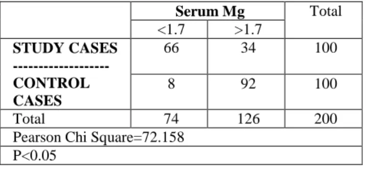
Maximum numbers of cases were in between age of 40-60 years (66%).The mean age of the diabetic patients was between 55.42±12.65 years and it was 55.58±12.84 years in the control group. They were 59% and 41% male and females in diabetes group and 61% and 39% males and females respectively in control group. Both cases were divided into, hypomagnesaemia group (serum Mg <1.7 mg/dl) and (serum Mg >1.7 mg/dl) as normal serum magnesium group. Hypomagnesaemia was present in 66 (66%) diabetic cases and 8(8%) in control group. In the normomagnesaemia level there were 34(34%) diabetic and 92(92%) in control group. (Table 1.).
Table 1: Serum Magnesium in Relation to Cases and Controls
Serum Mg Total
<1.7 >1.7
STUDY CASES ---CONTROL CASES
66 34 100
8 92 100
Total 74 126 200
Pearson Chi Square=72.158 P<0.05
The mean serum magnesium level in diabetic and non-diabetic was 1.67±0.36 mg/dl and 2.03 ±0.40 mg/dl respectively. Hypomagnesaemia was statistically significant with T2DM. (p<0.05).
In the hypomagnesaemia group 2 (3%) cases were less than 40 years, 46 (69.6%) cases were between 40-60 years and 18 (27.2%) cases were > 60 years. In the normomagnesaemia group 2 (5.8%) cases
were ≤ 40 years, 20 (58.8%) cases were between 40-60 years and 12 (35.2%) cases were > 60 years. Majority of patients were > 40 years of age. Thus, hypomagnesaemia was not significant with age. (p>0.05).There were 41(41%) males and 59(59%) female cases. The mean serum magnesium in males and females were 1.719±0.38 and 1.595±0.34 respectively. There was no significant difference in incidence of hypomagnesaemia with sex. (p>0.05). In this study the mean + SD of serum magnesium level was 1.6±0.36 and the mean + SD of FBS was 230.100±88.03.The value of serum magnesium was inversely related to FBS. There was a statistically significant fall in serum magnesium levels with higher levels of FBS. (p<0.05).There were 6 (9%) cases in the hypomagnesaemia group with 2 hr PPBS < 200mg/dl, and 60 (90.9%) cases with PPBS ≥ 200mg/dl. There were 8 (23.5%) cases with PPBS <200mg/dl and 26 (76.4%) cases with > 200mg/dl in normomagnesaemia. (Table.2.) Thus, higher 2 hr PPBS was statistically significantly associated with low serum magnesium levels. (p<0.05).
Table 2: Serum mg Levels in Relation to 2 hour PPBS
Serum Mg(mg/dl) Total <1.7 >1.7
2 hr PPBS (mg/dl)
<200 6 8 14
>200 60 26 86
Total 66 34 100
Pearson Chi-Square=3.885 P value<0.05
According to HbA1c the hypomagnesaemic T2DM cases were divided into 3 groups, HbA1c <7% (good control), HbA1c between 7-9 % (moderately control) and HbA1c >9 %( poorly control). In 24 (36.3%) patients HbA1C was 7-9% and in 42
(63.6%) cases HbA1c was >9%. In the normal
serum magnesium group 22 (64.7%) patients had HbA1C <7%, and 6 (17.6%) had HbA1c 7-9% and 6
(17.6%) had HbA1C >9%. (Table.3). There was
Dr Aradhana Sahoo et al JMSCR Volume 07 Issue 05 May 2019 Page 210
Table 3 Serum Mg Levels in Relation to HbA1c (%)
Serum Mg(mg/dl)
Total
<1.7 >1.7
HbA1c (%) <7 0 22 22
7-9 24 6 30
>9 42 6 48
Total 66 34 100
Pearson chi-square(x²)=72.158 P value<0.05
In the hypomagnesaemia group 14 (21.2%) cases had normal serum Triglyceride (TG) levels ≤150Mg/dl, whereas 52 (78.7%) patients had serum TG ≥ 150mg/dl. In the normal serum magnesium level 32 (94.1%) cases had normal serum TG <150mg/dl and only 2 (5.8%) cases had ≥ 150mg/dl. Serum TG level was inversely correlated with serum magnesium levels which is statistically significant (p<0.05).
The mean ± SD values of high density lipoprotein (HDL), low density lipoprotein (LDL) and total cholesterol (TC) were 43.08±9.1 mg/dl,153.68±51.6 mg/dl and 222.78±13.99 respectively. The mean Mg level was 1.66±0.36 mg/dl. Serum LDL and TC was negative correlated and serum HDL was positive correlated with serum Mg levels. (p<0.05). In the hypomagnesaemia cases, 20(30.3%) had microalbuminuria (<300 mg/dl) and 46 (69.6%) had macroalbuminuria (>300 mg/dl). With normal serum Mg levels 33(97%) cases had microalbuminuria and 1(3%) case had macroalbuminuria. (Table 4). Thus higher 24 hour urinary protein excretion was statistically significant with low serum Mg. (p<0.05).cases.
Table 4: Serum Magnesium in Relation to 24 hour Urinaria Proteinuria
24 Hrs Urinary Protein
Total
<300 >300
Serum Mg
<1.7 20 46 66
>1.7 33 1 34
Total 53 47 100
Pearson Chi-Square=40.145 P<0.05
In T2DM patients with normal serum Mg levels, retinopathy was present in 10 (29.4%) cases absent in 24(70.6%) cases.(Table.5. In hypomagnesaemic
cases retinopathy was present in 53 (80.3%) cases and absent in 13 (19.7%) cases. Thus the prevalence of retinopathy is statistically significant with low serum Mg.
Table 5 Serum Mg Levels in Relation to Retinopathy
Serum Mg(mg/dl)
Total
<1.7 >1.7
Retinopathy
Absent 13 24 37
Present 53 10 63
Total 66 34 100
Pearson chi-square(x²)=24.99 P<0.05
Fifty (50%), 32 (32%) and 18(18%) patients were on insulin, oral antidiabetic drugs (OADs) alone and on both insulin + OADs respectively. In hypomagnesaemic cases 40 (60.6%), 8 (12.1%) and 18 (27.2%) cases were on insulin alone, OAD alone and combination of OADs+ insulin respectively. In the normal serum magnesium levels 10 (29.4%) cases were on insulin and 24 (70.6%) were on OADs. The mean serum Mg level in the OADs group, insulin + OADs and insulin alone group was 2.02mg/dl, 1.59 mg/dl and 1.25 mg/dl respectively. The serum Mg levels were significantly lower in the insulin required group compared to the OADs treated group. (p<0.05).
Discussion
Type 2 DM is characterized by insulin resistance and relative insulin deficiency. Magnesium is known to play important role in carbohydrate metabolism and its deficiency has been implicated in diabetes mellitus, as a cause and consequences.¹⁴
Dr Aradhana Sahoo et al JMSCR Volume 07 Issue 05 May 2019 Page 211
The average age of the cases and control was 55.4 and 55.6 years respectively. Maximum number of patients i.e. 66 (66%) cases of T2DM patients had serum Mg (<1.7mg/dl) which is statistically significant. In 1979 Mather HM et al, established a prevalence of 25% hypomagnesaemia in diabetics.¹⁷
In 2002 Guerrero-Romero et al demonstrated 65.6% prevalence of hypomagnesaemia in diabetes mellitus.¹⁸ In 2006 Corica et al reported 49.3% prevalence of hypomagnesaemia in type 2 DM patients.¹⁹ In 2007 Phuong-chi T e al, reported incidence of hypomagnesaemia in 13.5-47.7% of T2DM patients. ²⁰In 2008 Berhane Seyoum et al demonstrated incidence of 25-39% in diabetics.²¹In this study incidence of hypomagnesaemia was similar to the findings of other workers and statistically significant. The cause of hypomagnesaemia in diabetes mellitus is still unclear.²² Magnesium deficiency in experimental animals has been found to cause alteration in blood lipid composition. Magnesium depletion has atherogenic property and Mg deficiency triggers vasoconstriction, enhances vascular endothelial cell injury leading to atherosclerosis by promoting inflammation and oxidative stress.²³ In this study higher serum TG was statistically significant with low serum magnesium levels and there was an inverse correlation between serum Mg and serum LDL and TC level and a positive correlation between serum Mg and serum HDL. In 2006 Corica et all demonstrated the prevalence of dyslipidemia in diabetics with hypomagnesaemia.¹⁹ In 2002 Guerrero-Romero et al reported the components of metabolic syndrome with hypomagnesaemia. Inverse correlation of serum Mg with serum TG, LDL, and TC was also obtained by Mishra S et al.¹⁸
Glycosylated haemoglobin (HbA1c) levels correlate well with glycaemic levels over a period of 6-10 weeks. ²⁴ In 1998 Diamon M et all reported significant higher level of HbA1c in patients having low serum Mg.²⁵ In 2009 Sikaris K et al reported the increased level of HbA1c in type 2 DM patients with hypomagnesaemia.²⁶In our study statistically significant higher HbA1c levels was seen in hypomagnesaemia patients.
In a study conducted by Corsonello et al, diabetic subjects with microalbuminuria or clinical proteinuria showed a significant decrease in serum ionized magnesium in comparison to normal albuminuria patients. ²³ In our study serum Mg had inverse correlation with 24 hour urinary protein excretion which was statistically significant. Hypomagnesaemia and albuminuria individually or in conjunction serve as indicators for dysglycaemia and could be used as marker for the risk of development of diabetic nephropathy.²⁷
Our study also showed a statistically significant association of hypomagnesaemia with diabetic retinopathy. Nadler JL et al (1992) evaluated intracellular (erythrocyte) Mg concentration in diabetics which was significantly reduced as compared to healthy controls and studied the effects of intravenous 3 hour Mg drip or 8 weeks of 400mg /day oral Mg supplementation on intracellular Mg levels and platelet reactivity.²⁸’²⁹ Oral Mg restored RBC magnesium concentration to normal. Both intravenous and oral Mg supplementations markedly reduce platelet reactivity in response to thromboxane A2 analogue U46619.²⁸’³⁰’³¹ and they suggested oral Mg may ameliorate Mg deficiency or prophylactic oral Mg may help avoid or ameliorate complications associated with Mg deficiency such as arrhythmias, hypertension and sudden cardiac death and may improve the course of diabetes.²⁸ In our study the Insulin requiring T2DM patients were more hypomagnesaemic than controlled with OADs treatment, suggesting severe insulin deficiency associated with hypomagnesaemia.
Conclusions
Dr Aradhana Sahoo et al JMSCR Volume 07 Issue 05 May 2019 Page 212
potential in control and prevention of complications of type 2 DM. Therefore, study of magnesium supplementation for treatment of T2DM and prevention of its complications needs further study.
Acknowledgements
We are very much thankful to participant patients and the control volunteers who cooperated in the study and the laboratory technicians who helped in performing the different investigations in the Regional Diagnostic Centre, VIMSAR, Burla.
Declarations
Funding: Nil
Conflict of interest: None.
Ethical approval: Regdn, No.
ECR/861/Inst/OR/2016.VIREC Decision No.2016/I-F-CT-01/031.
References
1. Paolisso G, Scheen A, D’ Onfrio F, Lefebvre
P. Magnesium and glucose.
Diabetologia .1990; 33: 511-514.
2. Yajnik CS, Smith RF, Hockaday TD, Ward NI. Fasting plasma magnesium concentrations and glucose disposal in diabetes. BMJ. 1984; 288:1032-1034.
3. Mario Barbagallo, Ligia J, Dominguez L J. Magnesium and type 2 diabetes. An update. Diabetes and Cli Res.2015; Jan 22, 2:1-5. 4. Barbagallo M, Dominguez LJ, Galatto A,
Ferlisi A,Cani C et al. Role of magnesium in insulin action, diabetes and cardiometabolic syndrome X. Mol Aspect Med.2003;24:39-52.
5. Madon B, Siga E, Chabardes D, Firsov D, Roenel N, De Rouffignac C. Insulin stimulates Na+,Cl-,Ca++,and Mg++ transport in TAL of mouse nephron. Cross potentiating with AVP .Am J Physiol.1993:F361-369.
6. Barbagallo M, Di Bella G, Brucato V,D Angelo, Domiano P, et al. Serum ionized magnesium in diabetes older person.Metabolism,2014;63:502-509.
7. Grubbs RD (2002) Intracellular magnesium and magnesium buffering. Biometals 15, 251-259.
8. Quamme GA. Renal handling of magnesium .I. Massry and Glossack’s text book of nepgrology.4th edn .Edited by Massry SH, Glossack RJ, Baltimore, Lipincott, Williams and Wilkins:2001;p-344-350.
9. Barbagallo M, Dominguez LJ. Magnesium metabolism in type 2diabetes mellitus, metabolic syndrome and insulin resistance. Arch Biochem Biophys.2007; 458:40-47. 10.Sakaguichi Y, Shoji T, Hayashi T, Sujuki A,
Shimizu M, Mitsumoto K, Tsubakihara Y. Hypomagnesaemia in type diabetes mellitus nephropathy; a novel predictor of end stage renal disease: Diabetes Care.2012, July; 1591-1597.
11.Pham PC, Pham SV, Miller JM, Pham PT. Hypomagnesaemia in patient with type 2 diabetes. Cli J Am Soc Nephrol.2007; 1:366-73.
12.Pramod P Rao, Mohmad Ghouse Shariff. Serum magnesium level in type 2 diabetes patients with microalbuminuria and normoalbuminuria. Int J Seintific study.July 2015; 3:4:11-15.
13.Arundhati Dasgupta, Dipti Sarma, UmaKaimulsaikia. Hypomagnesaemia in type 2 diabetes mellitus.Indian J Endocr Metab.2012; 16:6.p-100-3.
14.Sjogren A, Floren CH, Nilsson A. Magnesium, potassium and zinc deficiency in subjects with type II diabetes mellitus. Acta Med Scand 1988; 224:461–6.
15.Nadler JL, Buchanan T, Natarajan R, Antonipillai I, Bergman R, Rude R. Magnesium deficiency produces insulin resistance and increased thromboxane synthesis. Hypertension 1993; 21: 1024-1029.
Dr Aradhana Sahoo et al JMSCR Volume 07 Issue 05 May 2019 Page 213
[ME Shils, JEOlson, M Shike and AC Ross, editors]. Baltimore: Williams & Wilkins. 17.Mather HM, Nisbet JA, Burton GH, Poston
GJ, Bland JM, Bailey PA, Pilkington TR. Hypomagnesaemia in Diabetes. Clin Chim Acta 1979; 95(2):235-24.
18.Rodriguez-Moran M & Guerrero-Romero F (2002) Low serum magnesium levels and foot ulcers in subjects with type 2 diabetes. Arch Med Res 32, 300-303.
19.Corica F, Allegra A, Di Benedetto A, Giacobbe MS, Romano G, Cucinotta D, et al. Effects of oral magnesium supplementation on plasma lipid concentrations in patients with non insulin dependent diabetes mellitus. Magnes-Res1994; 7:43-47.
20.Phuong-Chi T. Pham, Son V. Pham Jeffery M, Hypomagnesaemia in patients with Type 2 Diabetes, Clinical Journal of Nephrology2:366-373, 2007.
21.Berhane Seyoum, MD, MPH; Elias S. Siraj, MD, Hypomagnesaemia in Ethiopians with diabetes mellitus, Ethnicity & Disease, Volume18, Spring 2008.
22.Clinical Nephrology (2005), June; 63(6):429-436.
23.Corsenello A,Lentile R, Buei M, Cucinatto D, Mouro VN, Macaione S, Corcia F. Serum ionized magnesium levels in type 2 diabetes mellitus nephropathy;a novel predictor of end stage renal disease.Diabetes Care.2012,July;1591-1597.
24.Reinhart RA (1988) Magnesium metabolism. A review with special reference to the relationship between intracellular content and serum levels. Arch Intern Med 148, 2415-2420.
25.Diamon M,Susa S.Yamatani K, Manaka H. Hyperglycemia is a factor for increase in serum ceruloplasmin in type 2 diabetes, Diabetes care1998;21(9):1525-1528.
26.Sikaris K,The correlation of HbA1c to blood glucose, Journal of Diabetes Science and Technology2009;3(3):429-438.
27.Haenni A, Ohrvall M, Lithel H. Magnesium Homeostasis, Metabolism. 2001; 50:1147-51. 28.Nadler JL, Malayan S, Luong H, Shaw S,
Natarajan RD & Rude RK (1992) Intracellular free magnesium deficiency plays a key role in increased platelet reactivity in type II diabetes mellitus. Diabetes Care. 15, 835-841.
29.Nadler JL & Rude RK (1995) Disorders of magnesium metabolism. Endocrinol Metab Clin North Am24, 623-641.
30.P. K. Chandie Shaw, L. A. van Es, L. C. Paul, F. R. Roosendaal, J. H. M. Souverijn, J. P. Vandenbroucke. Renal disease in relatives of Indo-Asian Type 2 diabetic patients with end-stage diabetic nephropathy. Diabetologia (2003) 46:618-624.
31.Lal J, Vasudev K, Kela AK, Jain SK .Effect of oral magnesium supplementation on the lipid profile and blood glucose of patients with type 2 diabetes mellitus. J Assoc Physicians India 2003; 51: 37-42.